

Principles of LA-ICP-MS Imaging
Laser ablation inductively coupled plasma mass spectrometry (LA-ICP-MS) imaging occupies a unique niche in spatial biology: it is the only technique that maps endogenous and exogenous metals, metalloids, and selected nonmetals across whole tissue sections with subcellular resolution and parts-per-billion sensitivity — without labels, without antibodies, and without matrix application. Where MALDI-MSI detects lipids and metabolites and immunohistochemistry maps proteins, LA-ICP-MS answers the orthogonal question: where are the iron, copper, zinc, platinum, cadmium, and arsenic atoms? For a broader overview of how mass spectrometry imaging maps molecular distributions in tissue, see our spatial metabolomics guide.
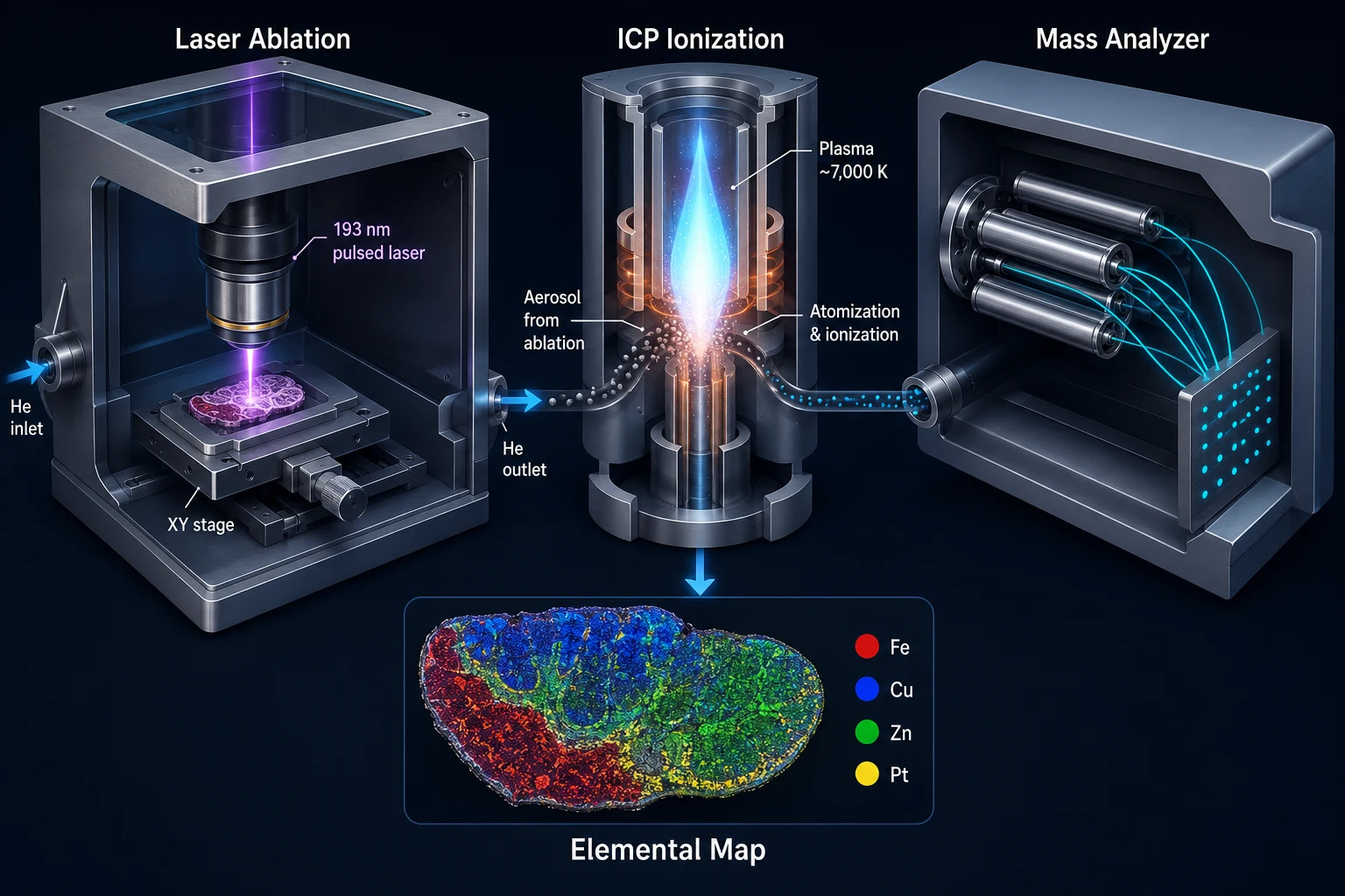 Figure 1: LA-ICP-MS Imaging Schematic — Instrument Cutaway. A 3D cutaway rendering of the LA-ICP-MS imaging workflow: a focused 193 nm ArF excimer laser beam (left) ablates a tissue section mounted on a glass slide, generating an aerosol plume of particles that is swept by helium carrier gas through a transfer tube into the ICP torch (center, 6,000-8,000 K plasma). The resulting ions are extracted through a sampler and skimmer cone interface into the mass analyzer (right) — shown as three interchangeable options: quadrupole (Q, sequential scanning), time-of-flight (TOF, full-spectrum simultaneous), and sector field (SF, highest mass resolution). A raster grid overlay on the tissue indicates the pixel-by-pixel sampling pattern, and a reconstructed multi-element heatmap (Fe, Cu, Zn, Pt) is displayed as the final output. Key parameters — laser wavelength, spot size, fluence, and carrier gas flow rate — are labeled with callouts.
The technique is conceptually straightforward. A pulsed laser beam — typically 193 nm (ArF excimer) or 213 nm (frequency-quintupled Nd:YAG) — is focused onto a tissue section mounted on a glass slide. Each laser pulse ablates a microscopic volume of tissue, generating an aerosol of particles that is swept by a helium carrier gas into an ICP torch (6,000-8,000 K), where the particles are atomized and ionized. The resulting ions are extracted into a mass analyzer — quadrupole (Q), time-of-flight (TOF), or sector field (SF) — for detection. By raster-scanning the laser across the tissue and recording an elemental spectrum at each pixel, a quantitative elemental image is reconstructed.
Laser Ablation Fundamentals
Laser wavelength, fluence (energy per unit area, J/cm²), and spot size collectively determine spatial resolution and ablation characteristics. At 193 nm, the photon energy exceeds the bond dissociation energy of most organic molecules, enabling direct bond cleavage and producing a fine, homogeneous aerosol with minimal elemental fractionation — the preferential vaporization of volatile elements. At 213 nm, ablation is partly thermal, which can introduce fractionation artifacts for elements with widely differing boiling points (e.g., Hg vs W). Spot sizes as small as 1-2 μm are commercially achievable at 193 nm, though practical bioimaging typically operates at 5-20 μm to balance resolution with signal intensity.
A 2025 Analytica Chimica Acta study by Boger et al. demonstrated quantitative single-cell zinc mapping at 5 μm resolution in human parietal cells, using AFM to verify ablation crater volumes and gelatin micro-droplets for external calibration. The study underscored that ablation yield varies with both laser parameters and tissue composition — a reminder that resolution numbers alone do not guarantee quantitative accuracy.
Mass Analyzer Selection: Q, TOF, or Sector Field
Quadrupole analyzers offer robustness, low cost, and excellent dynamic range, but scan sequentially — one m/z at a time — limiting the number of elements per pixel. TOF analyzers capture the entire mass spectrum simultaneously, making them ideal for multi-element imaging: a single laser shot records Li through U in microseconds. Sector field instruments provide the highest mass resolution (resolving Fe from ArO⁺ interference, critical for accurate Fe quantification in biological tissue) but at higher cost and lower scan speed.
The ICP-TOF-MS configuration has gained dominance for bioimaging because it captures quasi-simultaneous full-spectrum data without the spectral skew inherent in sequential quadrupole scanning. A 2026 Talanta paper by Nikolić et al. pushed TOF sensitivity further by applying detector overpulsing and isotope summation — improving limits of detection 2-5× for P, Fe, and Zn, and bringing phosphorus detection down to 31 fg per cell.
Sample Preparation for Quantitative Elemental Bioimaging
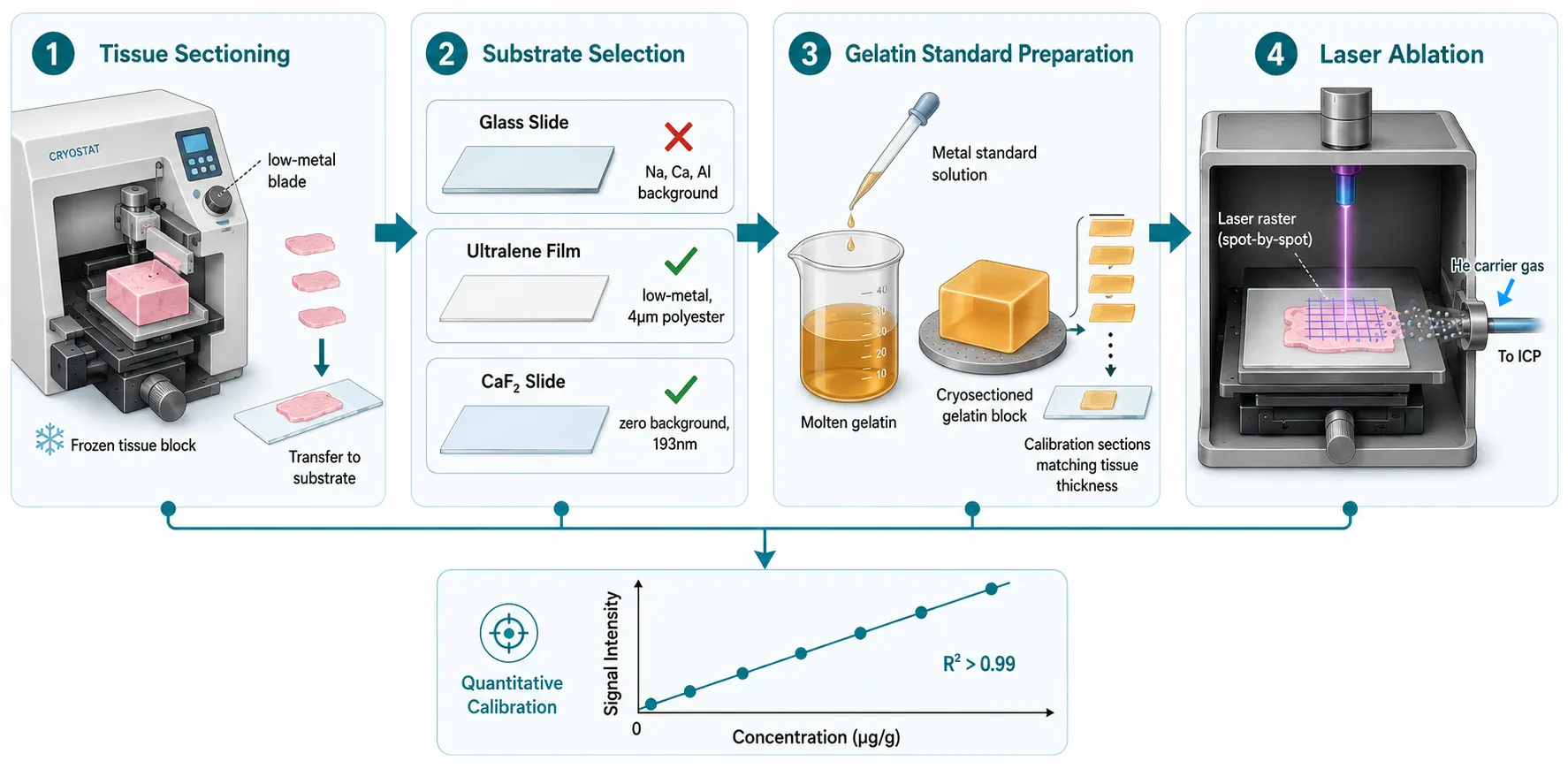 Figure 2: Sample Preparation Workflow for Quantitative LA-ICP-MS Bioimaging. A five-stage horizontal workflow diagram: (1) Tissue Harvest — snap-freezing in liquid nitrogen or isopentane/dry ice; (2) Substrate Selection — a decision panel showing glass slide (high metal background, Na/Ca/Al), Ultralene film (low-metal, 4 μm polyester), Kapton film (polyimide), and CaF₂/quartz slides (optimal, low background); (3) Cryosectioning — tissue sectioning at -20°C with a ceramic blade to avoid stainless steel metal smearing, annotated with recommended section thickness (10-20 μm); (4) Gelatin Calibration Standards — illustration of metal-spiked gelatin blocks cryosectioned at matching thickness for matrix-matched calibration; (5) MSI-Ready Slide — the final prepared slide with tissue and adjacent gelatin calibration spots. Inset callout highlights wavelength-specific gelatin optimization: at 193 nm, raw gelatin ablates like tissue; at 266 nm, UV-absorbing additives (L-tryptophan, gallic acid) are required.
Sample preparation is where most LA-ICP-MS imaging projects succeed or fail. Unlike MALDI-MSI, which requires matrix deposition, LA-ICP-MS works on native tissue — but that does not mean it works on any tissue section without forethought.
Substrate Selection
The tissue support substrate must satisfy two contradictory requirements: it must be transparent for optical microscopy (tissue inspection, cell localization) yet must not contribute background elemental signals. Standard glass microscope slides contain Na, Ca, Al, Si, and B at percent levels, producing enormous background during laser ablation. Ultralene film (4 μm polyester) and Kapton film (polyimide) are low-metal alternatives, though Ultralene absorbs slightly at 193 nm and can warp under prolonged laser exposure. Calcium fluoride (CaF₂) and quartz slides offer the best optical transparency with minimal elemental background for 193 nm systems, while specially coated glass slides with low-metal formulations are emerging for 213 nm work.
Metal delocalization during tissue sectioning is a second concern. Standard cryostat blades are stainless steel and can smear Fe, Cr, and Ni across the tissue surface. Ceramic or coated blades are recommended for studies quantifying these specific metals.
Matrix-Matched Calibration: Gelatin Standards
Quantification in LA-ICP-MS imaging requires external calibration standards that ablate identically to tissue. Gelatin — a protein-based hydrogel with similar carbon content and water affinity to biological tissue — has become the standard calibration matrix. Metal standards are spiked into molten gelatin at known concentrations, the gelatin is cryosectioned at the same thickness as the tissue, and calibration curves are constructed from the ablated gelatin sections.
However, gelatin is not a universal solution. Lockwood et al. demonstrated in a 2025 Talanta paper that raw gelatin does not ablate like tissue at 266 nm because it lacks aromatic amino acids and DNA bases that absorb UV at this wavelength. Adding 4 g/L L-tryptophan or 3 g/L gallic acid matched gelatin ablation to murine tissue at 266 nm. At 193 nm, gelatin absorbs uniformly and requires no additives — which is why 193 nm remains the preferred wavelength for quantitative bioimaging.
Model Organism Workflows
A 2026 bioRxiv protocol by Reynolds et al. details an end-to-end LA-ICP-MS workflow for C. elegans, addressing the specific challenges of whole-organism elemental imaging: embedding orientation for reproducible cross-sections, washing protocols to remove external metals from the cuticle, and registration with fluorescence microscopy for tissue-specific elemental quantification. The protocol achieves 5 μm resolution across whole worms, mapping Zn in intestinal granules, Fe in body-wall muscle, and Mn in the pharyngeal grinder — demonstrating that even millimeter-scale model organisms contain micrometer-scale elemental heterogeneity.
Quantification Strategies
Single-Cell Detection Limits
Quantifying elements in single cells by LA-ICP-MS pushes against the technique's fundamental sensitivity limits. A landmark 2026 Talanta paper by Foels et al. from the Koellensperger lab proposed a new LOD framework specifically for single-cell LA-ICP-TOFMS — part of a broader methodological push toward rigorous quantitative mass spectrometry imaging that spans calibration strategies from off-tissue spotting to CNN-based concentration prediction. Instead of reporting a single instrument LOD (which assumes a uniform pixel), their method estimates isotope- and cell-size-specific LODs by combining gelatin micro-droplet calibration with bootstrapped signal distributions from blank gelatin. The key insight is that a larger cell integrates more pixels — and therefore has a lower effective LOD — than a smaller cell at the same spatial resolution. Absolute elemental amounts are reported in femtograms of metal per cell, enabling direct cross-study comparison.
The approach generalizes to any ROI-based quantification: a tissue region, a cell cluster, or a subcellular compartment can all be treated as pixel-integrated entities with size-dependent LODs. For researchers pushing spatial resolution to the subcellular frontier, our single-cell spatial metabolomics guide covers platforms including t-MALDI-2, NanoSIMS, and TEMI tissue expansion. This is a methodological advance that moves LA-ICP-MS quantification from "semi-quantitative" to genuinely quantitative at the single-cell level.
XFM Cross-Validation
Synchrotron X-ray fluorescence microscopy (XFM or μXRF) provides an independent, matrix-effect-free reference method for validating LA-ICP-MS quantification. Because XFM excites elements via core-electron ejection rather than laser ablation, it is not subject to the same fractionation and transport-efficiency uncertainties. A 2026 JAAS study by Zee et al. demonstrated that LA-ICP-MS and XFM produce concordant Fe, Cu, and Zn concentrations in mouse brain when gelatin-based calibration is properly wavelength-matched, with deviations below 15% across all three elements. XFM validation should be considered the gold standard for any new LA-ICP-MS quantification protocol.
Hybrid Elemental + Molecular Imaging
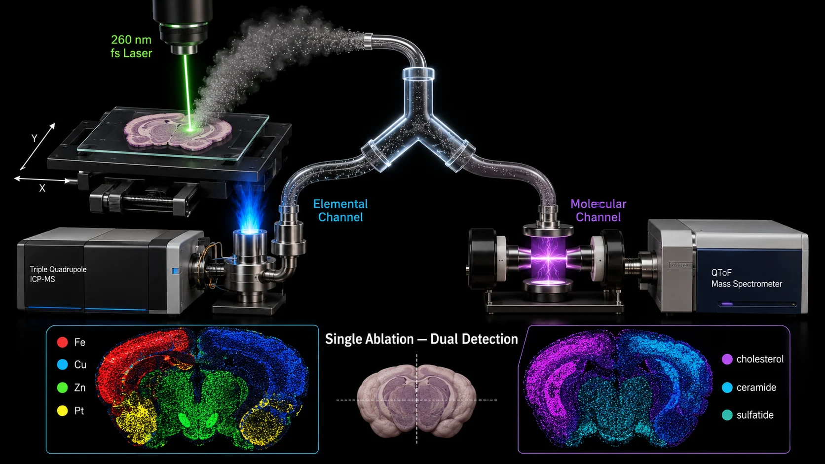 Figure 3: Hybrid Elemental + Molecular Imaging — Split-Flow LA-ICP-MS + DBDI-MS. Schematic of the Khoo et al. (2025) split-flow dual-mass-spectrometer system: a femtosecond Yb:KGW laser (260 nm, 290 fs) with galvo-mirror beam steering ablates the tissue, and the aerosol is divided by a Y-piece connector into two parallel streams. The left stream flows to an ICP-MS (triple quadrupole, NexION 5000) for elemental detection (Mg, P, Fe, Cu, Zn, Mo). The right stream enters a dielectric barrier discharge ionization (DBDI) source coupled to a QToF mass spectrometer for molecular detection (cholesterol, ceramides, sulfatides, adenine). Both streams generate perfectly co-registered images from a single ablation raster. Below the instrument schematic, a paired image panel shows an elemental map (Fe distribution, left) side-by-side with a molecular map (sulfatides, right) from the same mouse brain section, with a merged overlay in the center.
The most significant instrumentation advance in spatial metallomics since 2025 is the emergence of hybrid platforms that capture both elemental and molecular information from a single laser ablation event.
Split-Flow LA-ICP-MS + DBDI-MS
In a June 2025 JAAS paper, Khoo, Kubota, Matsukawa, and Hirata at the University of Tokyo demonstrated a split-flow laser ablation system using a femtosecond Yb:KGW laser (260 nm, 290 fs) with galvo-mirror beam steering. The ablation aerosol is split by a Y-piece connector into two streams: one directed to an ICP-MS (triple quadrupole, NexION 5000) for elemental detection, the other to a dielectric barrier discharge ionization (DBDI) source coupled to a QToF mass spectrometer for molecular detection.
The result is simultaneous, perfectly co-registered elemental and molecular images from a single tissue section. Applied to mouse brain, the system generated elemental maps for Mg, P, Fe, Cu, Zn, and Mo alongside molecular maps for cholesterol, ceramides, sulfatides, and adenine — all from the same ablation raster. This eliminates the registration ambiguity that plagues serial-section multi-modal workflows and represents the current state of the art in integrated spatial metallomics. The molecular channel data integrates naturally with dedicated lipidomics workflows, enabling deeper annotation of the lipid species that co-localize with specific metal distributions.
MALDI-MSI as an Emerging Elemental Imaging Tool
In a striking 2025 development, MALDI-MSI — long considered exclusively a molecular imaging technique — has been demonstrated as a viable platform for direct elemental imaging. Stopka et al. (Analytical Chemistry, 2025) showed that high-resolution MALDI-FTICR-MS can detect and spatially map endogenous metals (Fe, Ca) and exogenous elements (Gd from contrast agents, Pt from cisplatin) without chelation or derivatization, coining the term "spatial elementomics." The approach was validated in murine models of hemochromatosis and Wilson's disease, where hepatic Fe and Cu distributions matched histochemical staining patterns.
Simultaneously, Jia et al. (Analytical Chemistry, 2025) introduced 5,6-diamino-1,10-phenanthroline (DAP) hydrochloride as a bifunctional MALDI matrix: in positive ion mode, DAP chelates free metal ions (Fe²⁺/Fe³⁺, Cu⁺/Cu²⁺, Zn²⁺, Cd²⁺, Mn²⁺, Pb²⁺) with detection limits as low as 5 attomoles for Cu²⁺; in negative ion mode, it functions as a conventional matrix for endogenous metabolites. Applied to a copper-exposure mouse model, the matrix revealed simultaneous Cu accumulation and depletion of taurine, ascorbic acid, and glutathione in kidney — linking metal dysregulation directly to metabolic perturbation in a single experiment.
These advances do not mean MALDI will replace LA-ICP-MS for elemental imaging. LA-ICP-MS retains superior sensitivity for most metals, established quantification protocols, and better dynamic range. But MALDI-MSI now offers something LA-ICP-MS cannot: simultaneous elemental and molecular imaging from a single tissue section with no hardware modifications beyond matrix selection — making it a powerful complementary tool. For a detailed walkthrough of MALDI-MSI experimental design and data analysis, see our MALDI imaging workflow resource.
Key Biological Applications
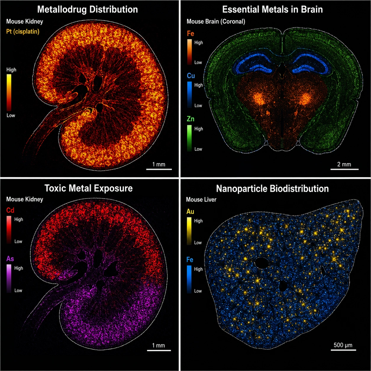 Figure 4: Elemental Mapping Gallery — Biological Applications of LA-ICP-MS Imaging. A 2×2 gallery panel illustrating four key application domains: (top-left) Metallodrug Imaging — Pt-195 map of cisplatin distribution in mouse kidney showing selective accumulation in renal cortex and corticomedullary junction, with proximal tubule damage correlated to high-Pt regions; (top-right) Neurodegeneration — Fe-56 map of human substantia nigra showing elevated iron in the pars compacta relative to pars reticulata, with a callout noting the Parkinson's disease iron dysregulation hypothesis; (bottom-left) Nanoparticle Biodistribution — Au-197 map of gold nanoparticle accumulation in a glioblastoma xenograft, distinguishing tumor core vs. invasive margin vs. contralateral normal brain; (bottom-right) Metal-Induced Metabolic Perturbation — paired Cd-114 and Zn-64 maps from a cadmium-exposure mouse model showing Cd accumulation in renal cortex with concurrent Zn depletion. Each panel includes a grayscale H&E inset for anatomical reference.
Metallodrug Imaging
LA-ICP-MS is the reference method for imaging platinum-based chemotherapeutics (cisplatin, carboplatin, oxaliplatin) in tumor and kidney tissue. Pt-195 detection at μg/g sensitivity reveals drug penetration depth, tumor heterogeneity in drug uptake, and nephrotoxic accumulation in proximal tubules. Beyond platinum, the technique is increasingly applied to ruthenium-based anticancer agents (e.g., KP1019, NAMI-A) and gold-based antirheumatic drugs, where Ru and Au are detected at comparable sensitivity to Pt. A single tissue section can simultaneously map the drug (e.g., Pt-195), endogenous metals displaced by the drug (e.g., Zn-64 depletion in tumor regions), and tissue architecture via Fe-56 and S-34 — providing a systems-level view of metallodrug action without requiring radiolabels or fluorescent tags. The technique also maps gadolinium deposition from MRI contrast agents in brain tissue — a topic of significant clinical interest since the 2014 discovery of Gd retention in the dentate nucleus.
Essential and Toxic Metals
Endogenous metals — Fe, Cu, Zn, Mn — are mapped in the context of neurodegenerative disease (elevated Fe in Parkinson's substantia nigra, Cu dysregulation in Alzheimer's hippocampus, Mn accumulation in the globus pallidus). Toxic metals — Cd, Pb, As, Hg — are mapped in environmental exposure studies, with LA-ICP-MS providing spatial context that bulk tissue digestion cannot: for instance, revealing that Cd concentrates specifically in the renal cortex while Pb deposits preferentially in bone marrow. For research teams studying metal-induced metabolic perturbation without direct LA-ICP-MS access, untargeted metabolomics can characterize the downstream metabolic consequences of metal exposure from tissue homogenates, complementing targeted elemental analysis.
Nanoparticle Biodistribution
Engineered nanoparticles (Au, Ag, TiO₂, iron oxide) are increasingly used as drug delivery vehicles, imaging contrast agents, and theranostic platforms. LA-ICP-MS imaging tracks their biodistribution at the organ and cellular level without requiring fluorescent or radioactive labeling, providing quantitative mass-per-tissue data that electron microscopy alone cannot deliver. The quantitative capability is particularly valuable: unlike fluorescence or TEM, which require assumptions about label stability and sampling representativeness, LA-ICP-MS directly measures the mass of the elemental tag (Au, Ag, Ti) per tissue region. Recent work has applied this to track gold nanoparticle penetration across the blood-brain barrier in glioblastoma models, quantifying both particle accumulation in tumor core vs. invasive margin and clearance kinetics in liver and spleen — all from a single ablation run per time point.
LA-ICP-MS vs. Other Elemental Imaging Methods
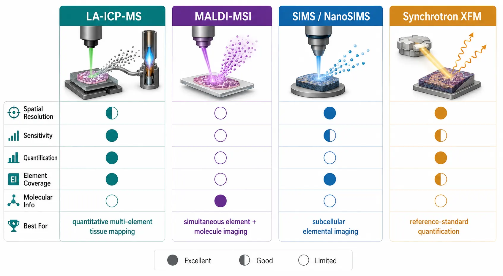 Figure 5: LA-ICP-MS vs MALDI vs SIMS vs XFM — Technique Comparison for Elemental Imaging. A head-to-head visual comparison across four elemental imaging techniques arranged as a quadrant infographic: (top-left) LA-ICP-MS — 193 nm laser on tissue with helium carrier gas to ICP, annotated as "Quantitative, 1-20 μm, ng/g sensitivity, most metals"; (top-right) MALDI-MSI (elemental) — matrix-coated tissue with laser desorption into FTICR-MS, annotated as "Simultaneous molecular+elemental, 5-50 μm, emerging"; (bottom-left) SIMS/NanoSIMS — focused Bi₃⁺ or Cs⁺ ion beam sputtering surface, annotated as "50 nm-1 μm, surface-sensitive, small FOV"; (bottom-right) Synchrotron XFM — X-ray beam exciting core electrons with fluorescence detector, annotated as "0.1-5 μm, matrix-effect-free, reference method, beamline access required." A radar chart at center compares the four techniques across five axes: spatial resolution, sensitivity, elemental coverage, quantitative accuracy, and molecular capability.
The table below provides a head-to-head comparison of the four principal techniques for spatial elemental analysis in biological tissue.
| Technique | Spatial Resolution | Sensitivity | Elements Detected | Quantitative? | Molecular Info? | Key Limitation |
|---|---|---|---|---|---|---|
| LA-ICP-MS | 1-20 μm | ng/g-μg/g | Li-U (most metals) | Yes (with standards) | No | Destructive; glass background |
| MALDI-MSI (elemental) | 5-50 μm | μg/g (emerging) | Fe, Cu, Zn, Pt, Gd, Cl, Ca | Semi-quantitative | Yes (same section) | Sensitivity lower than LA-ICP |
| SIMS / NanoSIMS | 50 nm-1 μm | μg/g | All elements | Semi-quantitative (matrix effects) | Fragment ions only | Extreme surface sensitivity; small FOV |
| Synchrotron XFM | 0.1-5 μm | μg/g (sub-ppm for some) | K-edge accessible (Ca-U); lighter with vacuum | Yes (reference method) | No | Beamline access; radiation damage |
All spatial metallomics and elemental imaging metabolomics services described in this article are provided for Research Use Only (RUO). These workflows are not intended for diagnostic, therapeutic, or clinical decision-making purposes.
FAQ
Q: What is the detection limit of LA-ICP-MS imaging?
A: Detection limits range from ng/g to low μg/g depending on the element, mass analyzer, and laser spot size. For single-cell analysis, a 2026 Talanta study reported isotope- and cell-size-specific LODs in the femtogram range per cell. Light elements (P, S) and elements with high ionization potentials (Se, As) have higher LODs than transition metals (Fe, Cu, Zn).
Q: Can LA-ICP-MS and MALDI-MSI be performed on the same tissue section?
A: Yes. A 2025 Analytical and Bioanalytical Chemistry study by Sarretto et al. validated a sequential workflow: AP-MALDI-MSI for lipids first on a matrix-coated section, followed by LA-ICP-MSI for elements on the same section. Matrix coating slightly suppresses elemental signals but spatial distributions remain intact.
Q: How is LA-ICP-MS imaging quantified?
A: The gold standard is matrix-matched external calibration using gelatin standards spiked with known metal concentrations, cryosectioned at the same thickness as the tissue. Gelatin requires wavelength-specific optimization — at 193 nm, raw gelatin ablates like tissue; at 266 nm, UV-absorbing additives (L-tryptophan, gallic acid) are needed.
Q: Can Creative Proteomics perform LA-ICP-MS elemental imaging?
A: Creative Proteomics' spatial omics platform currently focuses on molecular imaging via MALDI-Imaging Lipidomics and MS-based Spatial Proteomics. For elemental imaging needs, our Untargeted Metabolomics service can characterize metal-induced metabolic changes in tissue, and our scientific team can coordinate LA-ICP-MS access through collaborative networks for projects requiring direct elemental mapping. Contact our team to discuss multi-modal spatial metallomics workflows.
Q: What is the difference between LA-ICP-MS and LIBS for elemental imaging?
A: Both techniques use laser ablation, but they differ fundamentally in ionization and sensitivity. LA-ICP-MS uses an ICP torch for ionization, achieving ng/g detection limits across most metals. LIBS (laser-induced breakdown spectroscopy) detects optical emission from the laser-induced plasma directly, without an ICP — making it faster and suitable for field deployment, but with μg/g detection limits that are 100-1,000× higher than LA-ICP-MS. For biological tissue imaging where trace metals are the target, LA-ICP-MS is the preferred method. LIBS is better suited for geological samples and industrial quality control where speed matters more than sensitivity.
Q: Can LA-ICP-MS detect non-metals like phosphorus and sulfur?
A: Yes, but with important caveats. Phosphorus and sulfur are detectable by LA-ICP-MS and are biologically significant — P marks cell nuclei (DNA) and S marks protein-rich regions. However, these elements have higher first ionization potentials than transition metals, reducing sensitivity. A 2026 Talanta study by Nikolić et al. achieved phosphorus detection down to 31 femtograms per cell using detector overpulsing and isotope summation on an ICP-TOF-MS. For routine P and S imaging, sector field instruments provide better sensitivity than quadrupole instruments by resolving polyatomic interferences (e.g., ³¹P from ¹⁵N¹⁶O⁺).
References:
- Khoo HH, Kubota A, Matsukawa T, Hirata T. Dual mass spectrometers with plasma-based ion sources for multiplex imaging of elements and biomolecules. Journal of Analytical Atomic Spectrometry. 2025;40:2016-2025. doi:10.1039/d5ja00039d
- Foels E, Braun G, Molitor C, et al. Evaluating limits of detection for single-cell elemental bioimaging by LA-ICP-TOFMS. Talanta. 2026;305:129671. doi:10.1016/j.talanta.2026.129671
- Stopka SA, et al. Spatial profiling of elements through matrix-assisted laser desorption ionization mass spectrometry imaging. Analytical Chemistry. 2025;97:25334-25345. doi:10.1021/acs.analchem.5c05632
- Jia X, et al. MALDI mass spectrometry imaging of metal ions and metabolites in tissues using a bifunctional matrix. Analytical Chemistry. 2025;97(39):21384-21391. doi:10.1021/acs.analchem.5c02995
- Sarretto T, et al. Evaluation of combined workflows for multimodal mass spectrometry imaging of elements and lipids from the same tissue section. Analytical and Bioanalytical Chemistry. 2025;417:705-719. doi:10.1007/s00216-024-05696-w
- Boger V, Pirkwieser P, Orth N, Koehler M, Somoza V. AFM-optimized single-cell level LA-ICP-MS imaging for quantitative mapping of intracellular zinc concentration in immobilized human parietal cells using gelatin droplet-based calibration. Analytica Chimica Acta. 2025;1355:343999. doi:10.1016/j.aca.2025.343999
- Lockwood TE, Bordin DCM, Westerhausen MT, Bishop DP. Preparation of gelatine calibration standards for LA-ICP-MS bioimaging with 266 nm laser ablation systems. Talanta. 2025;283:127150. doi:10.1016/j.talanta.2024.127150
- Nikolić M, Lores Padín A, Dejonghe R, De Wever O, Vanhaecke F. Boosting the sensitivity of ICP-TOF-MS for single-cell metallome profiling via overpulsing and isotope summation. Talanta. 2026;307:129895. doi:10.1016/j.talanta.2026.129895
- Zee DZ, Ahn SH, Crawford AM, et al. Substrate and standard evaluation for correlative elemental mapping of biological samples by X-ray fluorescence microscopy and laser ablation ICP-MS. Journal of Analytical Atomic Spectrometry. 2026;41(2):708-721. doi:10.1039/d5ja00371g
- Körber A, Heeren RMA. Mass spectrometry imaging. Analytical Chemistry. 2025;97(29). doi:10.1021/acs.analchem.4c05249
- Doble PA, et al. Laser ablation-inductively coupled plasma-mass spectrometry imaging in biology. Chemical Reviews. 2021;121:11769-11822. doi:10.1021/acs.chemrev.0c01219














