Equilibrium Dialysis LC-MS/MS Service for Protein & Tissue Binding

Accelerate your lead optimization with our high-throughput Equilibrium Dialysis LC-MS/MS service.

Recognized globally as the gold standard for determining the true fraction unbound (fu) of drugs, our platform expertly overcomes non-specific binding challenges. Whether you are screening highly lipophilic compounds or exploring diverse tissue matrices, we partner with you to deliver the highly precise, reproducible thermodynamic binding data you need to move forward with confidence.

Key Advantages:

  • High-throughput RED format for rapid analog screening.
  • Unmatched LC-MS/MS sensitivity for >99% bound drugs.
  • Rigorous mass balance and recovery QC reporting.
  • Broad matrix support: plasma, microsomes, and tissue homogenates.
Scientific illustration comparing single dialysis tube to high-throughput 96-well RED plate.
What is ED-MS Binding The Creative Proteomics Advantage Applications & Matrices Workflow & QC Technology Comparison Deliverables & Demo Case Study FAQ

What is Equilibrium Dialysis-MS Binding?

Equilibrium dialysis combined with Mass Spectrometry (ED-MS) is the foundational method for measuring how much of a drug remains free and active in the bloodstream or within specific organs. When a drug enters a biological system, it naturally binds to proteins in the plasma or tissues. However, only the "free" or unbound portion of the drug can cross cell membranes, interact with therapeutic targets, and drive biological efficacy.

Equilibrium dialysis measures this critical "fraction unbound" (fu) by placing a biological sample (like plasma) and a clean buffer solution into two separate chambers, divided by a semi-permeable membrane. Over time, only the free, unbound drug molecules can pass through the membrane's microscopic pores. Once the concentration of free drug stabilizes equally on both sides—a state known as thermodynamic equilibrium—we extract the buffer and use highly sensitive LC-MS/MS to precisely quantify the free drug concentration. Because this is a label-free, true physiological simulation, it provides the most accurate and reliable binding data available in early drug discovery.

The Creative Proteomics Advantage: Solving the "Sticky" Problem

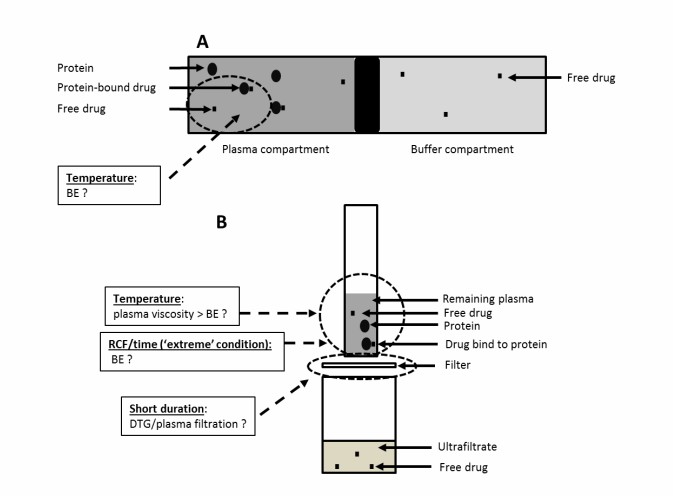
Every drug discovery team eventually encounters the "sticky" compound problem. As medicinal chemists design more complex, highly lipophilic (fat-soluble) molecules to hit challenging targets, these compounds tend to stick to everything—plastic tubes, assay plates, and filtration membranes. In standard assays, this Non-Specific Binding (NSB) leads to artificially low recovery rates, skewed binding data, and ultimately, incorrect dosing predictions. When hydrophobic interactions dominate, standard screening methods simply cannot be trusted.

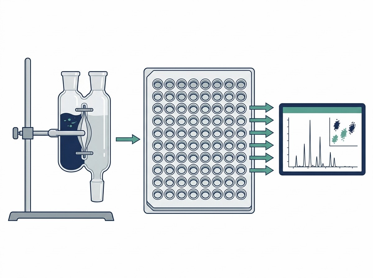
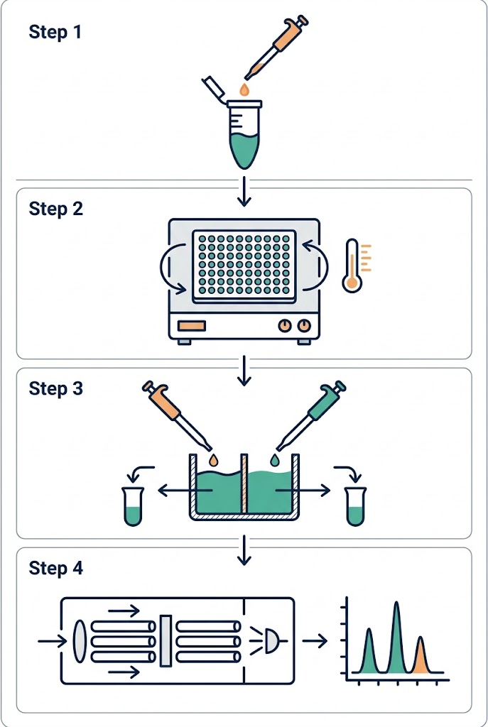
We understand how frustrating it is to synthesize a promising analog, only to have the data ruined by poor assay methodology. At Creative Proteomics, our ADME and pharmacokinetics team specializes in rescuing these difficult projects. We utilize the industry-leading Rapid Equilibrium Dialysis (RED) format, utilizing 96-well plates equipped with specialized low-binding regenerated cellulose membranes. This high-throughput setup minimizes the surface area-to-volume ratio where drugs can get trapped, significantly reducing NSB risks while maximizing sample recovery. By closely monitoring the thermodynamics of the system, we ensure that your compound remains in the solution phase where it belongs.

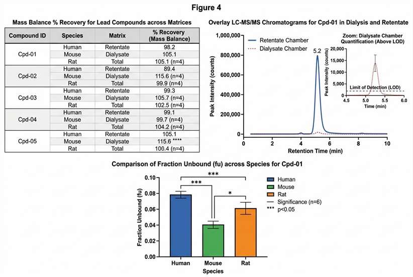
Furthermore, accurately calculating the fu of a drug that is >99% bound requires extreme analytical sensitivity, because the amount of free drug floating in the buffer chamber is minuscule. Our platform leverages state-of-the-art QQQ and Q-TOF mass spectrometers, allowing us to accurately detect and quantify trace levels of your compound that other laboratories might miss entirely. If your project is still in the earliest stages of hit identification and you need to screen thousands of compounds before optimizing for ADME properties, you can also explore our high-throughput Affinity Selection Mass Spectrometry (ASMS) solutions to rapidly filter your initial compound libraries.

Applications & Supported Matrices

Translational success requires understanding drug distribution beyond just the bloodstream. Different tissues express different proteins, which drastically alters local drug binding. To support a complete pharmacokinetic profile, our Equilibrium Dialysis LC-MS/MS service covers a comprehensive range of biological matrices.

  • Plasma Protein Binding (PPB): The standard starting point for understanding systemic drug exposure across various animal models. Essential for calculating safety margins and initial dosage models.
  • Tissue Binding (Brain, Liver, Lung): Critical for understanding whether your drug is actually penetrating the blood-brain barrier or accumulating in target organs. Brain homogenate binding (fu,brain) is particularly vital for central nervous system (CNS) therapeutics.
  • Microsomal Binding: Essential for determining the fraction of unbound drug available for metabolic enzymes in the liver, ensuring accurate intrinsic clearance (CLint) and hepatic extraction ratio calculations.

Sample Requirements

Matrix TypeRequired Volume / AmountDrug ConcentrationBuffer RequirementsShipping & Storage
Plasma (Human/Animal)1–2 mL per compound1-10 µM (Standard)K2EDTA or HeparinDry ice, avoid freeze-thaw
Tissue Homogenates0.5–1 g wet weightOptimization requiredPBS (pH 7.4)Dry ice
Microsomes2-5 mg proteinClient specifiedStandard MS compatibleDry ice

Standardized Workflow & Rigorous QC Checkpoints

We believe that reliable data is born from total workflow transparency. We do not treat your samples as a "black box." Our standardized, high-throughput protocol includes strict Quality Control (QC) checkpoints at every step to ensure the integrity of your thermodynamic data.

1

Sample Preparation & Spiking

Your compound is spiked into the chosen biological matrix at pre-defined concentrations. We carefully control pH and temperature from the very first step to mimic physiological environments and prevent early degradation.

2

37°C RED Plate Incubation

The spiked matrix and blank buffer are loaded into adjacent chambers of our high-throughput RED plates. The plate is sealed and incubated at 37°C on an orbital shaker. The continuous agitation speeds up the diffusion process, ensuring true equilibrium is achieved efficiently within 4 to 6 hours.

3

Matrix Matching & Extraction

Once equilibrium is reached, aliquots are removed from both the matrix and buffer chambers. To ensure our mass spectrometers read both samples accurately without matrix-induced ion suppression, we perform "matrix matching"—adding blank buffer to the plasma samples, and blank plasma to the buffer samples.

4

LC-MS/MS Quantification & Mass Balance Check

The samples undergo protein precipitation and are analyzed via targeted LC-MS/MS. Crucial QC Checkpoint: We actively calculate the total Mass Balance (Recovery Rate). If the total amount of drug recovered from both chambers falls outside the accepted 80%–120% range, it flags a severe NSB or stability issue, prompting our scientists to optimize the assay rather than handing you flawed data.

Equilibrium dialysis LC-MS workflow diagram

Standardized Equilibrium Dialysis workflow with integrated QC checkpoints.

Technology Comparison: Why Equilibrium Dialysis?

Choosing the right in vitro binding assay can save months of wasted optimization time and thousands of dollars in misdirected synthesis. While other methods exist, they each come with distinct physiological and physical limitations that can derail a preclinical program. Here is how Equilibrium Dialysis stacks up against alternative approaches.

DimensionEquilibrium Dialysis (ED)Ultrafiltration (UF)Ultracentrifugation (UC)
Scientific PrinciplePassive diffusion across a semi-permeable membrane to reach thermodynamic equilibrium.Forcing sample through a size-exclusion filter using centrifugal force.Spinning the sample at extreme speeds to physically pellet heavy protein-drug complexes.
ThroughputHigh (using 96-well RED plates).Medium to High.Extremely Low (requires specialized tubes and long spin times).
Non-Specific Binding (NSB) RiskLow to Moderate (easily controlled with optimized RED plate materials).Very High (lipophilic drugs readily stick to the filter membranes under pressure).Zero (no membranes or filters are used in the process).
Sample Volume RequiredLow (as little as 50-100 µL per well).Moderate.Very High (requires large volumes for the centrifuge rotors).
Best Used ForGold standard for routine fu calculation, highly bound drugs, and varied tissue matrices.Quick screening of highly soluble, non-sticky compounds only.Highly unstable compounds that degrade during ED, or exceptionally large molecular targets.

Solution Selection Strategy:

  • Choose Equilibrium Dialysis as your baseline, gold-standard method. It provides the most mathematically sound fu for routine PPB and small molecules, successfully balancing high throughput with rigorous accuracy. It is the only method broadly trusted by regulatory agencies for definitive free-fraction submissions.
  • Avoid Ultrafiltration if you are working with "sticky" or highly lipophilic compounds. The centrifugal pressure forces the drug into the filter membrane, causing concentration polarization and massive drug loss. This results in inaccurate free fraction readings that underestimate your drug's true availability.
  • Opt for Ultracentrifugation only as a last resort when testing highly unstable compounds that cannot survive a 4-hour 37°C incubation, or if you need to bypass membranes entirely due to unique macromolecular interactions.
  • If you are looking for methodologies that involve tethering a target to a solid support rather than measuring solution-phase equilibrium, you can explore our target immobilization LC-MS services.

Deliverables & Representative QC Data

We empower your decision-making by providing comprehensive, presentation-ready data packages. Our reporting goes beyond a simple percentage; we provide the visual and statistical evidence required for rigorous preclinical pathway planning and internal chemistry discussions.

Equilibrium dialysis fraction unbound mass spectrometry demo data

Representative data deliverables

Composite dashboard including mass balance tracking and precise fraction unbound calculation.

When you partner with us, your final report will feature:

  • Mass Balance & Recovery Tracking: Detailed tables ensuring that overall compound recovery stays within the target 80-120% range, proving to your team that no severe NSB or degradation compromised the assay.
  • High-Sensitivity LC-MS/MS Traces: Clear chromatograms demonstrating our Limit of Quantitation (LOQ) capability. We visually prove that we can accurately detect the minimal free drug present in the buffer chambers for >99.5% bound drugs, ensuring no signal is lost in the baseline noise.
  • Fraction Unbound (fu) Cross-Species Plots: Visual bar chart comparisons of your drug's binding variations across human, rat, and dog plasma, providing immediate insight into species-specific binding differences necessary for accurate allometric scaling and safety toxicology species selection.

Case Study: Reliable Assessment of Unbound Dolutegravir by Equilibrium Dialysis LC-MS/MS

Comparing ultrafiltration and equilibrium dialysis to measure unbound plasma dolutegravir concentrations based on a design of experiment approach

Background

Accurate measurement of plasma protein binding is especially important for highly bound drugs because the pharmacologically active portion is the unbound fraction rather than the total drug concentration. Dolutegravir is a highly plasma protein-bound antiretroviral drug, with binding reported to be greater than 99%, meaning that small analytical differences in the measured free fraction can materially affect interpretation of active exposure. In this study, the authors focused on validating a reliable strategy for measuring unbound dolutegravir in plasma and emphasized that equilibrium dialysis is considered the gold standard for evaluating the unbound form of a drug.

Methods

Researchers compared equilibrium dialysis (ED) and ultrafiltration (UF) for measuring unbound plasma dolutegravir concentrations, with LC–MS/MS used for quantification of both unbound and total drug concentrations. The study evaluated three UF parameters—temperature, centrifugation time, and relative centrifugal force (RCF)—and benchmarked UF results against ED performed at controlled conditions. The LC–MS/MS workflow included matrix-appropriate analysis of plasma, dialysate, and ultrafiltrate samples, with reported precision and accuracy suitable for quantitative bioanalysis.

Results

The study showed that UF results were highly condition-dependent, whereas ED provided the reference standard for interpreting unbound dolutegravir exposure. Temperature influenced measured unbound concentrations in both ED and UF, and the interaction between 2000 g and 20 min significantly affected UF-derived results. The authors identified 37 °C, 1000 g, and 20 min as the UF condition that produced results most comparable to ED. When this optimized UF condition was applied to HIV patient plasma samples, free fraction results did not differ significantly from ED results, supporting the use of ED as the benchmark and demonstrating how orthogonally validated workflows can improve confidence in unbound drug measurement.

Conclusions

This study demonstrates that equilibrium dialysis combined with LC–MS/MS provides a robust reference framework for measuring unbound concentrations of a highly bound drug. It also shows that high-throughput or clinically practical alternatives such as ultrafiltration must be validated against ED before results are interpreted for pharmacokinetic or therapeutic decision-making. For a service page, this case effectively supports the value of a rigorously controlled Equilibrium Dialysis LC-MS/MS workflow for generating reliable unbound exposure data in protein binding studies.

Comparison of unbound dolutegravir measurements generated by equilibrium dialysis and ultrafiltration under different analytical conditions.

Figure 1. Comparison of unbound dolutegravir results obtained by equilibrium dialysis and ultrafiltration across different analytical conditions. Equilibrium dialysis served as the reference method, while ultrafiltration performance varied with temperature, centrifugation time, and relative centrifugal force, highlighting the importance of validating alternative workflows against an equilibrium dialysis LC-MS/MS standard.

FAQ

Frequently Asked Questions

Q: What is the typical incubation time, and how do you ensure compound stability?

Most small molecules reach thermodynamic equilibrium within 4 to 6 hours using our 96-well RED plates on an orbital shaker at 37°C. To ensure stability, we continuously monitor the total mass balance. If a compound shows significant degradation during the standard incubation time, we can optimize the assay by utilizing alternative buffers, adjusting the pH, or employing time-course sampling to pinpoint the specific degradation window before equilibrium is compromised.

Q: How do you handle extremely lipophilic compounds that stick to the dialysis membrane?

For highly lipophilic "sticky" compounds, we first utilize specialized low-binding Teflon base plates and optimized regenerated cellulose membranes. If NSB remains high (indicated by poor mass balance recovery), we can implement troubleshooting protocols such as adding specific surfactants or proteins to the receiver buffer, altering the solvent concentrations, or pre-conditioning the membranes to block hydrophobic binding sites without disrupting the equilibrium exchange.

Q: Can you perform equilibrium dialysis on brain tissue homogenates?

Yes. Measuring the true unbound fraction in the brain (fu,brain) is critical for neuro-therapeutics crossing the blood-brain barrier. We routinely process complex tissue homogenates, including brain, liver, and lung. We carefully optimize the homogenization buffer (typically PBS) to maintain tissue protein integrity while ensuring the biological mixture remains fluid enough to allow for proper, unhindered drug diffusion across the dialysis membrane.

Q: Do you correct for volume shifts during the dialysis process?

Absolutely. During dialysis, the osmotic pressure generated by the high concentration of plasma proteins can cause water to migrate from the buffer chamber into the matrix chamber. This changes the volumes and can artificially alter the calculated drug concentrations. We meticulously weigh the plates before and after incubation to calculate the exact volume shift and apply rigorous mathematical volume shift corrections to ensure your final fu calculation is flawlessly accurate.

Q: Can equilibrium dialysis be used for irreversible binders?

If your drug discovery program is focused on compounds that form permanent covalent bonds with their targets (which cannot be measured by reversible equilibrium methods), please view our specialized covalent inhibitor profiling services instead.

Disclaimer: All services and products offered by Creative Proteomics are for Research Use Only (RUO). They are not intended for use in diagnostic procedures, clinical decision-making, or any therapeutic applications. We do not provide medical advice or clinical diagnostic conclusions.

Plan an ED-MS campaign with the MassTarget™ team

Share your compound details and our scientists will design a tailored Equilibrium Dialysis screening strategy for your discovery program.

Online Inquiry

Please submit a detailed description of your project. We will provide you with a customized project plan to meet your research requests. You can also send emails directly to for inquiries.

* Email
Phone
* Service & Products of Interest
Services Required and Project Description