HDX-MS and HDX-driven Epitope Mapping Services

Secure your intellectual property and gain comprehensive structural insights with our advanced HDX-MS epitope mapping services. Understanding the exact binding interface of your therapeutic candidate is a critical step for robust patent protection and precise mechanism-of-action validation.

By integrating fully automated labeling, sub-zero chromatography, and multi-protease digestion strategies, we deliver near amino-acid resolution of highly complex conformational epitopes. Our platform analyzes your target in its completely native solution state. Whether you are mapping novel monoclonal antibodies, deconvoluting polyclonal mixtures, or analyzing difficult membrane proteins, we capture the true dynamic structural changes of your complex—succeeding where traditional mutagenesis and rigid crystallography frequently fall short.

  • Label-free, 100% solution-phase conformational mapping
  • Automated sub-zero workflow minimizing back-exchange
  • High-resolution 3D structural data models for IP support
HDX-MS and HDX-driven Epitope Mapping Services
Decoding Conformational EpitopesService OverviewTechnology ComparisonWorkflow & QCDemo ResultsSample RequirementsCase StudyBioinformaticsFAQ

Decoding Conformational Epitopes with HDX-MS

Hydrogen-Deuterium Exchange Mass Spectrometry (HDX-MS) is a powerful, label-free analytical technique that measures the rate at which amide hydrogens on a protein backbone exchange with deuterium atoms in a surrounding heavy water solvent. This rate depends entirely on the protein's native three-dimensional folding and solvent accessibility.

When developing a new biologic therapeutic, knowing exactly where your antibody binds to its target is critical for both patent protection (IP) and mechanism-of-action validation. However, many critical binding sites are not simple, linear strings of amino acids. Instead, they are highly complex "conformational epitopes"—three-dimensional shapes formed only when the protein folds into its native, functional state. When an antibody or ligand binds to the target, it physically shields this specific binding site from the heavy water, dramatically slowing the deuterium exchange rate in that exact localized area.

By measuring this mass difference over various time points using high-resolution mass spectrometry, we can precisely map the exact footprint of your antibody. Because this entire process occurs in a natural physiological liquid buffer, we capture the true dynamic structural state of your proteins without forcing them into unnatural, rigid crystals or introducing artificial chemical cross-linkers. This provides the most biologically relevant snapshot of target engagement available in modern structural biology.

Service Overview & Target Capabilities

We recognize that modern drug discovery pipelines often focus on incredibly complex molecules that defy conventional structural characterization. Our HDX-MS platform is deliberately built to handle challenging targets that standard methods simply cannot process. We help you map binding interfaces, uncover hidden allosteric changes, and rigorously prove structural comparability for complex biosimilars.

Targets We Routinely Analyze:

Monoclonal & Bispecific Antibodies

As antibody engineering becomes more complex, simply knowing that an antibody binds is insufficient. We precisely map the binding interfaces to differentiate your novel clones from existing patents, strengthening your intellectual property claims. For bispecifics, we can track avidity effects and confirm that binding at one arm does not negatively induce allosteric distortion at the second binding interface.

Polyclonal Antibodies & Vaccine Sera

Unlike monoclonals, polyclonal mixtures bind to multiple regions simultaneously. We deconvolute these highly complex immune responses to identify immunodominant regions, providing critical structural data for vaccine development, formulation stability, and batch-to-batch consistency evaluations.

Membrane Proteins & Flexible Targets

Integral membrane proteins (like GPCRs and ion channels) and Intrinsically Disordered Proteins (IDPs) possess large flexible regions and often outright refuse to crystallize. HDX-MS tames this flexibility, allowing us to map epitopes on targets embedded in lipid nanodiscs or detergent micelles without losing the structural context of the membrane environment.

Highly Glycosylated Proteins

Heavy glycosylation (such as on viral envelope spikes or heavily modified receptors) physically shields the protein backbone from standard pepsin digestion, often resulting in massive data gaps. We circumvent this by utilizing specialized multi-protease strategies (incorporating proteases like Type XIII or Nepenthesins) and optimized quench buffers to digest heavily modified proteins, ensuring we never miss critical binding regions hidden beneath complex glycans.

If you have already confirmed the macroscopic binding affinity and kinetics of your compound using our Surface Plasmon Resonance (SPR) platform, HDX-MS provides the perfect, high-resolution structural complement, showing you exactly where the interaction occurs at the amino-acid level.

Technology Comparison: Why HDX-MS Outperforms Traditional Mapping?

Choosing the right method to map your epitope is a critical strategic decision that can save you months of frustrating setbacks and misdirected chemistry efforts. Traditional approaches often force discovery teams into a difficult compromise: you must either sacrifice structural resolution, or you must sacrifice the natural, physiological state of your protein.

FeatureHDX-MSAlanine Scanning (Mutagenesis)Cryo-EM / X-ray Crystallography
Conformational EpitopesExcellent detectionPoor (often destroys native folding)Excellent (if it can be visualized)
Native Solution StateYes (100% physiological buffer)Yes (but relies on mutated sequences)No (requires freezing or crystallization)
Risk of False NegativesVery LowHigh (mutations can cause global misfolding)High (flexible regions become invisible)
Sample ConsumptionLow (microgram scale)Varies by assay setupHigh (requires high concentration and purity)

Our Solution Selection Strategy:

  • Choose HDX-MS if you need to rapidly map complex, conformational epitopes without chemically or genetically altering your protein's natural state. It is the most reliable, court-tested way to secure intellectual property for novel binding mechanisms. Furthermore, it excels at detecting allosteric changes that occur far away from the primary binding pocket.
  • Choose Alanine Scanning only if you are strictly looking for simple linear epitopes, do not mind missing complex conformational sites, and have the extensive time and resources required to synthesize, express, and purify large mutant libraries. Be aware that mutating a structural residue to alanine can collapse the entire protein fold, leading to false-negative binding results that mislead your research.
  • Choose Cryo-EM if you require absolute atomic coordinates and your target complex is highly stable, massive, and can be safely frozen in vitreous ice without falling apart. However, be prepared that Cryo-EM often averages out the highly flexible loop regions of proteins—which are exactly where many neutralizing antibodies prefer to bind.

Fully Automated HDX-MS Workflow & Sub-Zero QC

The absolute biggest enemy of any HDX-MS experiment is "back-exchange"—the rapid loss of the heavy deuterium label back to normal hydrogen before the sample reaches the mass spectrometer for detection. If back-exchange is not rigorously controlled, your signal disappears, and the structural data is ruined. To solve this, we have engineered a highly controlled, fully automated workflow that protects your data integrity from the first second of labeling to the final mass detection.

1

Project Initiation & Quality Control

Before initiating any deuterium exchange, we run an intact mass check using Native ESI-MS for noncovalent complexes to strictly verify the purity, stoichiometry, and proper native folding of your starting materials.

2

Automated D2O Labeling

Utilizing advanced LEAP PAL robotics, we initiate the hydrogen-deuterium exchange with exact, reproducible timing intervals. We typically measure uptake across a logarithmic time scale (e.g., 10 seconds, 1 minute, 10 minutes, 1 hour, 4 hours) to capture both extremely fast solvent-exposed binding events and the slow breathing dynamics of deeply buried protein cores.

3

Rapid Quenching

The labeling reaction must be instantly stopped. We achieve this by robotically dropping the pH to exactly 2.5 and plummeting the temperature to near freezing (0°C). This specific thermodynamic state securely locks the deuterium atoms in place on the protein backbone.

4

Online Digestion & Sub-Zero Separation

The quenched sample is pumped through an immobilized pepsin column for rapid digestion. The resulting peptides are then separated using a specialized sub-zero UPLC system (maintained strictly at 0°C inside a thermal chamber) to prevent the deuterium from escaping back into the solvent during chromatography.

5

High-Resolution MS Detection & QC

The eluting peptides are immediately analyzed on a high-resolution, high-mass-accuracy mass spectrometer. We continuously monitor fully deuterated reference standards during the run to ensure that back-exchange remains consistently minimized and mathematically correctable throughout the entire campaign.

6

Data Delivery

We compile the complex mass shifts into a comprehensive, easy-to-read structural report.

Automated HDX-MS workflow emphasizing precise labeling, rapid quenching, and sub-zero LC separation

Demo Results: Visualizing Epitope Binding & Structural Dynamics

We understand that raw mass spectrometry data—consisting of thousands of isotopic envelopes and shifting m/z ratios—is incredibly difficult to interpret for teams outside of analytical chemistry. Our dedicated bioinformatics team transforms these massive data arrays into beautiful, highly intuitive visual models that you can drop straight into your patent filings, board presentations, or structural data packages.

Woods plots highlighting precise binding regions

Butterfly and Woods Plots

These mirrored bar charts provide a crystal-clear, sequence-level view of your protein from the N-terminus to the C-terminus. They plot the exact difference in deuterium uptake (usually in Daltons) between the free antigen and the antibody-bound antigen. A sharp, statistically significant drop in a specific peptide region instantly and definitively pinpoints your epitope.

Sequence coverage maps showing temporal deuterium incorporation dynamics

Deuterium Uptake Heatmaps

We provide detailed sequence coverage maps showing exactly how fast different regions of your protein absorb deuterium over time. By looking at the temporal dynamics, we can help differentiate between the direct, immediate shielding of a binding site and the slower, subtle allosteric stabilization happening elsewhere on the protein.

Direct projection of uptake data onto 3D protein models

3D Epitope Mapping

If a PDB crystal structure, Cryo-EM map, or high-confidence AlphaFold model of your target exists, we will project our HDX peptide data directly onto the 3D topology. We use a high-contrast red and blue color scale to visually highlight the shielded binding pockets (blue) and regions of induced flexibility or exposure (red), providing an instant, intuitive understanding of the binding architecture.

Sample Requirements & Preparation

To guarantee the highest possible sequence coverage and the sharpest structural resolution, we need your samples to arrive in optimal condition. Contaminants can severely suppress mass spectrometry signals and alter native folding. Please follow these guidelines:

Sample TypeRecommended Amount / ConcentrationBuffer MatrixPurity
Antigen (Target)1-2 mg (> 20 μM)PBS, Tris, or standard native buffer. Please strictly avoid primary amines if possible.> 90%
Antibody (Ligand)2-5 mgPBS, Tris, or standard native buffer.> 90%

Case Study: Polyclonal Antibody Epitope Mapping via HDX-MS

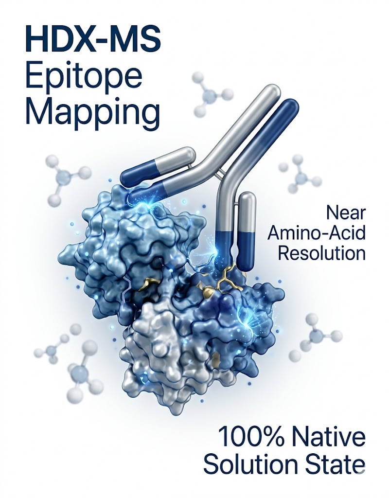
Epitope Mapping of Human Polyclonal Antibodies to the fHbp Antigen of a Neisseria Meningitidis Vaccine by Hydrogen-Deuterium Exchange Mass Spectrometry (HDX-MS). https://www.sciencedirect.com/science/article/pii/S1535947624000240

Background

Mapping epitopes for polyclonal antibodies (pAbs)—such as those generated by vaccines or found in convalescent plasma—is notoriously difficult for the biopharmaceutical industry. Because the human immune response produces a highly heterogeneous mixture of antibodies that bind to many different regions of an antigen simultaneously, traditional monoclonal mapping methods (like obtaining a single co-crystal) are completely ineffective. Understanding exactly which parts of a vaccine antigen are targeted by the immune system is vital for evaluating vaccine efficacy and ensuring batch-to-batch structural consistency.

Methods

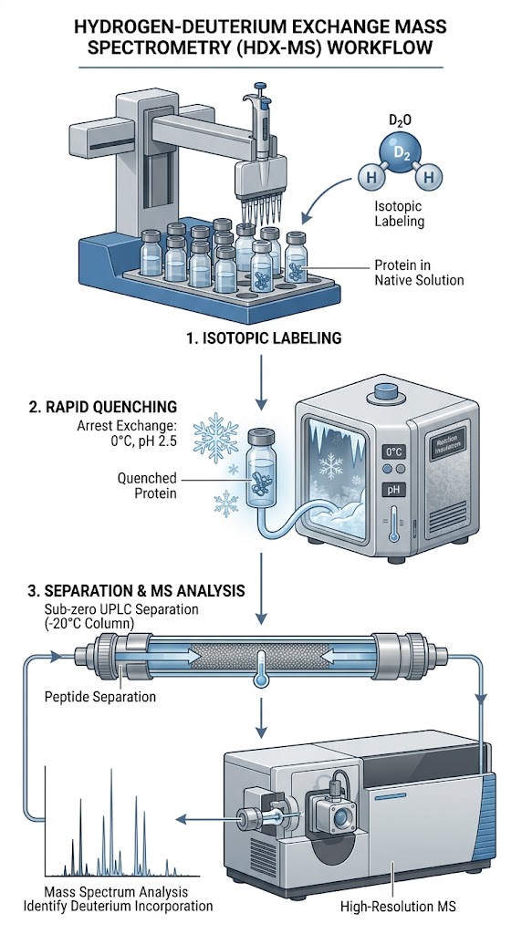
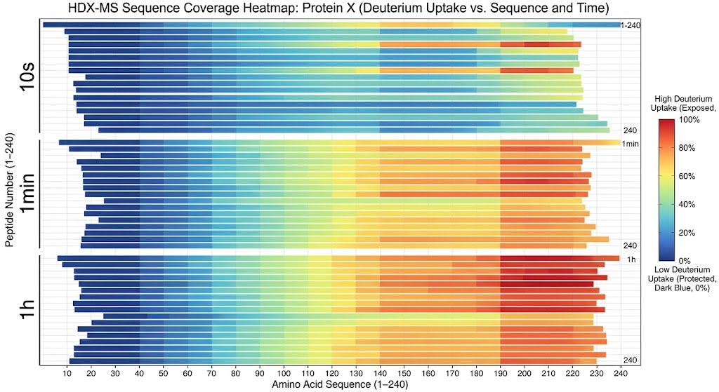
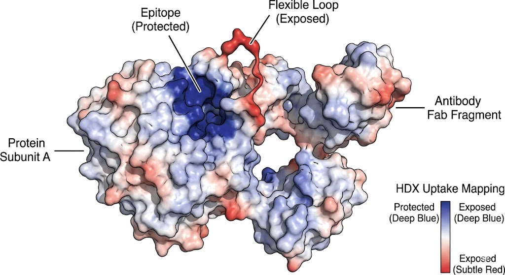
To overcome this massive analytical hurdle, researchers utilized HDX-MS to map the binding interfaces of human pAbs against the fHbp (factor H binding protein) antigen, a critical component of a Neisseria meningitidis vaccine. The recombinant fHbp antigen was incubated with various purified pAb fractions derived from vaccinated individuals. The antigen-antibody complexes were then subjected to timed deuterium exchange (ranging from 10 seconds to 10,000 seconds), rapidly quenched to pH 2.5 to halt the exchange, digested online with pepsin, and analyzed via high-resolution MS to track the localized mass shifts across the entire protein sequence.

Results

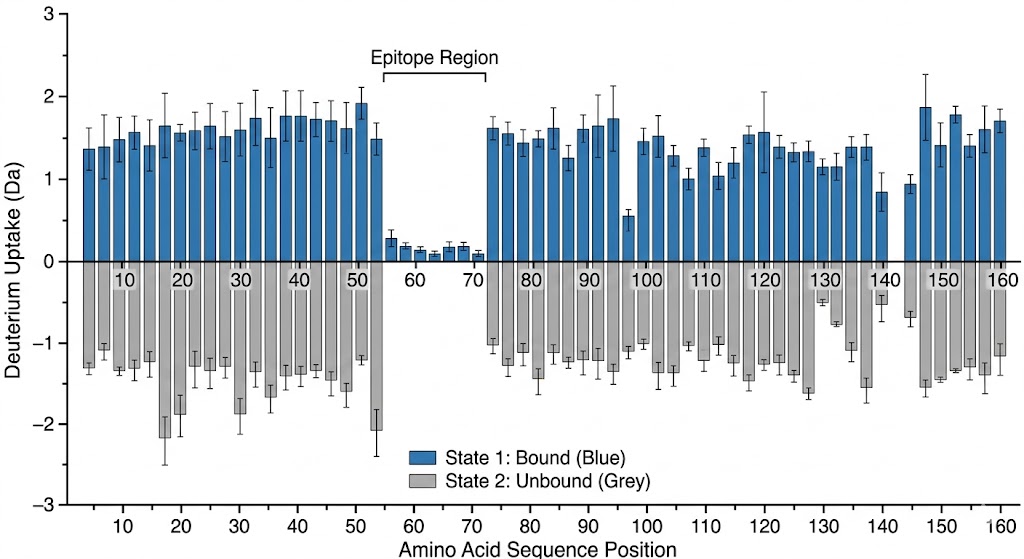
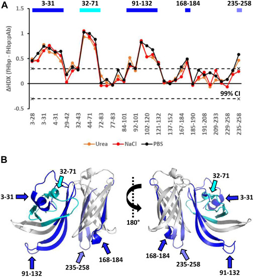
As shown in the graphical data representations from the published study, HDX-MS successfully managed the complexity of the polyclonal mixture. It identified highly specific, localized decreases in deuterium uptake on the fHbp antigen upon pAb binding. The high-resolution mass spectrometry data, coupled with rigorous statistical difference testing, clearly distinguished the immunodominant conformational epitopes across different vaccine recipient samples. Furthermore, it revealed that the polyclonal response was directed at several distinct, spatially separated patches on the folded protein surface.

Conclusion

HDX-MS proved to be a highly effective, label-free method for profiling the complex polyclonal epitope landscape. It provided critical, high-resolution structural insights for vaccine evaluation and development that would be impossible to capture using standard mutagenesis, ELISA, or crystallography. This powerful application highlights HDX-MS as an essential tool not just for monoclonal antibody discovery, but for advanced vaccine characterization and immune response profiling.

HDX-MS identifying binding interfaces of polyclonal antibodies on the fHbp antigen

Representation of HDX-MS capturing localized mass shifts on antigen targets derived from complex polyclonal responses.

Bioinformatics Data Deliverables (IP-Ready Data Packages)

When you partner with us for structural analysis, you do not just receive raw instrument files. You receive a complete, publication-grade data package engineered from the ground up to support your intellectual property claims, defend your patents against challengers, and satisfy rigorous data reviews.

Minimum Deliverables:

  • A comprehensive peptide coverage map. We consistently aim for >85% to 90% overlapping sequence coverage to ensure no potential binding site goes unmonitored.
  • Detailed deuterium uptake plots (kinetics curves) for every analyzed peptide across all time points, complete with standard deviations from technical replicates.
  • Statistical difference plots (Woods/Butterfly plots) highlighting the exact binding regions with strict confidence intervals (e.g., 98% or 99% significance thresholds for uptake differences).
  • A full methodology and QC report detailing instrument parameters, digest conditions, and back-exchange calculations.

Optional Add-ons:

  • High-Resolution 3D Structural Mapping: We map the binding data onto PyMOL or Chimera 3D models for stunning visual evidence, delivering high-resolution rendering files ready for journal submission or patent inclusion.
  • Orthogonal Validation: We can combine your HDX-MS data with Cross-Linking Mass Spectrometry (XL-MS) to provide exact atomic distance constraints, giving you a complete, multi-angled view of your protein complex's architecture.
FAQ

Frequently Asked Questions

Q: What peptide sequence coverage can I expect for my target?

With our optimized online digestion workflows, we routinely achieve over 85% to 90% overlapping sequence coverage for standard protein targets. The redundancy of overlapping peptides allows us to pinpoint binding interfaces with much higher resolution than the length of a single peptide. For highly complex, disulfide-rich, or heavily glycosylated proteins, we utilize multi-protease strategies to maintain maximum coverage and ensure no critical epitopes are missed.

Q: How do you control and minimize hydrogen-deuterium back-exchange?

We combat back-exchange through strict thermodynamic control and automation. We use robotic systems for millisecond-precise timing, instantly quench the reaction at exactly pH 2.5 (the absolute minimum point of intrinsic exchange rate), and perform the entire liquid chromatography separation in a specialized sub-zero chamber to keep the deuterium securely locked onto the backbone amides until it enters the high-vacuum of the mass spectrometer.

Q: Can HDX-MS map epitopes for heavily glycosylated proteins?

Yes, absolutely. While heavy glycosylation can shield the underlying protein backbone from standard pepsin digestion (resulting in large coverage gaps), we have deep experience designing custom digestion protocols for viral spikes and heavily modified receptors. By using alternative acid-stable proteases or performing rapid online deglycosylation within the sub-zero workflow, we can successfully map epitopes on highly modified targets that defeat standard HDX setups.

Plan an HDX-MS Epitope Mapping campaign with the MassTarget™ team

Share your target details and our scientists will design a custom HDX-MS strategy to deliver IP-ready structural evidence for your discovery program.

Online Inquiry

Please submit a detailed description of your project. We will provide you with a customized project plan to meet your research requests. You can also send emails directly to for inquiries.

* Email
Phone
* Service & Products of Interest
Services Required and Project Description