CE-MS Affinity Screening Services: True Label-Free Kd Determination

Overcome the limitations of surface-based binding assays. Our CE-MS affinity screening platform provides true in-solution, label-free Kd determination with ultra-low sample consumption. Ideal for intrinsically disordered proteins (IDPs), RNA targets, and challenging fragments, we deliver high-resolution mass spectrometry evidence to accelerate your hit-to-lead pipeline and help you make confident decisions.

  • 100% label-free, native-state analysis
  • Nanoliter-scale sample consumption
  • Eliminates surface-induced false positives
CE-MS Affinity Screening Services platform featuring true in-solution and label-free mass spectrometry detection
What Is CE-MS Key Benefits Service Overview Workflow & QC Applications Comparison Sample Requirements Deliverables Case Study FAQ

What Is CE-MS Affinity Screening?

Capillary Electrophoresis-Mass Spectrometry (CE-MS) affinity screening is a highly advanced analytical technique that allows our scientists to measure how strongly a drug candidate binds to a target molecule directly in its native biological environment. Unlike older biophysical methods that require you to tether, immobilize, or chemically label your protein on a sensor chip, CE-MS takes place entirely in a free-flowing physiological solution.

The process is elegantly straightforward yet analytically rigorous. We mix your target protein and compound library together in a precisely calibrated liquid buffer. After the molecules achieve thermodynamic binding equilibrium, we apply an electric field. The capillary electrophoresis component gently separates the intact protein-ligand complexes from the unbound, free compounds based on their unique charge-to-size ratios. Immediately following this separation, a high-resolution mass spectrometer detects and weighs these molecules with extreme precision using electrospray ionization (ESI). This continuous, fully integrated workflow gives you a clear, direct readout of the interaction without ever altering your target's natural three-dimensional shape or biological function.

Key Benefits of CE-MS for Drug Discovery

When you are working with hard-to-drug targets or complex molecular glues, traditional screening methods often hit a technological wall. Optical interference, target protein precipitation, and surface crowding can ruin your early-stage data. Our CE-MS platform is specifically engineered to bypass those roadblocks through four core advantages:

Ultra-Low Sample Consumption

We understand that expressing and purifying complex proteins—especially membrane proteins, transcription factors, or large multi-domain complexes—is incredibly difficult, time-consuming, and expensive. CE-MS requires only nanoliter injection volumes. This means we can execute comprehensive screening campaigns using just 10 to 50 micrograms of your total protein, saving you months of upstream protein production time and thousands of dollars in cell culture costs.

True Native-State Analysis

Because there is absolutely no surface immobilization involved, your target protein remains completely free in solution. This preserves its natural folding, conformational flexibility, and active site integrity, ensuring that the binding data you receive accurately reflects real physiological biology.

Zero Surface Interference

We eliminate the false positives and false negatives caused by compounds sticking non-specifically to dextran matrices, gold sensor chips, or plastic multi-well plates. If a compound shows binding in our system, it is because it is genuinely interacting with your protein, not the assay hardware.

High-Resolution Sensitivity

Our mass spectrometry readout does not rely on generic mass-density changes. It identifies the exact isotopic fingerprint of your molecules. It easily distinguishes between closely related compound analogs, identifies specific binding stoichiometries (e.g., 1:1 vs. 2:1 binding ratios), and captures weak, transient interactions that traditional optical methods completely miss.

Service Overview: Our CE-MS Screening Modes

We know that every drug discovery program faces unique challenges depending on the target class and the specific stage of preclinical development. Our team offers highly tailored screening modes to match your specific pipeline needs:

MODE 1

Small-Molecule Hit Validation

Primary high-throughput screens (HTS) often generate hundreds of initial hits, many of which are false positives driven by compound aggregation, auto-fluorescence, or assay interference. We provide fast and highly accurate validation of these hits. By moving your candidates into our solution-phase CE-MS assay, we quickly weed out the artifacts and provide highly accurate binding constants (Kd) to help your medicinal chemistry team rank and prioritize the best molecules for optimization.

MODE 2

Fragment-Based Screening (FBLD)

Fragment libraries are notoriously difficult to screen. Because fragments are so small (typically under 300 Daltons), their binding affinities are extremely weak, often residing in the high micromolar to millimolar range. To see these interactions in traditional assays like NMR or SPR, you must use very high concentrations of the fragment, which frequently leads to concentration-dependent aggregation and non-specific binding. Our high-resolution MS easily detects these subtle, weak interactions without requiring massive compound concentrations. Explore our fragment-based drug discovery services to see how we integrate this specific CE-MS approach into broader fragment-to-lead pipelines.

MODE 3

Difficult Targets: IDPs and RNA

Intrinsically Disordered Proteins (IDPs) and dynamic nucleic acids (like structured RNA motifs) do not have a single, rigid 3D shape; they exist as an ensemble of rapidly changing conformations. When you attach an IDP to a solid surface for SPR, you artificially restrict its radius of gyration and impose a massive entropic penalty on the molecule, fundamentally changing how drugs bind to it. Our in-solution CE-MS environment is the ideal—and sometimes the only—way to study these highly flexible targets in their active states without biasing the binding pocket.

MODE 4

Custom Assay Development

If your target requires a highly specific physiological environment—such as a specific volatile buffer system, essential metal cofactors, or unique temperature controls to maintain stability—our expert analytical chemists will build and optimize a bespoke CE-MS method from the ground up to ensure robust, artifact-free data collection.

CE-MS Screening Workflow & Quality Control

Our workflow is built for transparency, absolute accuracy, and strict reproducibility. We implement rigorous quality control (QC) checkpoints at every phase to ensure your data is undeniably reliable. The standard workflow operates seamlessly from the initial target incubation step, through the precision CE separation, directly into the MS detection chamber, culminating in advanced data analytics.

1

Target and Library Incubation

We carefully mix your protein target and compound libraries in an optimized, mass-spectrometry-compatible volatile buffer (such as ammonium acetate). We allow sufficient time for the system to reach true thermodynamic equilibrium.

2

Capillary Electrophoresis Separation

Using high-voltage capillary electrophoresis, we gently separate the intact protein-ligand complexes from any unbound compounds. QC Checkpoint: We meticulously verify separation resolution, baseline stability, and ensure the electric field does not induce complex dissociation before proceeding.

3

High-Resolution MS Detection

The separated molecules transition directly into the high-resolution mass spectrometer via a specialized electrospray interface. This provides a direct, label-free readout of the exact molecular weights of the complex.

4

Data Analytics and Kd Calculation

Our informatics team processes the raw mass spectra, performing mass deconvolution to identify the precise species. We then use non-linear regression models on the titration data to calculate precise binding affinities (Kd) and verify complex stoichiometry.

CE-MS Affinity Screening Workflow featuring Target Incubation, Capillary Electrophoresis Separation, and MS Detection

Applications: Solving Traditional Screening Bottlenecks

You should strongly consider partnering with us for CE-MS affinity screening when your laboratory encounters the following persistent challenges:

When Functional Assays Do Not Exist

If your target is a scaffolding protein, a transcription factor, or an adaptor protein that lacks a measurable enzymatic activity, traditional biochemical assays are useless. CE-MS provides a direct physical binding readout so you can start your screening campaigns immediately without spending months inventing a complex reporter assay.

When Proteins Precipitate or Lose Activity

If your target aggregates on SPR chips, denatures at room temperature, or dies when subjected to chemical cross-linking tags, our solution-phase approach keeps it stable, hydrated, and biologically active.

When You Need Unbiased Orthogonal Validation

If you have an exciting list of hits from an AI-driven virtual screen or a high-throughput fluorescence assay, CE-MS serves as the ultimate "truth test." It strips away the fluorescent tags and computational assumptions, confirming which molecules genuinely bind to the target in reality.

Technology Comparison: CE-MS vs. SPR vs. ITC

Selecting the right biophysical tool is critical for your project's timeline, budget, and ultimate success. Here is a transparent look at how our CE-MS platform compares to other common interaction analysis techniques.

Comparison DimensionCE-MS (Our Platform)SPR (Surface Plasmon Resonance)ITC (Isothermal Titration Calorimetry)
Core PrincipleSeparation of complexes in a free solution followed by highly accurate mass detection.Real-time optical detection of mass-density changes on a functionalized gold chip.Measurement of micro-heat changes (released or absorbed) during a binding event.
Key StrengthsTrue in-solution equilibrium (no immobilization); ultra-low sample consumption; completely label-free; high mass accuracy.Excellent for determining binding kinetics (on-rates and off-rates); extremely high throughput for stable proteins.Provides a complete thermodynamic profile (enthalpy and entropy); label-free; directly measures stoichiometry.
Key LimitationsRequires optimization for MS-compatible volatile buffers (highly saline physiological buffers need careful handling).Covalent immobilization can alter protein shape; highly prone to non-specific chip binding and surface crowding.Requires massive amounts of highly purified protein (milligram scale); very low throughput.

Solution Selection Strategy:

  • Choose SPR when you are conducting primary high-throughput screens of highly stable, easily expressed proteins, and where real-time kinetic rates are strictly required for your SAR (Structure-Activity Relationship) modeling.
  • Choose CE-MS when your protein sample is extremely scarce, when the target is highly dynamic (like IDPs or RNA), or when you must eliminate surface-induced artifacts to get a true, solution-phase equilibrium Kd.
  • Choose ITC only when you need deep thermodynamic insights into the mechanism of action and possess abundant quantities of stable protein.

Sample Requirements

To ensure the highest quality data and minimize assay optimization time, we recommend the following parameters for your shipped samples. Our team will work closely with you to accommodate specific biological constraints.

Sample TypeRequired AmountConcentrationPurityBuffer Conditions / Notes
Protein Target50–200 µg1–10 µM≥90% preferredMS-compatible (no glycerol, low detergents). Provide sequence, tags, and known ligands.
Protein Complex100–300 µg1–5 µM≥85%Native buffer preferred. Indicate expected stoichiometry.
Membrane Protein200–500 µg1–5 µM≥80%DDM/LMNG acceptable. Provide stabilization conditions.
Small Molecule Library1–5 mg or 10 mM stock-≥90%DMSO. Can screen unpurified libraries.
Fragment Library1–5 mg-≥95%DMSO. Provide SDF if available.

Typical Deliverables & Data Interpretation

We believe in delivering data that you can immediately present in your internal pipeline meetings and use to make critical Go/No-Go decisions. At the conclusion of your CE-MS project, you will receive a comprehensive, audit-ready data package. This includes all raw data files, processed deconvoluted spectra, and a summarized hit-ranking table highlighting the most promising candidates.

The core of our scientific reporting focuses on the direct visualization of complex formation. You will receive Extracted Ion Electropherograms (EIE). These chromatograms clearly show the baseline separation of the compound-bound target peak from the unbound target peak, proving that the complex survived the separation intact.

Alongside the EIEs, we provide classical non-linear regression binding titration curves. By plotting the calculated fraction of the bound target against the varied ligand concentrations, we map the exact saturation point. This combined view not only provides you with highly accurate Kd calculations but also serves as undeniable visual proof of target engagement, completely free from the surface artifacts that plague other methods.

Extracted Ion Electropherogram and binding titration curve calculating in-solution Kd

Representative CE-MS Data Output

Case Study: Profiling Binding Interactions via Affinity Separation-MS

Reference Publication
https://doi.org/10.3389/fimmu.2024.1347871

Background

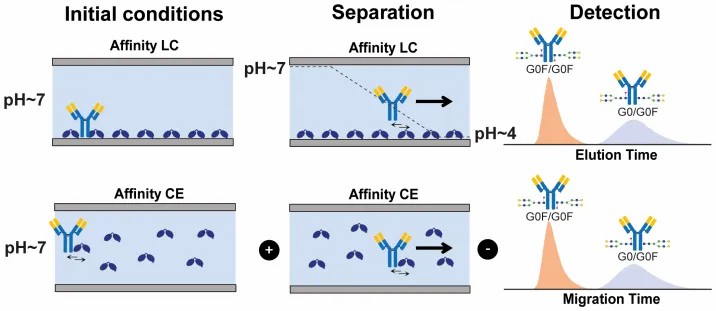
In early-stage biologics and drug discovery, understanding how distinct isoforms or closely related biological species interact with a target is notoriously difficult. Bulk biophysical methods often average out the signals, masking the subtle but critical binding differences between these species. For example, the precise glycosylation state of an antibody can drastically alter its binding affinity to effector receptors, dictating its clinical efficacy and influencing Antibody-Dependent Cellular Cytotoxicity (ADCC).

Methods

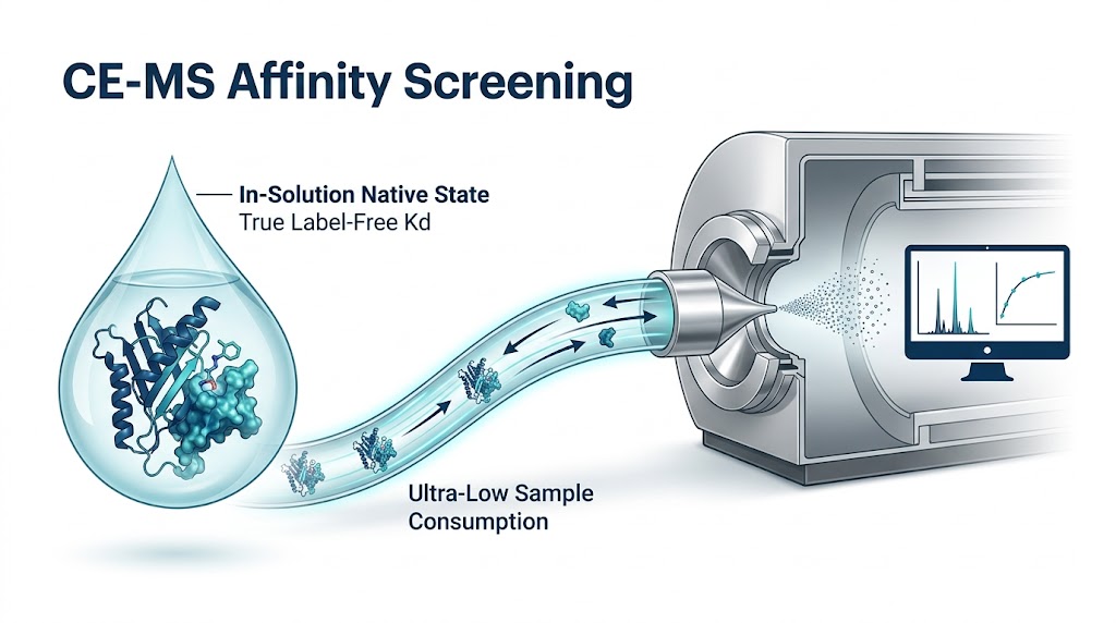
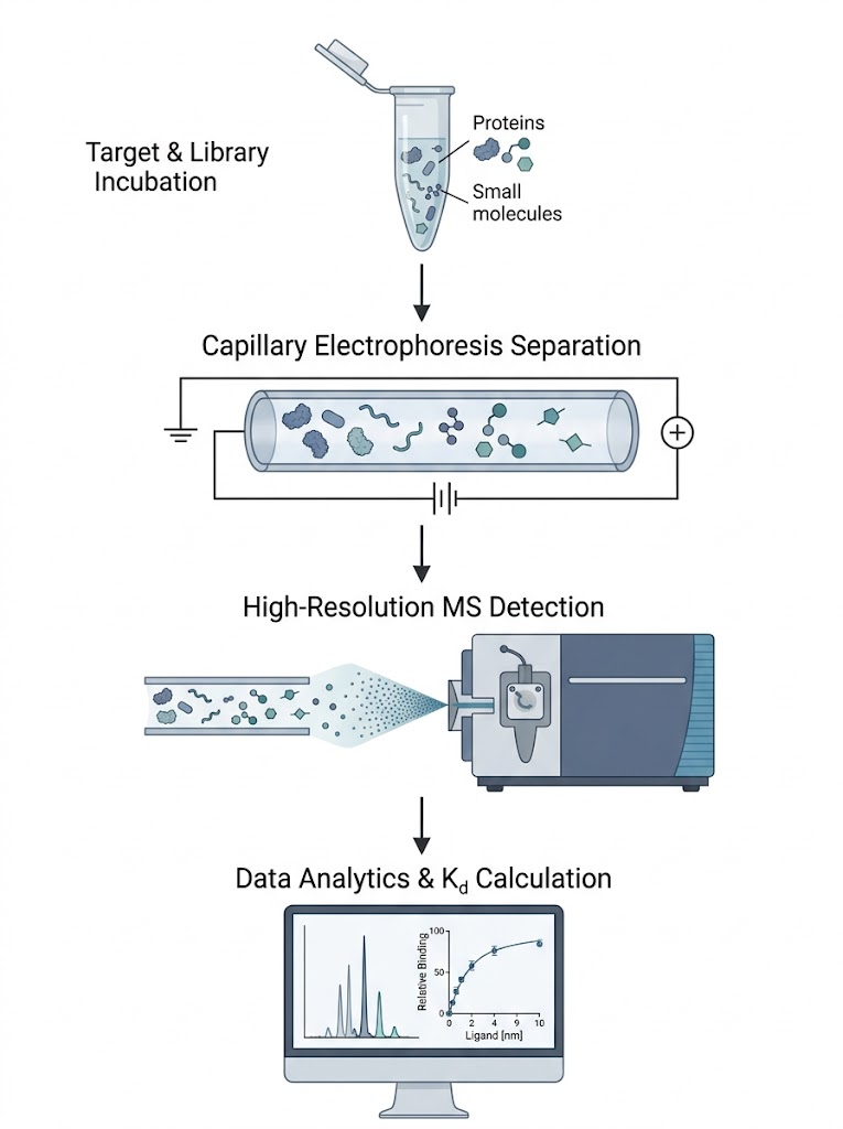
To resolve this, an affinity separation-mass spectrometry workflow was deployed to study the interaction between varying antibody glycoforms and the FcγRIIIa receptor. The target receptor and the heterogeneous antibody ligands were incubated naturally in-solution, allowing them to reach true thermodynamic equilibrium without the steric hindrance of surface tethering. The intact complexes were then separated using high-efficiency capillary techniques. Crucially, they were subsequently analyzed using a high-resolution mass spectrometer to directly resolve the interactions based on exact mass.

Results

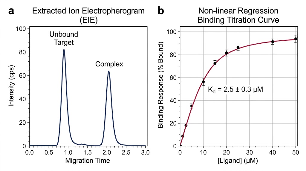
The high-resolution MS readout provided clear, reviewable signal evidence of the distinct interaction profiles without requiring any target modification or fraction purification. Because the mass spectrometer can easily detect the exact mass difference of specific sugar residues (for example, the ~146 Da mass shift indicating the presence of a fucose residue, or the precise mass of terminal galactose units), it could distinguish which specific glycoforms were actively binding to the receptor in real-time. As demonstrated in Figure 1 of the published methodology, the extracted ion chromatograms (EICs) successfully differentiated the specific binding signatures of these closely related species. The data clearly showed that fully fucosylated species had different binding profiles compared to hemi-fucosylated species, and even differentiated binding based on the specific F158/V158 receptor variants, showcasing the platform's unparalleled resolution.

Conclusion

This approach proves that high-resolution affinity separation-MS is highly effective not just for simple binary screening, but for dissecting complex molecular interactions in their native state. By directly observing the exact mass of the binding partners, it provides discovery teams with robust, artifact-free data to prioritize candidates based on highly specific structural nuances that govern in vivo efficacy.

Extracted ion chromatograms differentiating specific binding signatures in an Affinity Separation-MS workflow

Affinity separation-MS workflow resolving binding interactions.

FAQ

Frequently Asked Questions

How does CE-MS differ fundamentally from traditional SPR or BLI assays?

The most critical difference is the physical environment of the assay. SPR and BLI require you to use chemical linkers to tether your protein to a solid gold sensor surface or a fiber-optic tip. This immobilization can restrict the protein's natural movement, force it into an unnatural orientation, or even chemically block the very binding pocket you are trying to target. CE-MS mixes the protein and drug in a free-floating liquid solution, fully preserving the natural biology and preventing false results caused by the sensor chip itself.

What is the typical sample consumption for a CE-MS binding assay?

One of our greatest technical advantages is extreme sample efficiency. While ITC might require milligrams of protein, we typically only need between 10 to 50 micrograms of your target protein for a complete, multi-point screening campaign. This makes CE-MS an ideal choice when your protein is incredibly difficult, time-consuming, or expensive to express and purify, allowing you to conserve precious reagents for downstream functional testing.

Can CE-MS screen compound mixtures or only single purified compounds?

We can screen both highly purified singles and complex mixtures. CE-MS is highly effective at screening pooled compound libraries. Because the high-resolution mass spectrometer acts as an incredibly powerful identification tool, we can incubate your target with a pool of 10 to 50 fragments simultaneously. The MS easily distinguishes between the different compounds in the same batch based on their unique, exact molecular weights, allowing for higher throughput without sacrificing data quality.

Plan a CE-MS campaign with the MassTarget™ team

Share your target and library details and our scientists will design a tailored CE-MS screening strategy for your discovery program.

Compliance & Disclaimer: Creative Proteomics operates under an ISO 17025 quality management system. All services, workflows, and data deliverables provided by MassTarget™ are intended for Research Use Only (RUO). Our services do not provide medical advice, nor are they intended for use in clinical diagnosis, human treatment, or any diagnostic procedures.

Online Inquiry

Please submit a detailed description of your project. We will provide you with a customized project plan to meet your research requests. You can also send emails directly to for inquiries.

* Email
Phone
* Service & Products of Interest
Services Required and Project Description