NADP+/NADPH Analysis Service
Creative Proteomics provides precise NADP+/NADPH analysis services using advanced HPLC and LC-MS technologies. We help researchers quantify NADP+ and NADPH levels, assess redox balance, evaluate enzymatic activities, and study metabolic pathways, offering reliable results for applications in cancer, neurodegenerative diseases, and drug development.
Submit Your Request Now
×
- What We Provide
- Technology Platform
- Advantages
- Sample Requirements
- Demo
- FAQs
- Publications
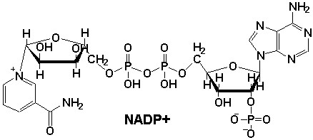
What are NADP+/NADPH?
Nicotinamide adenine dinucleotide phosphate (NADP+) and its reduced form (NADPH) are essential cofactors in a variety of biochemical processes. These molecules are crucial in cellular metabolism, particularly in redox reactions that maintain the cellular balance of oxidative stress and antioxidant defenses. The proper regulation of NADP+/NADPH levels is integral to energy production, cell signaling, and overall cellular health.
NADP+ plays a pivotal role in biosynthetic reactions, acting as a cofactor in various enzymes like dehydrogenases and reductases. NADPH, the reduced form of NADP+, serves as a key electron donor in anabolic reactions, including fatty acid synthesis, nucleic acid synthesis, and detoxification processes.
What Specific Projects Does Creative Proteomics Offer?
Creative Proteomics offers a comprehensive suite of services for NADP+/NADPH analysis, specifically tailored to address both basic and advanced research needs. Our NADP+/NADPH quantification service utilizes state-of-the-art equipment and scientific expertise to ensure precise and reproducible results.
NADP+/NADPH Quantification: Accurate measurement of NADP+ and NADPH levels in various biological samples, such as blood, tissues, and cell cultures.
Enzymatic Activity Assessment: Quantification of NADP+/NADPH-dependent enzymatic activities, including NADPH-dependent reductases and dehydrogenases.
Redox Status Evaluation: Analysis of cellular redox balance, crucial for studying oxidative stress, aging, and disease conditions like cancer and neurodegeneration.
Metabolic Profiling: Monitoring NADP+/NADPH dynamics to investigate metabolic pathways and biosynthetic processes.
Time-Dependent Analysis: Tracking NADP+/NADPH fluctuations over time to understand metabolic changes in response to experimental conditions or treatments.
List of NADP+/NADPH We Can Analyze
| Compound | Detection Method | Typical Biological Samples |
|---|---|---|
| NADP+ (Nicotinamide Adenine Dinucleotide Phosphate) | High-performance liquid chromatography (HPLC), Mass Spectrometry | Blood, tissue, cell lysates |
| NADPH (Nicotinamide Adenine Dinucleotide Phosphate, Reduced Form) | HPLC, Enzyme-Linked Immunosorbent Assay (ELISA) | Blood, tissue, cell lysates |
| NADH (Nicotinamide Adenine Dinucleotide, Reduced Form) | HPLC, Fluorescence Detection | Blood, tissue, cell lysates |
| NAD+ (Nicotinamide Adenine Dinucleotide) | Mass Spectrometry | Blood, tissue, cell lysates |
Methods for NADP+/NADPH Detection
High-Performance Liquid Chromatography (HPLC)
NADP+ and NADPH are separated using a chromatography column, and their absorbance differences are detected at 260 nm using a UV detector.
Instrument Models: Agilent 1260 Infinity II HPLC: Equipped with a quaternary pump, auto-sampler, and Diode Array Detector (DAD).
Detection Parameters:
- Mobile phase: Phosphate buffer (pH 6.0) and methanol gradient elution (flow rate: 1.0 mL/min).
- Retention times: NADP+ ~8.2 minutes, NADPH ~9.5 minutes.
- Sensitivity: Detection limit (LOD) 0.05 μM, Quantification limit (LOQ) 0.2 μM.
Liquid Chromatography-Mass Spectrometry (LC-MS)
Mass spectrometry's high resolution is used to differentiate NADP+ (molecular weight 743.41 Da) and NADPH (molecular weight 745.42 Da). Quantification is performed using Multiple Reaction Monitoring (MRM) mode.
Instrument Models:
Thermo Scientific Q Exactive™ HF-X: Equipped with an electrospray ionization (ESI) source in positive ion mode.
Sciex Triple Quad 6500+: MRM mode quantifies ion pairs m/z 744.3→506.2 (NADP+) and 746.3→508.2 (NADPH).
Detection Parameters:
- Chromatography column: HILIC column (2.1×100 mm, 1.7 μm).
- Mobile phase: Acetonitrile-water (with 0.1% formic acid) gradient elution.
- Sensitivity: LOD 0.02 μM, Linear range 0.05–10 μM (R² >0.995).

Agilent 1260 Infinity II HPLC (Fig from Agilent)

Q Exactive™ HF-X Hybrid Quadrupole-Orbitrap™ Mass Spectrometer (Fig from Thermo Fisher)

Sciex Triple Quad 6500+ (Fig from Sciex)
Advantages of Our NADP+/NADPH Detection
- Dual-Technology Validation: Combines HPLC (Agilent 1260 Infinity II) and LC-MS/MS (Thermo Q Exactive HF-X) for cross-verified accuracy.
- Ultra-High Sensitivity: LC-MS/MS achieves a detection limit of 0.02 μM, ideal for rare samples (e.g., biopsies or CSF).
- Broad Dynamic Range: Quantifies 0.05–100 μM concentrations, covering physiological to pathological levels.
- Absolute Quantification: Uses isotope-labeled internal standards (¹³C-NADP⁺, D₄-NADPH) for<5% CV reproducibility.
Sample Requirements for NADP/NADPH Analysis
| Sample Type | Minimum Sample Volume | Storage Conditions | Recommended Sample Preparation |
|---|---|---|---|
| Blood (Plasma/Serum) | 50 µL | Store at -80°C | Collect in EDTA or heparin tubes, centrifuge immediately to separate plasma/serum. |
| Tissue (Frozen) | 100 mg | Store at -80°C | Homogenize tissue samples in cold buffer. |
| Cell Lysates | 50 µL | Store at -80°C | Lyse cells in appropriate buffer and centrifuge. |
| Cell Culture Supernatant | 100 µL | Store at -80°C | Centrifuge to remove cell debris before collection. |
| Urine | 100 µL | Store at -80°C | Collect in clean, sterile containers. |
Applications of NADP/NADPH Assay
Metabolic Pathway Analysis
Study of NADPH-dependent biosynthetic processes, including lipid and nucleotide synthesis.
Oxidative Stress Assessment
Evaluate cellular redox balance and antioxidant defense mechanisms.
Cancer Research
Investigate NADP+/NADPH's role in tumor metabolism and cell proliferation.
Neurodegenerative Disease Research
Examine NADP+/NADPH levels in relation to oxidative damage in Alzheimer's and Parkinson's.
Drug Development
Analyze the impact of drugs on redox balance and metabolic pathways.
Enzyme Activity Profiling
Assess NADP+/NADPH-dependent enzyme activity and metabolic changes
Demo Result of Targeted Metabolomics Service
Figures come from (Li, Y.et.al, Sci Rep,2023)

Quickly show the distribution of samples across principal components.

OPLS-DA point cloud diagram
Illustrate the separation of sample groups in a multidimensional space.

Plot of multiplicative change volcanoes
Volcano plot depicting multiplicative changes in metabolite levels.

Sample Hierarchical Clustering
Each column in the figure represents a sample
FAQ of NADP/NADPH Analysis
Is an isotope internal standard necessary for mass spectrometry?
Yes. It is recommended to use stable isotope-labeled NADP+ (e.g., ¹³C-NADP+) as an internal standard to correct for ionization efficiency differences.
How to choose between HPLC and LC-MS?
If high sensitivity and multi-component analysis (e.g., simultaneous detection of glutathione) are required, choose LC-MS. For large sample volumes and limited budgets, HPLC is recommended.
What is the sample storage duration?
Ultrafiltered extracts can be stably stored at -80°C for 1 month (for LC-MS); untreated samples should be stored at -80°C for ≤72 hours.
Does mass spectrometry support absolute quantification? How to ensure accuracy?
Yes. We use stable isotope internal standard methods (e.g., ¹³C-NADP+ and ²H-NADPH) for absolute quantification. The internal standard behaves consistently with the target during extraction and ionization, correcting for matrix effects and ion suppression, ensuring data accuracy (RSD<5%).
Will extreme pH or high-salt samples (e.g., gastric fluid, urine) affect detection results?
Yes. High salt or extreme pH may cause column clogging or ion source contamination. Recommendations:
- Pre-treatment: Use ultrafiltration (10 kDa) or solid-phase extraction (SPE) to remove interferents.
- Special protocol: For extreme samples, customize buffer solutions to adjust pH to neutral.
Can NADP+/NADPH and other redox molecules (e.g., glutathione) be detected simultaneously?
Yes. Our LC-MS method supports multi-target analysis, for example:
- Simultaneous detection of NADP+/NADPH, NAD+/NADH, GSH/GSSG (linear range 0.05–20 μM).
- Use HILIC columns to separate polar molecules and achieve high specificity with MRM mode.
How to validate the specificity of the analytical method?
Specificity is validated through the following steps:
Blank matrix test: Add standards to samples without targets to confirm no background interference.
Spiked recovery: Add known concentrations of NADP+/NADPH to cell lysates; recovery should be 90–110%.
Mass spectrometry fragment analysis: Confirm characteristic fragment ions of targets (e.g., m/z 506.2 for NADP+) using high-resolution mass spectrometry (HRMS).
How to improve detection sensitivity for samples with extremely low NADPH content (e.g., mitochondrial extracts)?
The following strategies are recommended:
Enrichment: Use affinity columns (e.g., NADPH-specific antibody-conjugated magnetic beads) for pre-concentration.
Micro-volume injection: Use nanoflow LC-MS systems (e.g., Eksigent ekspert™ nanoLC 425) to reduce sample consumption and enhance sensitivity to fmol levels.
Is it necessary to provide metabolic state information (e.g., hypoxic/normal conditions) of the samples?
Yes. The NADP+/NADPH ratio is sensitive to cellular metabolic states. It is recommended to provide:
- Cell culture conditions (e.g., oxygen concentration, starvation treatment duration).
- Rapid quenching methods during sample collection (e.g., liquid nitrogen freezing or methanol/dry ice quenching).
What is included in the data report? Are raw data provided?
The standard report includes:
- Target concentrations (μM or pmol/mg protein), NADP+/NADPH ratio, and quality control sample results (CV values).
- Optional: Raw chromatograms/mass spectra, method validation report (compliant with FDA/EMA guidelines).
- Raw data (.raw or .mzML format) can be downloaded via a secure cloud platform.
Is large-scale cohort study support available (e.g., thousands of clinical samples)?
Yes. For high-throughput needs, we provide:
- Automated pre-processing: Hamilton STARlet liquid handling workstation with a daily processing capacity of up to 500 samples.
- Batch effect correction: Insert quality control (QC) and blank samples in each batch, and use the ComBat algorithm to eliminate batch differences.
How to address chlorophyll interference in plant samples (e.g., leaves)?
Chlorophyll can interfere with UV detection (HPLC). Solutions include:
- Solid-phase extraction (SPE): Use C18 columns to adsorb chlorophyll and elute the targets.
- Mass spectrometry avoidance: LC-MS excludes chlorophyll signals (m/z >900) through mass selectivity.
Learn about other Q&A.
NADP+/NADPH Analysis Case Study

Publications
Here are some publications in Metabolomics research from our clients:

- Characterization of a novel AraC/XylS-regulated family of N-acyltransferases in pathogens of the order Enterobacterales. 2020. https://doi.org/10.1371/journal.ppat.1008776
- Quantification of choline in serum and plasma using a clinical nuclear magnetic resonance analyzer. 2022. https://doi.org/10.1016/j.cca.2021.11.031
- Anti-inflammatory activity of black soldier fly oil associated with modulation of tlr signaling: A metabolomic approach. 2023. https://doi.org/10.3390/ijms241310634
- Untargeted metabolomics reveal sex-specific and non-specific redox-modulating metabolites in kidneys following binge drinking. 2023. https://doi.org/10.1530/REM-23-0005
- Non-invasive elevation of circulating corticosterone increases the rejection of foreign eggs in female American robins (Turdus migratorius). 2022. https://doi.org/10.1016/j.yhbeh.2022.105278