Digital & Molecular Pathology Service
Welcome to our comprehensive Digital & Molecular Pathology Service. We provide state-of-the-art tissue analysis and imaging solutions designed to accelerate your research, enhance biomarker discovery, and deliver precise, actionable data.
Whether you need foundational morphological assessments or highly complex spatial biology insights, our end-to-end services are tailored to meet the rigorous demands of modern scientific and preclinical studies. Explore our core capabilities below to find the right solution for your project.
Submit Your Request Now
×
- Our services
- Workflow
- Platform
- Applications
- Why Choose
- FAQs
Our core services
| Service | Description | Best For |
|---|---|---|
| Immunohistochemistry (IHC) & Immunofluorescence (IF) | Detect and localize specific proteins within your tissue sections with high sensitivity and specificity for routine expression analysis. | Biomarker discovery, target validation, and cellular localization. |
| Multiplex IHC (mIHC) & Multiplex IF (mIF) | Visualize and quantify multiple targets simultaneously within a single tissue section, unlocking deep insights into complex tissue microenvironments and cellular interactions. | Spatial phenotyping, immune profiling, and maximizing data from limited tissue. |
| In Situ Hybridization (ISH) | Precisely localize specific nucleic acid sequences (DNA or RNA) directly within their morphological context at the single-cell level. | Transcriptomics, viral genome detection, and gene amplification/deletion analysis. |
| Hematoxylin and Eosin (H&E) Staining | High-quality, reproducible, and automated H&E staining to serve as the foundational histological evaluation for your tissue samples. | High-quality, reproducible H&E staining providing essential morphological context, serving as both a foundational histological evaluation and a critical map for spatial omics workflows. |
| Confocal Microscopy for 3D Tissue Imaging | Advanced confocal microscopy for deep tissue imaging, precise co-localization studies, and detailed spatial reconstruction. | 3D tissue reconstruction, deep imaging of thick sections, and subcellular structural analysis. |
Workflow
Our streamlined process is designed to take your project from initial concept to actionable data with full transparency and rigorous quality control at every step.

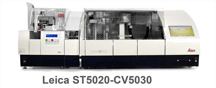
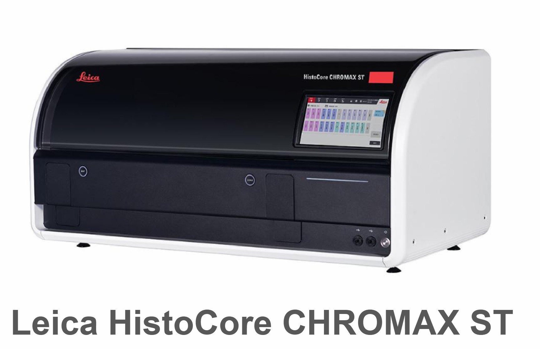
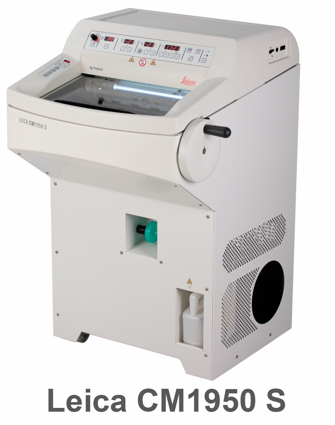
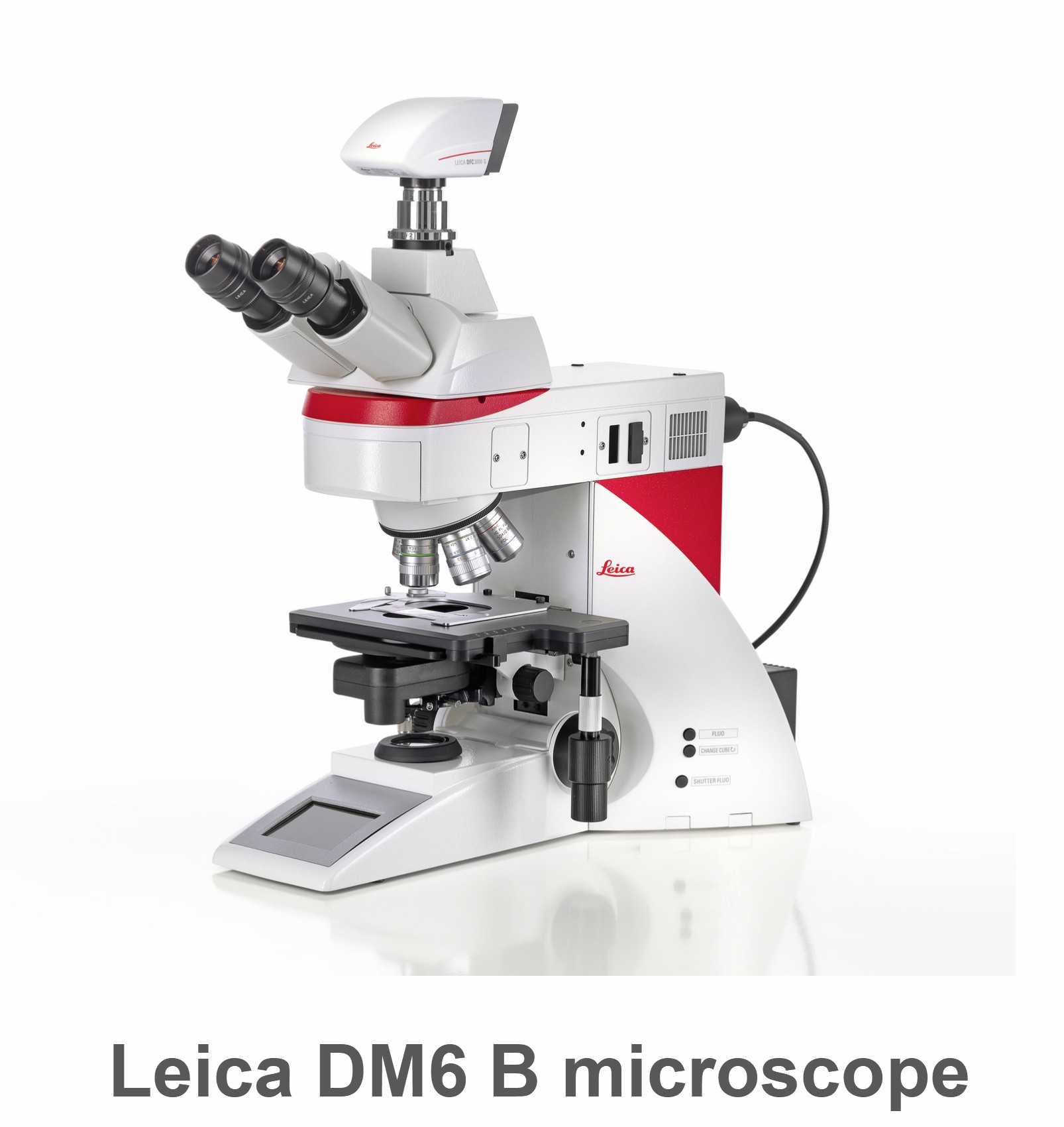
Platform

Key Applications
Our digital and molecular pathology solutions are designed to support a wide range of scientific disciplines and drug development phases. We frequently partner with researchers across the following areas:
- Oncology & Immuno-Oncology: Deeply profile the tumor microenvironment (TME), assess immune cell infiltration, and evaluate immune checkpoint expression using our integrated multiplexing and spatial analysis workflows.
- Neuroscience: Map neuroinflammation, characterize cellular phenotypes in neurodegenerative diseases, and perform high-resolution 3D reconstruction of neural tissue architectures.
- Drug Discovery & Toxicology: Accelerate your pipeline from early target validation and target engagement studies to late-stage tissue cross-reactivity (TCR) testing and safety profiling.
- Spatial Biomarker Discovery & Validation: Seamlessly translate the discovery of novel spatial signatures into robust, optimized assays tailored for advanced basic research and enterprise-scale tissue profiling.
Why Choose Our Pathology Services?
- Advanced Digital Scanning: Whole-slide imaging capabilities for easy remote viewing, annotation, and digital archiving of your slides.
- Expert Analysis: Access to experienced pathologists and image analysis scientists to help interpret complex spatial data.
- Customizable Workflows: From assay development and protocol optimization to full-scale staining and quantitative image analysis, we tailor our approach to your specific study goals.
FAQs
Do you offer custom antibody optimization and assay development?
Yes. If you are investigating a novel biomarker or using a proprietary antibody, our scientists will work with you to develop and fully validate a custom staining protocol. We ensure maximum sensitivity and specificity before applying the assay to your full study cohort.
Can I combine multiple services for a single project?
Absolutely. In fact, we highly recommend an integrated approach. For example, many clients utilize our H&E Staining Service to assess baseline morphology and select specific Regions of Interest (ROIs), which then guides the targeted analysis using our Multiplex IHC or ISH spatial profiling workflows.
What is the typical turnaround time for a project?
Turnaround times vary based on the scale and complexity of your study. For an accurate delivery timeframe, please contact our technical team for a consultation.
Learn about other Q&A.


