Disease Mechanism and Pathway Analysis Service

Multi-modal disease mechanism analysis integrating target engagement, signaling, metabolomics, chemical proteomics, and ADME characterization from the MassTarget platform.

Disease mechanisms operate across multiple biological dimensions — target binding, signaling modulation, metabolic reprogramming, and cellular phenotype. Understanding how a compound or genetic perturbation drives a disease phenotype requires evidence from each of these layers.

The MassTarget platform addresses this challenge by integrating seven complementary MS-based service categories into a coordinated disease mechanism analysis workflow. From affinity screening and thermal profiling to phosphoproteomics, metabolomics, chemical proteomics, and ADME characterization — each module provides a distinct piece of the mechanism puzzle.

Solution Highlights:

  • Seven integrated service categories covering the full mechanism analysis spectrum
  • Modular design — select the combination that matches your research question
  • Coordinated sample processing for consistent, comparable datasets
  • Cross-module integration with interpreted biological pathway reports
  • Direct pipeline connectivity to target discovery and validation services
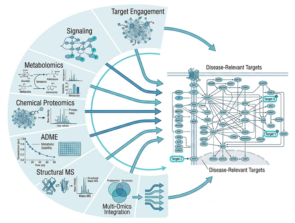
Disease mechanism analysis platform diagram showing seven integrated service categories converging from target engagement through signaling, metabolomics, chemical proteomics, and ADME to pathway interpretation.
Overview Research Questions Capabilities Strategy Workflow Sample Deliverables Case Study FAQ

Disease Mechanism Analysis Requires More Than One Assay

Your team has identified a compound of interest, a disease-relevant mutation, or a phenotypic hit — and now the real question emerges: how does it work? What cellular pathways does it engage? Is the observed phenotype driven by target engagement, signaling modulation, metabolic reprogramming, or a combination of these?

Answering these questions is the central challenge of preclinical drug discovery. Yet the complexity of disease mechanisms means that no single analytical readout can capture the full picture. A compound that inhibits a kinase may produce no change in that kinase's protein abundance — its effect is on phosphorylation state, not expression level. A metabolic shift observed in a disease model might be a primary driver or a secondary consequence of broader pathway dysregulation. Distinguishing cause from effect requires layered evidence from multiple experimental dimensions.

The MassTarget platform is designed specifically for this challenge. Rather than offering a single assay or omics modality, we integrate seven complementary categories of MS-based services — spanning target engagement, signaling analysis, metabolic profiling, chemical proteomics, ADME characterization, multi-omics integration, and high-throughput screening — into a coordinated disease mechanism analysis workflow. This enables your team to build a complete mechanistic picture from a single project engagement, with each layer providing orthogonal evidence that strengthens the conclusions drawn from the others.

Layered Research Questions We Address

Disease mechanism analysis in drug discovery typically progresses through a hierarchy of questions, each requiring a different analytical approach. The MassTarget platform addresses them systematically:

Target Engagement Level. Is the compound engaging its intended target in the disease-relevant cellular context? Which proteins does it bind beyond the primary target? Questions at this level are answered through affinity-based approaches — affinity selection MS (AS-MS), equilibrium dialysis–MS binding, and native ESI-MS for noncovalent complexes — combined with cellular thermal stability profiling such as thermal proteome profiling (TPP) and 2D-TPP, which report target engagement and off-target binding directly in live cells without requiring compound modification.

Signaling and PTM Level. Once target engagement is confirmed, the next question is which signaling pathways are modulated. Protein abundance changes alone rarely reveal pathway activation. Phosphoproteomics activation mapping captures phosphorylation events across thousands of sites, identifying kinase cascades and network-level signaling changes. Ubiquitinomics, acetylomics, and other PTM-focused analyses further refine the view.

Metabolic and Functional Level. What are the functional consequences of pathway modulation? Untargeted and targeted metabolomics capture the metabolic state of treated cells or tissue — shifts in energy metabolism, lipid homeostasis, nucleotide biosynthesis, and oxidative stress markers. Our untargeted metabolomics for MoA service provides a direct functional readout that complements proteomic and signaling data.

Chemical and Covalent Level. For covalent inhibitors, natural products, and probes that bind through reactive chemistry, understanding which residues are modified and which proteins are engaged requires dedicated chemical proteomic approaches. Activity-based protein profiling (ABPP), reactive residue profiling (Cys/Lys/Tyr/Ser), and photoaffinity labelling (PAL-MS) identify direct protein targets and modification sites with residue-level resolution.

ADME and Metabolite Level. Understanding the metabolic fate of a compound is essential for interpreting disease mechanism data. Our ADME/DMPK research platforms cover metabolic stability, metabolite identification (MetID), and drug permeability profiling, providing the pharmacokinetic context for mechanism-of-action studies.

Solution Capabilities — Seven Service Categories

The MassTarget platform organizes its disease mechanism analysis capabilities into seven service categories. Each category contains multiple individual service modules that can be selected, combined, or sequenced according to your specific research question.

CATEGORY 1

Target Engagement and Binding Evidence

MS-based and biophysical approaches for establishing whether a compound interacts with its proposed target. Modules include AS-MS, equilibrium dialysis–MS binding, native ESI-MS, BLI, SPR, HDX-MS, and thermal stability profiling (TPP, PISA, SPROX, 2D-TPP). Each provides a different type of binding evidence — affinity, kinetics, stoichiometry, or cellular occupancy — and the choice depends on target type, compound modality, and the evidence required for your project stage.

CATEGORY 2

Structural and Conformational MS

When the mechanism involves conformational changes — allosteric modulation, protein-protein interaction disruption, or epitope mapping — structural MS methods provide the necessary resolution. HDX-MS, FPOP, XL-MS, and limited proteolysis MS (LiP-MS) each report different aspects of protein conformation and dynamics in solution.

CATEGORY 3

Signaling and PTM Mapping

Understanding disease mechanism often requires tracking post-translational modifications beyond simple protein abundance. Phosphoproteomics, ubiquitinomics, and targeted PTM analysis reveal the signaling cascades and regulatory networks that drive disease phenotypes. Kinase enrichment analysis from phosphoproteomic data identifies upstream kinases responsible for observed phosphorylation changes.

CATEGORY 4

Metabolic and Functional Profiling

Untargeted metabolomics (HILIC + RP), targeted metabolomics panels, lipidomics, fluxomics (13C tracer), and cellular metabolomics screening provide a direct functional readout of the metabolic consequences of disease or drug treatment. These modules capture the metabolic phenotype that results from pathway modulation.

CATEGORY 5

Chemical Proteomics and Target Deconvolution

For covalent inhibitors, electrophilic fragments, natural products, and phenotypic hits with unknown targets, chemical proteomics provides the most direct route to target identification. ABPP (global, enzyme-family-specific, and competitive formats), reactive residue profiling, SuFEx chemoproteomics, PAL-MS, and covalent fragment screening cover the full spectrum of chemoproteomic needs.

CATEGORY 6

ADME/DMPK Characterization

Early understanding of metabolic stability, metabolite profiles, and drug permeability informs disease mechanism interpretation. Our ADME modules include metabolic stability (microsomes/S9/hepatocytes), metabolite identification (MetID), metabolic soft-spot analysis, plasma protein binding, and LC-MS/MS bioanalysis for PK curve generation.

When multiple omics datasets are generated from the same sample set, our seventh category — Multi-Omics Integration and Systems Analysis — correlates proteomic, phosphoproteomic, metabolomic, and lipidomic changes into unified pathway maps, adding a systems-level dimension to mechanism interpretation.

Technology Strategy — Which Module Fits Each Question

Selecting the right combination of analytical modules is the first step in designing an effective disease mechanism study. The table below maps common research questions to the most appropriate MassTarget service modules.

Research QuestionRecommended Module(s)Key AdvantageTypical Output
Does the compound bind the proposed target?AS-MS, Native ESI-MS, TPPLabel-free; no target modification requiredBinding confirmation, affinity rank, melt curve shift
Which off-targets does the compound engage?2D-TPP, ABPP-MS, PAL-MSProteome-wide coverage; no pre-selection biasOff-target list with confidence scores
Which signaling pathways are activated?Phosphoproteomics, Kinase enrichmentCaptures pathway modulation invisible to proteomicsPhosphosite table, kinase enrichment, signaling network
What are the metabolic consequences?Untargeted metabolomics, LipidomicsDirect functional readout; captures non-protein-mediated effectsDifferential metabolite table, pathway enrichment
What is the metabolic fate of the compound?Metabolic stability, MetIDEssential for PK/PD interpretation; identifies active metabolitesMetabolic stability t1/2, metabolite ID list
Is the compound a covalent modifier?ABPP, Reactive residue profiling, SuFExIdentifies exact modification sitesTarget list with residue-level modification sites
How do proteomic and metabolic changes connect?Multi-omics integration, Network pharmacologyOrthogonal cross-validation across datasetsIntegrated pathway map, prioritized target nodes

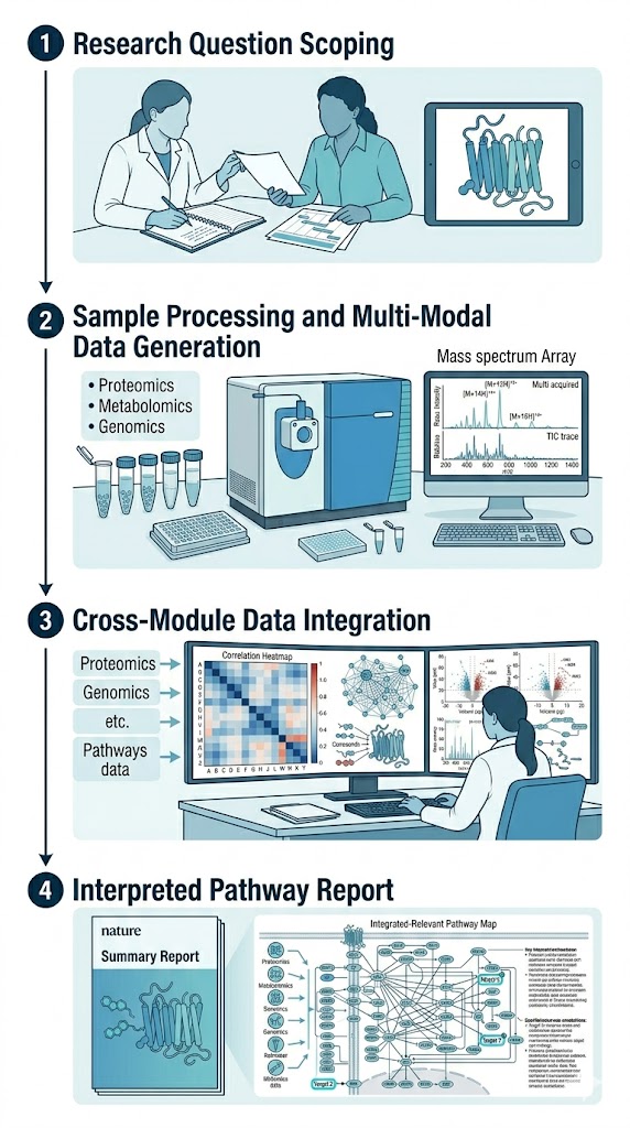
Our Workflow — From Research Question to Integrated Report

A structured four-stage process designed to match analytical depth to your project stage while maintaining data consistency across modules.

1

Research Question Scoping

We begin with a structured consultation to understand your compound, disease model, and primary research question. Based on this discussion, we recommend a tailored combination of service modules. The goal is to match the analytical depth to your project stage — not every mechanism study requires all seven categories, but the selected set must provide sufficient orthogonal evidence to support robust conclusions.

2

Sample Processing and Multi-Modal Data Generation

Samples are distributed across the selected service modules. Where possible, samples are split from a single intake to ensure matched biological and technical variability across datasets. Each module runs through its optimized, validated workflow — from sample preparation through MS data acquisition to initial quality control.

3

Cross-Module Data Integration

Data from each module are processed independently, then integrated through correlation-based and network-based approaches. For multi-omics projects, we use established integration frameworks to identify convergent evidence across modalities. For projects combining target engagement data with signaling or metabolic readouts, we map the relationships between binding events and downstream pathway perturbations.

4

Interpreted Pathway Report

You receive a comprehensive report that includes module-level QC metrics, differential expression or binding tables for each module, integrated pathway maps showing how the datasets converge on a unified mechanism, and a written biological interpretation with prioritized pathway nodes as candidate intervention points.

Four-stage workflow for disease mechanism analysis: research question scoping, multi-modal data generation, cross-module integration, and interpreted pathway report.

Sample Requirements and Project Intake

Sample requirements vary depending on the selected service modules. The following table provides general guidelines for the most common sample types.

Sample TypeMinimum per GroupRecommendedAmount per SampleFormat
Cell pellets (cultured)3 per condition5 per condition1 x 107 cellsDry pellet, snap-frozen
Tissue (snap-frozen)5 per group10-15 per group20-50 mgCryovial, LN2 snap-frozen
Plasma/serum10 per group20-30 per group50-100 microLClear aliquot, no hemolysis
CSF10 per group15-20 per group100-200 microLClear aliquot, frozen
Tissue (FFPE)10 per group20-30 per group5 x 10 microm sectionsUnstained slides
Purified protein--50-100 microg per cond.Buffer, flash-frozen

Note: For multi-modality projects combining target engagement, omics, and ADME analyses, sample requirements may vary. We provide a detailed intake checklist after the initial scoping consultation.

Bioinformatics Integration and Deliverables

Each project delivers module-level outputs tailored to the selected services, plus a comprehensive integration report. Standard deliverables include:

  1. Module-level QC reports — Performance metrics for each service module: protein identification depth, phosphosite localization confidence, metabolite annotation rate, thermal stability curve quality, binding assay reproducibility.
  2. Differential expression and binding tables — For each omics modality: fold changes, p-values, FDR. For binding modules: binding curves, affinity estimates, target lists with confidence scores.
  3. Pathway enrichment — KEGG, Reactome, GO, and WikiPathways enrichment analyses for each dataset, with cross-module comparison of enriched pathways.
  4. Integrated pathway map — Unified visualization of dysregulated pathways with annotations showing which module(s) contributed each piece of evidence.
  5. Written biological interpretation — A project-specific narrative connecting the experimental findings to your original research question, with prioritized pathway nodes and candidate follow-up targets.

Case Study: MS Proteomic Screening Reveals Parkinson's Disease Mechanism

Lee JH, et al. "Parkinson's disease-associated LRRK2-G2019S mutant acts through regulation of SERCA activity to control ER stress in astrocytes." Acta Neuropathologica Communications, 2019, 7, 68. DOI: 10.1186/s40478-019-0716-4 (CC BY 4.0).

Background

The LRRK2-G2019S mutation is one of the most common genetic causes of familial Parkinson's disease. Previous studies had linked LRRK2 to ER stress, but the molecular mechanism connecting the mutant protein to ER dysfunction remained unclear. This study aimed to define the mechanism by which LRRK2-G2019S accelerates ER stress and cell death in astrocytes — a key question for understanding the non-cell-autonomous mechanisms of PD pathogenesis.

Methods

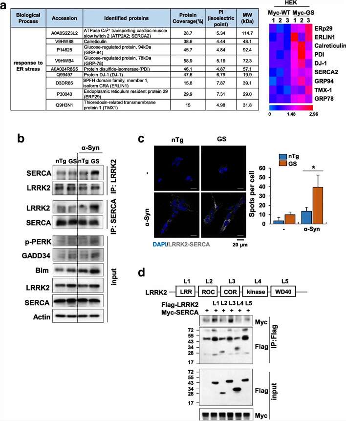
Using label-free MS proteomic screening, the team immunopurified LRRK2-associated protein complexes from HEK293T cells expressing either wild-type LRRK2 or the G2019S mutant, followed by LC-MS/MS identification of co-purifying proteins. Key steps included:

  • Co-transfection of HEK293T cells with siRNA targeting endogenous LRRK2 and 3xMyc-tagged wild-type or G2019S-mutated LRRK2.
  • Immunoprecipitation of LRRK2 protein complexes followed by label-free MS analysis (Fig. 3a).
  • Cytoscape network analysis and GO enrichment to identify enriched cellular components and biological processes.
  • Validation of candidate interactions via co-immunoprecipitation, proximity ligation assays (PLA), and truncated LRRK2 mutant constructs (Fig. 3b-d).

Results

The MS analysis identified 2,831 proteins, of which 232 showed enhanced binding to LRRK2-G2019S compared to wild-type LRRK2. Cytoscape network analysis revealed enrichment in ER and stress response pathways. Among the top hits was SERCA2 (sarco/endoplasmic reticulum Ca2+-ATPase 2), a critical regulator of ER calcium homeostasis. Follow-up validation confirmed that LRRK2-G2019S binds SERCA with higher affinity through its LRR and COR domains (Fig. 3b-d) and inhibits SERCA activity by stabilizing the interaction with its negative regulator phospholamban (PLN). This led to ER calcium depletion, PERK-CHOP pathway activation, ER-mitochondrial calcium transfer, and mitochondrial dysfunction.

Conclusions

The study established a complete mechanistic chain from the LRRK2-G2019S mutation to astrocyte cell death: enhanced LRRK2-SERCA binding → SERCA inhibition → ER calcium depletion → ER stress → mitochondrial dysfunction. This exemplifies how MS-based proteomic screening can identify unexpected disease-relevant protein interactions, enabling the construction of a full mechanistic model that would be impossible from transcriptomic or single-readout approaches alone.

LRRK2-G2019S MS proteomic screening workflow showing identification of SERCA as interacting protein leading to ER stress mechanism in Parkinson's disease.

Fig. 3a-d: MS proteomic screening identified SERCA as a LRRK2-G2019S interacting protein, with validation by co-IP, PLA, and truncated mutant mapping (Lee JH, et al. Acta Neuropathol Commun, 2019).

How to Choose the Right Strategy

Not every disease mechanism project requires all seven service categories. The right strategy depends on your project stage, compound modality, and the specific research questions you need to answer:

Hit-to-lead stage. If your priority is establishing that a compound engages its intended target and understanding the basic mechanism, start with target engagement (AS-MS or TPP) and global proteomics. Add phosphoproteomics if the target is a kinase or signaling protein.

Lead optimization stage. At this stage, off-target liability and metabolic stability become critical. Add chemical proteomics (ABPP or PAL-MS) for off-target profiling and ADME modules (metabolic stability, MetID) for PK characterization.

Natural products or phenotypic hits. If the target is unknown, begin with chemical proteomics (ABPP, PAL-MS, or reactive residue profiling) and untargeted metabolomics with GNPS molecular networking for mechanism deconvolution.

Late-stage candidate characterization. For compounds nearing preclinical nomination, a full multi-modal assessment combining target engagement, signaling, metabolic, chemical proteomic, and ADME modules provides the comprehensive evidence package needed for candidate selection decisions.

We will work with your team to design the optimal module combination and provide a detailed project proposal before any work begins.

FAQ

Frequently Asked Questions

Q: Why is a multi-modal approach better than single-omics for disease mechanism analysis?

Disease mechanisms operate across multiple biological dimensions — target binding, signaling, metabolism, and cellular phenotype. A single omics modality captures only one dimension. Multi-modal evidence from orthogonal approaches provides convergent validation and reduces the risk of interpreting correlative changes as causal.

Q: Can I start with one module and add others later?

Yes. Our workflow is designed for modular expansion. However, if you plan to integrate multiple modalities, we recommend coordinating sample collection and processing from the start to ensure matched biological material across all modules.

Q: How much sample is needed for a full multi-modal analysis?

The amount depends on the selected modules. A combined analysis of proteomics + phosphoproteomics + metabolomics typically requires 20-50 mg tissue or 1 x 10⁷ cells per condition. Adding target engagement modules may require additional material. We assess this during the scoping consultation.

Q: Can I use FFPE tissue for disease mechanism analysis?

Proteomics and phosphoproteomics can be performed on FFPE tissue. Metabolomics from FFPE is limited — fresh-frozen samples are strongly preferred. Target engagement methods like TPP require live or fresh-frozen cells and cannot be performed on FFPE.

Q: How does the MassTarget platform differ from using multiple individual CROs?

Using a single platform eliminates cross-vendor variability in sample handling, data formats, and interpretation frameworks. Our integrated pipeline ensures that all datasets are generated under consistent QC standards and interpreted within a unified mechanistic model, rather than requiring your team to reconcile disparate outputs from multiple providers.

References

  1. Lee JH, et al. "Parkinson's disease-associated LRRK2-G2019S mutant acts through regulation of SERCA activity to control ER stress in astrocytes." Acta Neuropathologica Communications, 2019, 7, 68. DOI: 10.1186/s40478-019-0716-4
  2. Savitski MM, et al. "Tracking cancer drugs in living cells by thermal profiling of the proteome." Science, 2014, 346(6205), 1255784. DOI: 10.1126/science.1255784
  3. Meissner F, et al. "The emerging role of mass spectrometry-based proteomics in drug discovery." Nature Reviews Drug Discovery, 2022, 21(9), 637-654. DOI: 10.1038/s41573-022-00409-3

Ready to map disease mechanisms with a multi-modal approach?

Contact our team to design the optimal module combination for your project.

Disclaimer: All products and services provided by Creative Proteomics are for research use only (RUO). They are not intended for use in diagnostic, therapeutic, or clinical procedures.

Online Inquiry

Please submit a detailed description of your project. We will provide you with a customized project plan to meet your research requests. You can also send emails directly to for inquiries.

* Email
Phone
* Service & Products of Interest
Services Required and Project Description