Patch-Clamp MS Coupling for Ion Channel Drug Binding Analysis

Integrating gold-standard electrophysiology with high-resolution mass spectrometry for simultaneous functional and molecular drug-target engagement evidence.

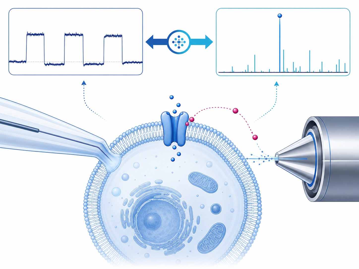
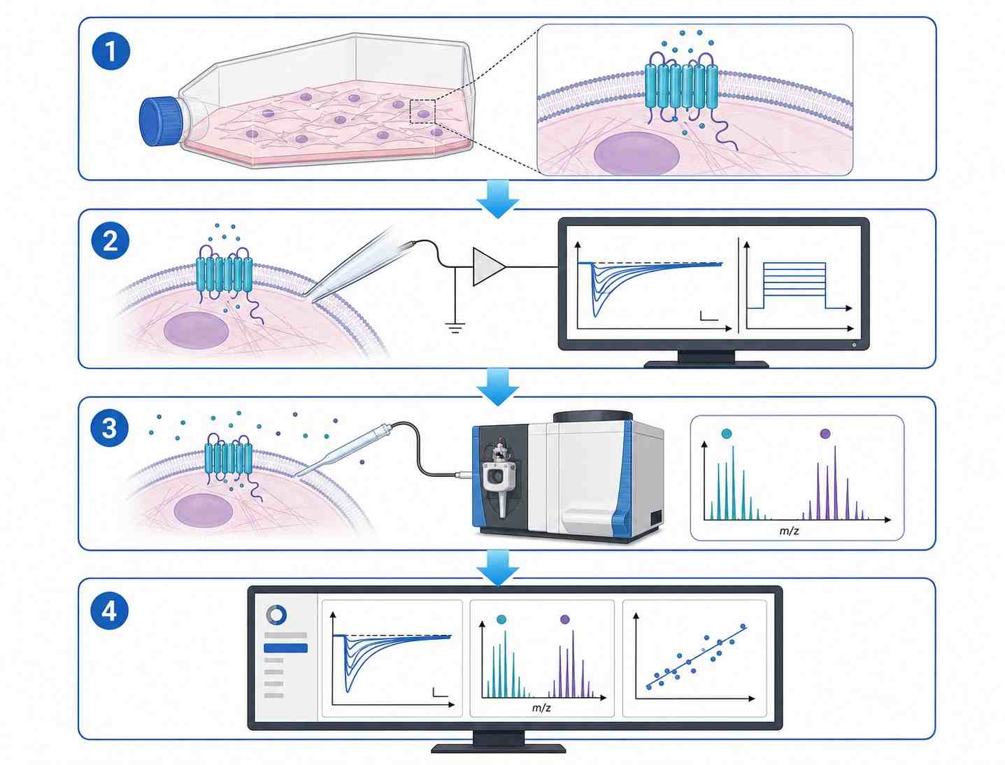
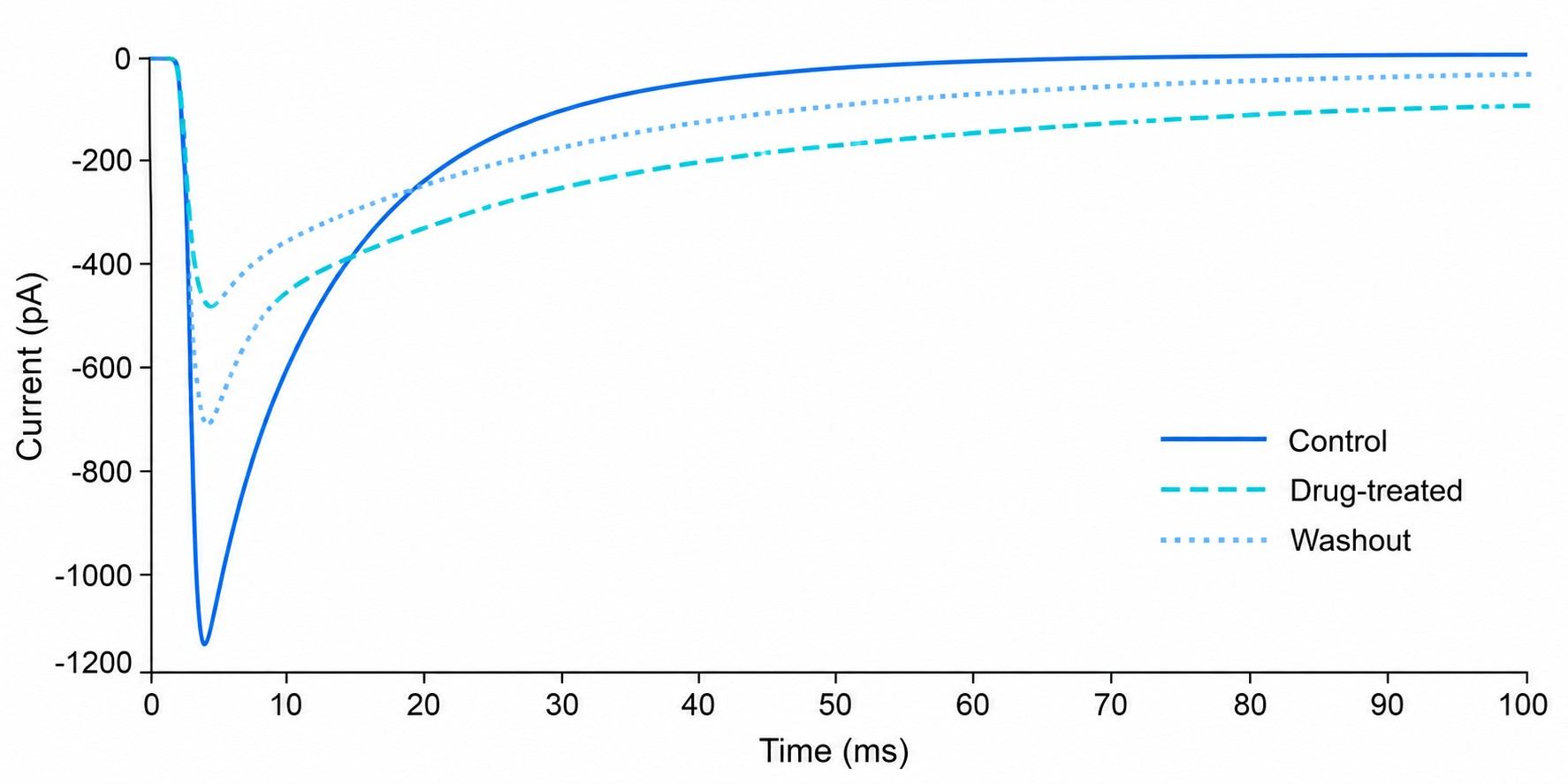
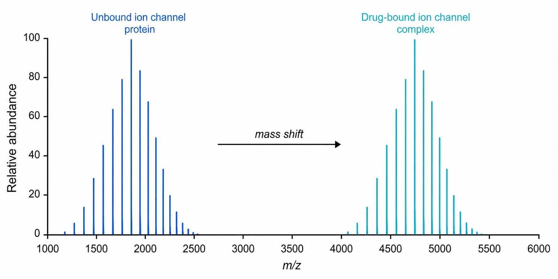
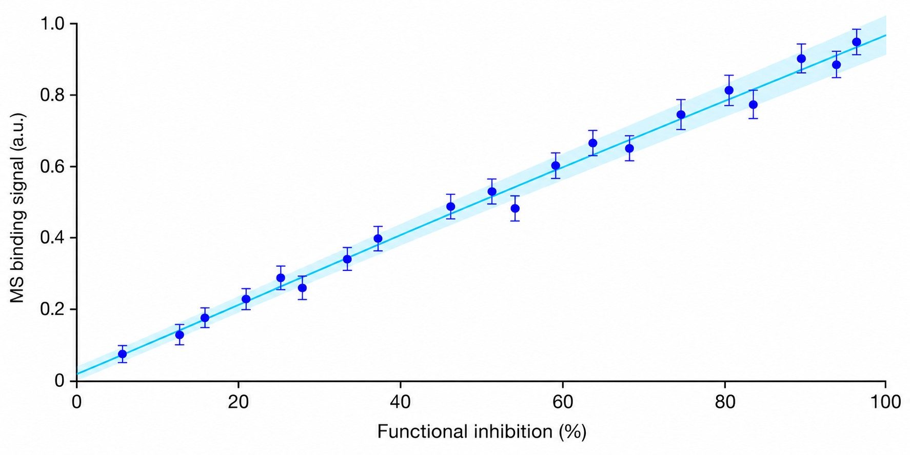
Patch-clamp MS coupling combines whole-cell patch-clamp electrophysiology with mass spectrometry-based detection to deliver concurrent functional activity and direct drug binding confirmation for ion channel targets. This integrated approach addresses a critical gap in ion channel drug discovery: traditional patch-clamp provides exquisite functional data but no molecular binding evidence, while standard binding assays lack functional context. By correlating channel activity with drug binding stoichiometry and chemical identity in a single experiment, researchers can make more informed decisions during lead optimization and candidate selection.

At Creative Proteomics, our Patch-Clamp MS Coupling service is designed to support ion channel drug discovery programs across pharmaceutical, biotech, and academic research settings. We provide end-to-end support from assay design through data integration, delivering orthogonal evidence that accelerates development timelines and reduces late-stage attrition.

Key Advantages:

  • Simultaneous electrophysiology and MS readout from the same cellular preparation.
  • Direct drug binding confirmation in native cell membrane environments.
  • Reduced false positives from functional-only screening approaches.
  • Orthogonal evidence for medicinal chemistry optimization.
  • Compatible with voltage-gated, ligand-gated, and other ion channel classes.
  • Custom assay design tailored to your specific ion channel target.
Patch-Clamp MS Coupling technology platform diagram integrating patch-clamp electrophysiology with high-resolution mass spectrometry for ion channel drug binding analysis.
What Is Patch-MS Service Overview Workflow Applications Comparison Sample Demo Case Study FAQ

What Is Patch-Clamp MS Coupling?

Patch-clamp MS coupling is an integrated analytical technique that combines whole-cell patch-clamp electrophysiology with high-resolution mass spectrometry to enable simultaneous measurement of ion channel functional activity and direct chemical detection of drug binding events.

The principle is straightforward: a cell expressing the ion channel of interest is approached with a patch-clamp micropipette to establish a gigaseal and whole-cell configuration. Once stable electrophysiological recording is established, the drug compound is applied, and ion channel currents are monitored in real time to assess functional modulation. Simultaneously or sequentially, the cellular contents or membrane-associated material are sampled and introduced into a mass spectrometer for direct detection and quantification of the bound drug molecule.

This dual-readout approach provides two complementary data streams from the same biological preparation: (1) functional electrophysiology data showing how the drug modulates channel activity (activation, inhibition, gating kinetics), and (2) molecular binding data confirming drug-target engagement and providing stoichiometric information. The correlation of these two data types within a single experiment eliminates the uncertainty inherent in comparing results from separate functional and binding assays.

For related MS-based binding approaches, explore our Affinity Selection MS (AS-MS) service.

Key Benefits of Integrated Patch-Clamp MS Analysis

Dual functional and molecular readout

Obtain electrophysiological activity data and direct drug binding confirmation from the same experiment, eliminating cross-assay variability.

Native membrane context

Drug-target interactions are studied in intact cell membranes, preserving the native lipid environment, channel conformation, and regulatory protein interactions.

Reduced false positives

Functional-only screens can produce false positives from assay interference or off-target effects. MS binding confirmation provides orthogonal validation.

Mechanistic differentiation

Correlate functional effects (activation, inhibition, modulation) with binding parameters (stoichiometry, residence time) to distinguish orthosteric vs. allosteric mechanisms.

Our Patch-Clamp MS Coupling Service Workflow

Our service workflow is designed to integrate seamlessly with your ion channel drug discovery pipeline, from assay design through data delivery.

1

Target expression and cell preparation

Ion channel of interest is expressed in a suitable cell line (HEK293, CHO, or other). Cells are cultured, harvested, and prepared for patch-clamp recording under optimized conditions.

2

Whole-cell patch-clamp recording

A gigaseal is established, and whole-cell configuration is achieved. Baseline channel activity is recorded, followed by drug application. Current traces are acquired under voltage-clamp or current-clamp mode as required.

3

Drug binding sampling and MS analysis

Following functional recording, bound drug molecules are sampled from the cellular preparation and introduced into a high-resolution mass spectrometer (QToF or Orbitrap) for detection and quantification.

4

Data integration and reporting

Electrophysiology traces and MS spectra are aligned, correlated, and presented in an integrated report with functional-binding correlation plots, binding stoichiometry, and statistical analysis.

Patch-Clamp MS Coupling service workflow diagram showing four steps from cell preparation through patch-clamp recording, MS analysis, and data integration.

Ion Channel Targets and Applications

Our Patch-Clamp MS Coupling service supports a broad range of ion channel classes and drug discovery applications. Below are representative target classes and application scenarios.

Voltage-gated sodium channels

Example targets: Nav1.1–Nav1.9

Applications: Pain therapeutics, epilepsy, cardiac arrhythmia

Confirm drug binding to sodium channel isoforms with simultaneous functional and molecular readout.

Voltage-gated potassium channels

Example targets: Kv1–Kv12, hERG (Kv11.1)

Applications: Cardiac safety (hERG), neurological disorders, immuno-oncology

Assess both functional hERG inhibition and direct drug binding for comprehensive safety profiling.

Voltage-gated calcium channels

Example targets: Cav1–Cav3

Applications: Hypertension, chronic pain, epilepsy

Differentiate orthosteric vs. allosteric calcium channel modulators with dual-readout evidence.

Ligand-gated ion channels

Example targets: GABA-A, nAChR, NMDA, AMPA, P2X

Applications: CNS disorders, pain, inflammation

Study neurotransmitter receptor drug binding in native membrane environments.

TRP channels

Example targets: TRPV1, TRPA1, TRPM8

Applications: Pain, inflammatory conditions, metabolic disease

Evaluate drug binding and functional modulation of thermosensory and nociceptive channels.

Chloride channels

Example targets: CFTR, ClC family

Applications: Cystic fibrosis, genetic channelopathies

Support rare disease drug discovery with integrated functional-binding characterization.

Key application areas:

  • Lead optimization: Confirm drug-target binding for lead series and differentiate mechanism of action.
  • hERG safety profiling: Assess both functional hERG inhibition and direct drug binding to the hERG channel.
  • Orthosteric vs. allosteric differentiation: Correlate functional modulation patterns with binding site occupancy.
  • Residence time analysis: Evaluate drug unbinding kinetics using MS-based washout detection.
  • Selectivity profiling: Test drug candidates across related channel subtypes with both functional and binding readouts.

For related high-throughput screening capabilities, explore our HTS-MS screening platform and RapidFire MS screening services.

Technical Comparison — Patch-Clamp MS vs. Conventional Approaches

DimensionPatch-Clamp MS CouplingPatch-Clamp AloneMS Binding Alone
Functional readout✓ Full electrophysiology✓ Full electrophysiology✗ Not available
Binding confirmation✓ Direct MS detection✗ Inferred only✓ Direct MS detection
Membrane context✓ Native cell membrane✓ Native cell membrane✗ Purified protein typically
ThroughputModerateLow–ModerateHigh
Data dimensionalityHigh (function + binding)Medium (function only)Low (binding only)
Orthogonal validationBuilt-inRequires separate assayRequires separate assay

For alternative MS-based binding approaches, our Native MS binding analysis and cell-based MS screening services provide complementary capabilities.

Sample Requirements

Sample TypeRecommended QuantityPreparationShipping ConditionsNotes
Suspended cells≥ 1 × 10⁶ cells per conditionCentrifuge at 400–1000g for 5–10 min, wash 2–3× with PBS, flash freeze in liquid nitrogenDry ice, overnight deliveryIt is recommended to prepare at least 2–3 tubes per sample, each containing > 50 μL cell pellet
Adherent cells≥ 1 × 10⁶ cells per conditionRemove medium, wash 3× with PBS, digest with trypsin, collect and wash with PBSDry ice, overnight deliveryRecord cell volume before freezing
Stable cell line (recommended)≥ 2 × 10⁶ cells per conditionCulture to 70–80% confluence, harvest, wash with PBS, flash freezeDry ice, overnight deliveryProvide cell line information, passage number, and culture conditions

General notes:

  • Samples should be collected, flash-frozen in liquid nitrogen, and stored at −80 °C immediately.
  • Avoid repeated freeze-thaw cycles — aliquot samples before freezing.
  • For projects with fewer than 10 samples, the laboratory does not perform quality control by default. If necessary, please note this when placing the order.
  • Under permissive conditions, it is recommended to prepare an extra sample as a backup.
  • For sample types not listed here or for specific inquiries regarding sample preparation, please contact our team for consultation.

For broader cellular drug binding applications, see our cell permeability MS service.

Demo Results

Our Patch-Clamp MS Coupling service delivers three complementary data types that together provide a complete picture of drug–ion channel interactions.

Electrophysiology current traces showing drug-induced modulation of ion channel activity with baseline, drug application, and washout phases.

Electrophysiology traces

Whole-cell current recordings showing baseline channel activity, drug-induced modulation, and recovery upon washout.

Mass spectrum showing drug-bound ion channel complex with labeled peaks for unbound and bound species.

MS binding spectra

High-resolution mass spectra confirming drug binding to the ion channel target, with clear separation of bound and unbound species.

Integrated correlation plot showing functional inhibition percentage versus MS binding signal for multiple drug concentrations.

Function–binding correlation

Integrated plot correlating functional inhibition (from electrophysiology) with drug binding signal (from MS) across concentration ranges.

Case Study: Automated Patch Clamp Protocol for Drug–Ion Channel Binding Dynamics

Lukacs P, Pesti K, Földi MC, Zboray K, Toth AV, Papp G, Mike A. "An Advanced Automated Patch Clamp Protocol Design to Investigate Drug—Ion Channel Binding Dynamics." Frontiers in Pharmacology, 2021, 12:738260. https://doi.org/10.3389/fphar.2021.738260

Background

Ion channel drug discovery requires methods that can assess both functional activity and binding dynamics of drug candidates. Standard automated patch clamp protocols often provide only endpoint measurements, missing detailed information about drug binding and unbinding kinetics. Lukacs et al. (2021) developed an advanced automated patch clamp protocol design using the IonFlux Mercury microfluidics-based system to investigate drug–sodium channel binding dynamics with high temporal resolution.

Methods

The authors designed a voltage protocol consisting of 17 pulses organized into three groups: SDO (slow depolarization offset), RFI (recovery from inactivation), and SSI (steady-state inactivation). This protocol was applied repeatedly throughout the experiment to track changes in channel properties during drug application. Four well-known sodium channel inhibitors were tested: lidocaine, riluzole, benzocaine, and bupivacaine. Automated fitting algorithms were used to extract Boltzmann and double-exponential parameters from each sweep, enabling real-time tracking of drug-induced changes.

Results

The advanced protocol successfully resolved compound-specific, concentration-independent biophysical properties of sodium channel inhibition. As shown in Figure 5 of the study, lidocaine caused a concentration-dependent slowing of recovery from inactivation, with the slow time constant component (A2) increasing from 8.8% in control to 68.8% at 1000 µM, while the slow time constant value itself remained concentration-independent. Lidocaine also induced a concentration-dependent hyperpolarizing shift in the voltage dependence of steady-state inactivation (V1/2). Riluzole showed a distinct profile with different effects on recovery kinetics and steady-state inactivation compared to lidocaine, demonstrating the protocol's ability to differentiate compound-specific mechanisms. The automated fitting approach achieved throughput of approximately 10 minutes per drug concentration, substantially faster than conventional manual patch clamp protocols.

Conclusions

The study demonstrated that advanced automated patch clamp protocols can provide detailed, mechanism-specific information about drug–ion channel binding dynamics that goes beyond simple endpoint measurements. This approach enables researchers to differentiate compound-specific biophysical signatures, assess concentration-dependent binding effects, and obtain mechanistic insights that support medicinal chemistry optimization.

Automated patch clamp protocol design for drug-ion channel binding dynamics showing voltage protocol schematic and concentration-dependent effects of lidocaine on sodium channel inactivation.

Figure 5 from Lukacs et al. (2021) showing concentration-dependent effects of lidocaine on sodium channel recovery from inactivation and steady-state inactivation parameters.

FAQ

Frequently Asked Questions

Q: What types of ion channels can be studied with patch-clamp MS coupling?

Our service supports a broad range of ion channel classes, including voltage-gated sodium (Nav), potassium (Kv, hERG), calcium (Cav) channels, ligand-gated channels (GABA-A, nAChR, NMDA), TRP channels, and chloride channels. The key requirement is stable expression in a cell line suitable for patch-clamp recording. Please contact us to discuss your specific target.

Q: How does the MS component detect drug binding to ion channels?

Following functional recording, bound drug molecules are sampled from the cellular preparation and analyzed by high-resolution mass spectrometry (QToF or Orbitrap). The MS detects the intact drug molecule based on accurate mass measurement and fragmentation patterns, providing direct chemical evidence of drug-target engagement. Quantification is achieved through comparison with internal standards.

Q: What is the minimum sample amount required for an experiment?

We recommend a minimum of 1 × 10⁶ cells per condition for suspended or adherent cell preparations. For stable cell lines, 2 × 10⁶ cells per condition is preferred. Samples should be flash-frozen in liquid nitrogen and stored at −80 °C. Please see our Sample Requirements section for detailed guidelines.

Q: Can this technique be used for hERG safety screening?

Yes. Our Patch-Clamp MS Coupling service can be applied to hERG (Kv11.1) channel safety profiling. The integrated approach provides both functional hERG inhibition data (IC50) and direct confirmation of drug binding to the hERG channel, offering a more complete safety assessment than functional data alone. This dual readout can help differentiate true hERG blockers from false positives caused by assay interference.

Q: How do you correlate electrophysiology data with MS binding data?

Data correlation is achieved by aligning the time-resolved electrophysiology recording with the MS binding measurement from the same cellular preparation. We generate integrated correlation plots showing functional modulation (e.g., percent inhibition, shift in V1/2) versus MS binding signal across multiple drug concentrations. This enables direct comparison of functional potency and binding affinity within a single experimental system.

Q: What is the typical turnaround time for a study?

Turnaround time depends on the complexity of the study design, number of compounds, and ion channel target. Typical single-compound studies can be completed within 2–4 weeks. Larger selectivity panels or multi-concentration studies may require 4–8 weeks. We will provide a detailed timeline during the project scoping phase.

References

  1. Lukacs P, Pesti K, Földi MC, Zboray K, Toth AV, Papp G, Mike A.An advanced automated patch clamp protocol design to investigate drug—ion channel binding dynamics. Front Pharmacol. 2021;12:738260.
  2. Aerts JT, Louis KR, Crandall SR, Govindaiah G, Cox CL, Sweedler JV.Patch clamp electrophysiology and capillary electrophoresis–mass spectrometry metabolomics for single cell characterization. Anal Chem. 2014;86(6):3203-3208.
  3. Zhu H, Li Q, Liao T, et al.Metabolomic profiling of single enlarged lysosomes. Nat Methods. 2021;18:788-798.
  4. Ahmadi S, Benard-Valle M, Boddum K, Cardoso FC, King GF, Laustsen AH, Ljungars A.From squid giant axon to automated patch-clamp: electrophysiology in venom and antivenom research. Front Pharmacol. 2023;14:1249336.

Advance your ion channel drug discovery program

Share your target and compound details, and our scientists will design a tailored Patch-Clamp MS Coupling strategy for your ion channel drug binding study.


For Research Use Only. Not for use in diagnostic or clinical procedures.

Online Inquiry

Please submit a detailed description of your project. We will provide you with a customized project plan to meet your research requests. You can also send emails directly to for inquiries.

* Email
Phone
* Service & Products of Interest
Services Required and Project Description