Digital Microfluidics MS for Automated Nanoscale Sample Processing

Program individual droplets on an electrode array — extract, derivatize, purify, and ionize at the nanoliter scale, all under software control, with direct mass spectrometric readout.

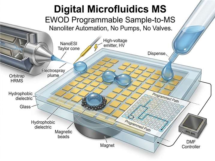
Digital microfluidics (DMF) manipulates discrete droplets on an open or covered array of insulated electrodes using electrowetting-on-dielectric (EWOD). Unlike channel-based microfluidics, DMF has no pumps, no valves, and no fixed fluid paths — every droplet is individually addressable by activating a sequence of electrodes. When integrated with electrospray ionization mass spectrometry, DMF becomes a programmable sample-to-MS platform: nanoliter-volume samples are dispensed, mixed, reacted, extracted, and delivered directly to the MS inlet under full software automation.

Our digital microfluidics MS service leverages this architecture for applications where manual sample preparation introduces variability, consumes precious sample, or limits throughput. We deliver integrated DMF-MS workflows for proteomics sample processing, on-chip chemical derivatization, magnetic bead-based solid-phase extraction, and multi-step reaction monitoring — each droplet a self-contained micro-laboratory with direct MS readout.

Digital microfluidics EWOD chip coupled to electrospray mass spectrometry platform diagram
What Is Digital MS Service Overview Technology Comparison Workflow Sample Demo Case Study FAQ

What Is Digital Microfluidics–Mass Spectrometry?

Digital microfluidics–mass spectrometry (DMF-MS) combines electrowetting-on-dielectric (EWOD) droplet actuation with electrospray ionization mass spectrometry. An EWOD chip consists of an array of independently addressable electrodes coated with a hydrophobic dielectric layer — typically a Parylene-C insulator topped with Teflon-AF. Applying an AC or DC voltage (50–200 VRMS) to an adjacent electrode asymmetrically modulates the droplet contact angle via the Lippmann-Young equation, generating a surface tension gradient that drives droplet motion. By sequentially activating electrodes under software control, droplets are dispensed from on-chip reservoirs, transported across the array, merged, mixed by bidirectional oscillation, split, and positioned at designated locations — the complete liquid-handling repertoire — without any moving mechanical parts.

The integration with MS is direct: a dedicated electrode on the chip periphery aligns the processed droplet with a nanoESI emitter or a liquid junction that feeds an ESI source. A small orifice in the chip cover plate or an open-edge geometry allows the droplet to contact the emitter capillary, and the electrospray potential (1.5–3 kV) is applied either through the emitter itself or through a dedicated electrode beneath the droplet. The droplet contents are sprayed directly into the mass spectrometer, yielding spectra within seconds of the final sample-preparation step. This closed-loop, software-driven architecture replaces an entire bench-top workflow — pipetting, incubation, centrifugation, transfer, and LC injection — with a single programmable chip that consumes 500–1,000 times less sample than conventional protocols. For discovery-stage drug research programs, this sample-conserving capability is especially valuable for treatment-versus-control experimental designs where biological material from sorted cell populations or microdissected tissue is inherently limited and conventional preparation workflows consume the entire sample.

Key Advantages of Digital Microfluidics MS

Programmable Automation Without Fluidic Hardware

Every liquid-handling step — dispensing, transport, merging, mixing, splitting — is executed by software-controlled electrode activation. No syringe pumps, no rotary valves, no tubing connections to leak or fail. Workflows are defined as electrode sequences in a script, enabling reproducible, unattended operation across hundreds of samples with the exact same timing for each droplet.

500–1,000× Reduction in Sample and Reagent Consumption

DMF operates on 100 nL–5 μL droplet volumes per step. A full reduction-alkylation-digestion-desalting proteomics workflow that consumes 50–100 μL in a tube consumes under 200 nL on-chip — an enzyme and reagent cost saving that compounds across large screening campaigns. Precious samples — laser-microdissected tissue, sorted cell populations, low-abundance protein isolates from co-immunoprecipitation — are conserved for downstream orthogonal analysis rather than consumed in preparation.

Multi-Step Sample Preparation With Online MS Readout

Because each droplet is individually tracked by capacitive position sensing, multi-step protocols execute sequentially with the final droplet delivered directly to the ESI emitter within a single chip layout. There is no off-line transfer step between sample preparation and MS analysis — eliminating sample loss to tube walls and pipette tips, airborne keratin contamination, and oxidation artifacts that compromise labile post-translational modifications such as methionine oxidation or cysteine sulfenylation.

Magnetic Bead Integration for On-Chip Solid-Phase Extraction

DMF chips integrate magnetic bead manipulation as a native operation: beads are suspended in a droplet, captured by an external neodymium magnet positioned beneath the chip, and the supernatant is moved away electronically by activating adjacent electrodes. This enables on-chip desalting, enrichment (e.g., phosphopeptide TiO2 beads, glycan HILIC beads), protein purification (Ni-NTA, streptavidin), and buffer exchange — all within the same platform that delivers the purified analyte directly to the MS. The magnetic capture is fully reversible, enabling serial elution and re-capture steps that are difficult to automate off-chip.

Service Overview

MODE 1

Automated Bottom-Up Proteomics Sample Preparation

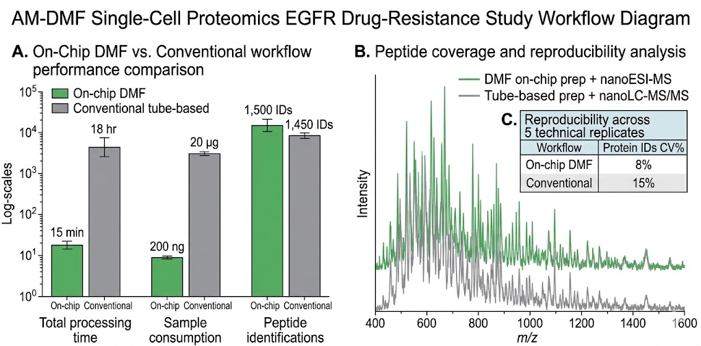
All steps performed on-chip in nanoliter droplets: protein reduction (DTT, 56 °C, 10 min), alkylation (IAA, dark, 20 min), tryptic/LysC digestion (37 °C, 30–45 min), and C18 desalting via magnetic beads. The purified peptide droplet is delivered directly to nanoESI-MS for shotgun proteomics. We routinely achieve >1,500 protein identifications from 100 ng of total protein input — comparable to conventional overnight tube-based protocols that consume 50–100× more sample. The entire process from sample loading to first MS spectrum is completed in under 45 minutes, eliminating manual pipetting variability and enabling cohort-scale studies with uniform preparation. This workflow integrates with our broader shotgun proteomics drug effect analysis service for treatment-response profiling.

MODE 2

On-Chip Chemical Derivatization & Labeling

Execute chemical labeling reactions — TMTpro 16/18-plex, iTRAQ 8-plex, reductive dimethylation, or custom derivatization chemistries — inside individually addressed droplets. Reagent addition, mixing by electrode oscillation, incubation under temperature control, and quenching are all programmed as electrode sequences. Direct MS infusion after on-chip derivatization avoids sample losses inherent to off-line SPE cartridge cleanup, enabling detection of low-stoichiometry labeled species that would otherwise fall below the MS detection limit after sample handling losses. Label incorporation efficiency is quantified for every droplet, providing per-sample QC that is impractical in batch-labeling workflows.

MODE 3

Magnetic Bead-Based On-Chip SPE & Enrichment

Functionalized magnetic beads (C18, TiO2, HILIC, Ni-NTA, streptavidin) are co-encapsulated with sample droplets. After binding, beads are immobilized by a magnet beneath the chip while the supernatant is moved away. Wash and elution droplets are delivered sequentially, with the final eluate routed to the MS emitter. Applied to phosphopeptide enrichment for kinase signaling studies, glycopeptide capture for biotherapeutic characterization, His-tagged protein purification, and detergent removal for membrane protein digests. For targeted phosphorylation analysis, this on-chip phosphopeptide enrichment couples directly with our phosphoproteomics activation mapping service to trace signaling network responses to drug treatment.

MODE 4

Multi-Step Reaction Monitoring & Kinetic Profiling

Program sequential reagent additions to a single droplet or droplet network and sample aliquots at defined time points for direct MS analysis. Monitor reaction progress, intermediate accumulation, and product formation in real time without quenching the entire reaction. Suitable for enzyme kinetics (Km, kcat determination), chemical reaction condition optimization, and compound stability studies where multiple time points are required from a single reaction mixture. The software-defined nature of DMF means that time-point sampling intervals can be tuned to the reaction kinetics — from seconds to hours — without changing any hardware configuration.

Workflow

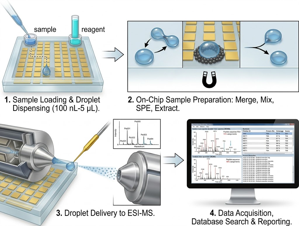
1

Sample Loading & Droplet Dispensing

Sample, reagent, and buffer reservoirs (5–10 μL each) are loaded onto the DMF chip via pipette-accessible ports. Electrode activation sequences dispense precisely metered droplets (100 nL–5 μL) from each reservoir, with real-time capacitive sensing to verify droplet volume (CV<5%) and XY position on the electrode grid.

2

On-Chip Sample Preparation Protocol

Droplets are merged, mixed by bidirectional electrode oscillation (5–20 Hz), incubated under Peltier temperature control (4–95 °C), and subjected to magnetic bead-based extraction steps according to the protocol script. Every electrode activation event is timestamped in the execution log for full traceability. The chip can process multiple samples in parallel, with independent droplet paths that never cross-contaminate.

3

Droplet Delivery to ESI-MS

The processed droplet is routed to the designated emitter electrode, where it contacts a nanoESI high-throughput MS capillary or a liquid-junction ESI source. Ionization proceeds directly from the droplet without intervening LC separation, yielding survey MS spectra within 2–5 seconds of delivery. For samples requiring chromatographic separation, the droplet can alternatively be injected into a nanoLC column via a switching valve.

4

Data Acquisition, Analysis & Reporting

High-resolution MS and MS/MS spectra are acquired in data-dependent acquisition (DDA), data-independent acquisition (DIA), or parallel reaction monitoring (PRM) mode. Data are processed for peptide/protein identification (database search with FDR control), label incorporation efficiency, or kinetic parameters. The final report includes annotated spectra, the complete DMF protocol execution log, and QC metrics for each on-chip step.

Digital microfluidics MS workflow from sample loading to ESI-MS analysis

Platform Instrumentation

ModuleInstrument / SystemCore Capability
DMF Chip & ControllerCustom-fabricated EWOD chips (glass substrate, Cr/Au electrodes, Parylene-C dielectric, Teflon-AF hydrophobic layer); FPGA-based multi-channel voltage controller100–120 individually addressable electrodes; 100 nL–5 μL droplet range; 0.1–10 mm/s transport velocity; capacitive droplet position sensing
Magnetic Bead ModuleNeodymium magnet array with programmable vertical positioning beneath chipBead capture, wash, and release in nanoliter droplets; compatible with commercial magnetic bead chemistries (C18, TiO2, HILIC, Ni-NTA, streptavidin)
Temperature ControlIntegrated Peltier element with Pt100 sensor feedback; 4–95 °C range, ±0.5 °C accuracyOn-chip incubation for enzymatic digestion, chemical derivatization, and binding reactions at controlled temperature
Ionization InterfaceNanoESI capillary emitter (1–10 μm tip ID) with DMF-to-ESI liquid junction; optional sheath-flow ESI for higher flow ratesDirect droplet-to-MS ionization; no LC column or switching valve between chip and MS for rapid survey analysis
Mass SpectrometryOrbitrap Exploris 480, Q Exactive HF-X, timsTOF Pro 2High-resolution MS and MS/MS; DDA, DIA, PRM acquisition modes; intact mass analysis under native conditions; ion mobility separation (timsTOF)

Technology Comparison

FeatureDigital Microfluidics MS (EWOD)Channel-Based Microfluidics MSConventional Bench-Top Prep + LC-MS
Fluid ManipulationElectrode-actuated discrete droplets; software-reconfigurable pathsPressure-driven continuous flow in fixed channelsManual pipetting, centrifugation, vacuum manifold
Sample Volume Range100 nL–5 μL per droplet1–100 μL/min continuous flow10–500 μL per preparation
Protocol FlexibilityFully programmable; any droplet can follow any electrode path; new protocol = new script, no hardware changeFixed channel layout; protocol changes require new chip fabricationManual or limited liquid-handler automation; protocol changes require re-programming and re-plumbing
Multi-Step Prep IntegrationAll steps on single chip including magnetic bead SPERequires multi-chip or multi-module integration with interconnectsMultiple instruments and manual transfers between tube, plate, and cartridge formats
Online MS CouplingDirect droplet-to-ESI from chip emitter; optional nanoLC injectionDirect ESI from channel outlet; or post-columnLC column between prep and MS; 5–60 min per sample gradient
Sample LossMinimal (closed droplet, single surface, no transfers)Low–moderate (channel wall adsorption, interconnection dead volume)Moderate–high (tube/plate transfers, pipette tip retention, SPE cartridge breakthrough, vial adsorption)
Sample Throughput5–15 min per complete prep+MS cycle; parallel droplet processingContinuous; 1–10 samples/h depending on protocol complexityHours to overnight per batch of 12–96 samples
TraceabilityComplete electrode actuation log with ms timestampsFlow rate and pressure sensor logsManual lab notebook entries; limited liquid-handler logs

Sample Requirements

Sample TypeRequired AmountCompatible Conditions
Protein / Proteomics Sample1–20 μg total protein; 100–500 ng per on-chip preparationMS-compatible buffer (ammonium bicarbonate, triethylammonium bicarbonate); avoid PEG-based surfactants, glycerol (>1%), and high concentrations of non-volatile salts (>50 mM NaCl)
Small-Molecule / Metabolite Extract100 ng–10 μg total; 0.1–1 μg per DMF preparationDried extract reconstituted in aqueous buffer; organic solvent ≤50% (v/v) compatible with EWOD actuation on Teflon-AF surface
Peptide / PTM Enrichment Sample100 ng–5 μg digested peptidesLow-salt loading buffer for magnetic bead binding; TFA-free for optimal EWOD droplet movement (TFA reduces surface hydrophobicity and impairs actuation)
Single-Cell / Low-Input Sample1–1,000 cells; 10 ng–1 μg total proteinCell lysis buffer compatible with downstream proteolysis and MS; surfactant (e.g., RapiGest SF, n-dodecyl-β-D-maltoside) preferred for membrane protein solubilization

Deliverables

  • Complete data package: raw MS and MS/MS files (.raw, .d, or .mzML format), processed peak lists, and database search results with FDR-controlled protein/peptide identifications
  • DMF protocol execution log: timestamped record of every droplet movement, merge, split, and magnetic bead operation with electrode activation trace — equivalent to an electronic lab notebook for the entire sample preparation sequence
  • QC metrics report: droplet volume consistency (CV%), magnetic bead capture efficiency, on-chip digestion/derivatization efficiency, carryover assessment between sequential droplets, and MS system suitability tests
  • For proteomics: protein/peptide identifications with FDR, sequence coverage, label incorporation efficiency (for TMT/iTRAQ workflows), and PTM site localization scores with annotated MS/MS spectra
  • For reaction monitoring: kinetic curves with extracted ion chromatograms at each sampled time point, fitted rate constants (kobs, kcat, Km) where applicable, and intermediate species identification
  • Written interpretation of results with recommendations for protocol optimization on subsequent runs, including suggested adjustments to incubation time, reagent stoichiometry, or bead-to-sample ratio

Representative Demo Data

On-chip proteomics sample preparation compared to conventional tube-based protocol MS spectra

Case Study

Active-Matrix DMF Single-Cell Proteomics Identifies Drug-Resistance Signature in EGFR-Mutant NSCLC Cells

Yang et al. (2024) developed an active-matrix digital microfluidic single-cell proteomics (AM-DMF-SCP) platform and applied it to characterize the proteomic landscape of EGFR inhibitor resistance at single-cell resolution. The study was published in JACS Au (DOI: 10.1021/jacsau.4c00027).

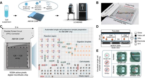
Study design: An active-matrix DMF chip — where each electrode is individually addressable via thin-film transistor (TFT) switching, analogous to an LCD display — integrated all sample preparation steps (cell lysis, protein reduction, alkylation, digestion, and peptide cleanup) within nanoliter-volume droplets. The processed peptides were analyzed by data-independent acquisition (DIA) mass spectrometry using a 15-minute LC gradient. Single HeLa cells yielded an average of 2,258 protein groups identified per cell. The platform was then applied to compare three human tumor cell lines — HeLa (cervical adenocarcinoma), A549 (lung adenocarcinoma), and HepG2 (hepatocellular carcinoma) — with machine learning classification identifying cell-line-specific proteomic features that distinguished each lineage at the single-cell level.

Drug-resistance application: To demonstrate translational utility, the AM-DMF-SCP platform profiled NCI-H1975 non-small cell lung cancer cells harboring the EGFR L858R/T790M double mutation, comparing parental cells against a derived line resistant to the third-generation EGFR inhibitor ASK120067 (67R cells). Single-cell proteomic analysis revealed elevated vimentin (VIM) expression in the resistant 67R population — a mesenchymal marker consistent with epithelial-to-mesenchymal transition (EMT)-associated drug resistance — alongside alterations in metabolic enzymes and cytoskeletal proteins. These single-cell-resolved proteomic signatures were concordant with bulk-sample analyses, validating the platform's quantitative fidelity while additionally resolving cell-to-cell heterogeneity masked in bulk measurements.

Relevance to DMF-MS services: This study demonstrates that active-matrix DMF platforms achieve single-cell proteomic depth and quantitative accuracy comparable to established microfluidic systems, while offering greater programmability through TFT-based electrode addressing. For drug discovery programs seeking to characterize resistance mechanisms, assess target engagement heterogeneity, or profile precious clinical specimens at the single-cell level, our DMF-MS platform provides the same core architecture — programmable nanoliter sample preparation with direct MS readout — adapted to your specific experimental design and throughput requirements.

AM-DMF single-cell proteomics EGFR drug-resistance study workflow diagram

References

  1. Yang Z, Jin K, Chen Y, et al. AM-DMF-SCP: Integrated Single-Cell Proteomics Analysis on an Active Matrix Digital Microfluidic Chip. JACS Au. 2024;4(5):1811–1823. doi:10.1021/jacsau.4c00027
  2. Steinbach MK, Leipert J, Matzanke T, Tholey A. Digital Microfluidics for Sample Preparation in Low-Input Proteomics. Small Methods. 2025;9(1):e2400495. doi:10.1002/smtd.202400495
FAQ

Frequently Asked Questions

Q: What types of samples and workflows are compatible with the DMF-MS platform?

The platform accepts purified proteins, cell lysates, peptide mixtures, metabolite extracts, and small-molecule reaction mixtures — all in aqueous or mixed aqueous-organic buffers compatible with EWOD actuation. Workflows we routinely execute on-chip include reduction-alkylation-digestion for bottom-up proteomics, TMT/iTRAQ labeling, phosphopeptide enrichment with TiO2 magnetic beads, glycopeptide HILIC enrichment, His-tagged protein purification, detergent removal, and multi-step chemical derivatization. If your workflow involves sequential liquid-handling steps at the microliter scale, it is likely adaptable to the DMF format. We offer a free feasibility assessment: send us your protocol, and our development team will evaluate DMF compatibility and propose an on-chip adaptation strategy.

Q: How does DMF-MS compare to the microfluidic chip–MS approach?

Microfluidic chip–MS uses fixed-channel architectures where fluids flow continuously through pre-defined paths. DMF-MS instead manipulates discrete droplets on an open electrode grid, enabling fully reconfigurable fluid routing without hardware changes — a new protocol is a new software script, not a new chip. DMF also integrates magnetic bead-based SPE natively via a magnet beneath the chip, whereas channel-based chips require packed beds or monoliths for solid-phase extraction. For workflows requiring multi-step sample preparation with individual droplet tracking and full automation traceability, DMF-MS offers greater protocol flexibility; for continuous-flow, high-throughput screening where the same operation is repeated thousands of times, channel-based chip-MS may be more suitable. We provide both platforms and recommend the optimal approach based on your specific workflow requirements.

Q: What is the typical throughput of the DMF-MS proteomics workflow?

A complete reduction-alkylation-digestion-desalting-MS cycle for a single sample takes approximately 5–15 minutes on-chip (trypsin digestion at 37 °C is the rate-limiting step; this can be accelerated to <5 min by increasing enzyme-to-substrate ratio or operating at elevated temperature with thermostable proteases). By parallelizing digestion incubation across multiple droplets — each droplet occupies a distinct electrode zone and proceeds independently — we process 6–12 samples per hour in batch mode. This is 10–20 times faster than conventional overnight tube-based protocols and eliminates sample-to-sample variability from manual pipetting. For large cohort studies (>100 samples), the protocol script is executed identically for every sample, ensuring batch-effect-free data suitable for quantitative proteomics.

Q: Can the DMF-MS platform handle membrane proteins or detergent-containing samples?

Yes — and detergent removal is one of DMF's strongest capabilities. Membrane protein samples solubilized in MS-incompatible detergents (SDS, Triton X-100, NP-40) can be processed on-chip: after digestion, magnetic C18 beads capture peptides while the detergent-containing supernatant is moved away electronically by activating adjacent electrodes. Wash and elution droplets complete the cleanup, and the purified peptides are delivered directly to the MS. This workflow eliminates the precipitation, centrifugation, and offline desalting steps that often cause membrane protein sample losses exceeding 50% in conventional protocols. We have successfully applied this approach to GPCRs, ion channels, and transporter proteins solubilized in a range of detergent systems.

Q: What is the minimum sample amount required for DMF-MS proteomics?

We have successfully processed samples containing as little as 100 ng of total protein on the DMF chip, with approximately 10–50 ng consumed per MS analysis. This sensitivity makes DMF-MS particularly valuable for laser-microdissected tissue sections (LCM), fluorescence-activated cell sorting (FACS)-isolated cell populations, and low-abundance immunoprecipitation eluates. For single-cell proteomics applications, the DMF platform processes individual cells captured by micromanipulation or limiting dilution, with typical proteomic coverage of 1,500–2,500 protein groups per cell depending on cell type and MS acquisition mode. For optimal sequence coverage and PTM detection sensitivity, we recommend 1–5 μg of starting material when sample availability permits.

Q: Can I access the raw DMF protocol files for reproducibility?

Yes. Every DMF-MS run generates a complete electrode actuation log — equivalent to an electronic lab notebook entry — that we include in the deliverables. The log records droplet dispensing volumes, merge/split events, incubation durations, magnetic bead capture steps, and MS acquisition triggers with millisecond timestamps. This traceability supports troubleshooting, protocol transfer between laboratories, and regulatory documentation requirements. For integration with your broader screening or characterization program, we coordinate with our microfluidics and emerging MS platforms suite to ensure consistent data formats and reporting standards across all microfluidic service engagements.

Automate Your Nanoscale Sample Processing With Digital Microfluidics MS

From single-cell proteomics sample preparation and on-chip peptide labeling to magnetic bead-based enrichment and multi-step reaction monitoring, our DMF-MS platform replaces manual workflows with programmable, nanoliter-scale automation. Contact our team to discuss your sample preparation challenge, target analytes, and desired MS readout — we will propose a tailored DMF protocol and provide a feasibility assessment based on your specific workflow.

Online Inquiry

Please submit a detailed description of your project. We will provide you with a customized project plan to meet your research requests. You can also send emails directly to for inquiries.

* Email
Phone
* Service & Products of Interest
Services Required and Project Description