Drug–Protein Adduct MS: Detect Covalent Binding & Reactive Metabolites with High-Resolution LC-MS/MS

De-risk your drug candidates early by identifying and characterizing drug–protein adducts with atom-level resolution.

Drug-induced toxicity remains one of the leading causes of attrition in drug development. Many small-molecule drugs undergo cytochrome P450-mediated bioactivation to form reactive metabolites that covalently modify proteins — generating drug–protein adducts that can trigger hepatotoxicity, hypersensitivity reactions, and idiosyncratic adverse drug responses. Detecting these adducts early in discovery is critical for selecting safer candidates before committing to costly preclinical and clinical development.

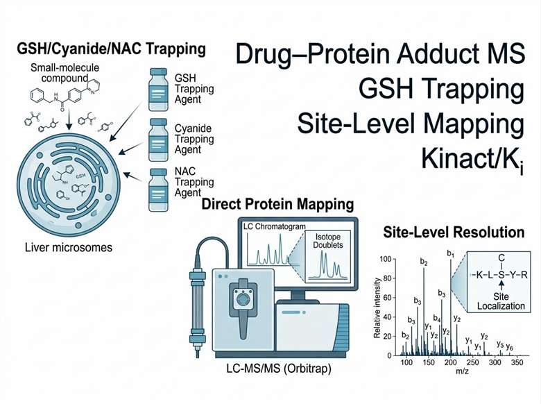
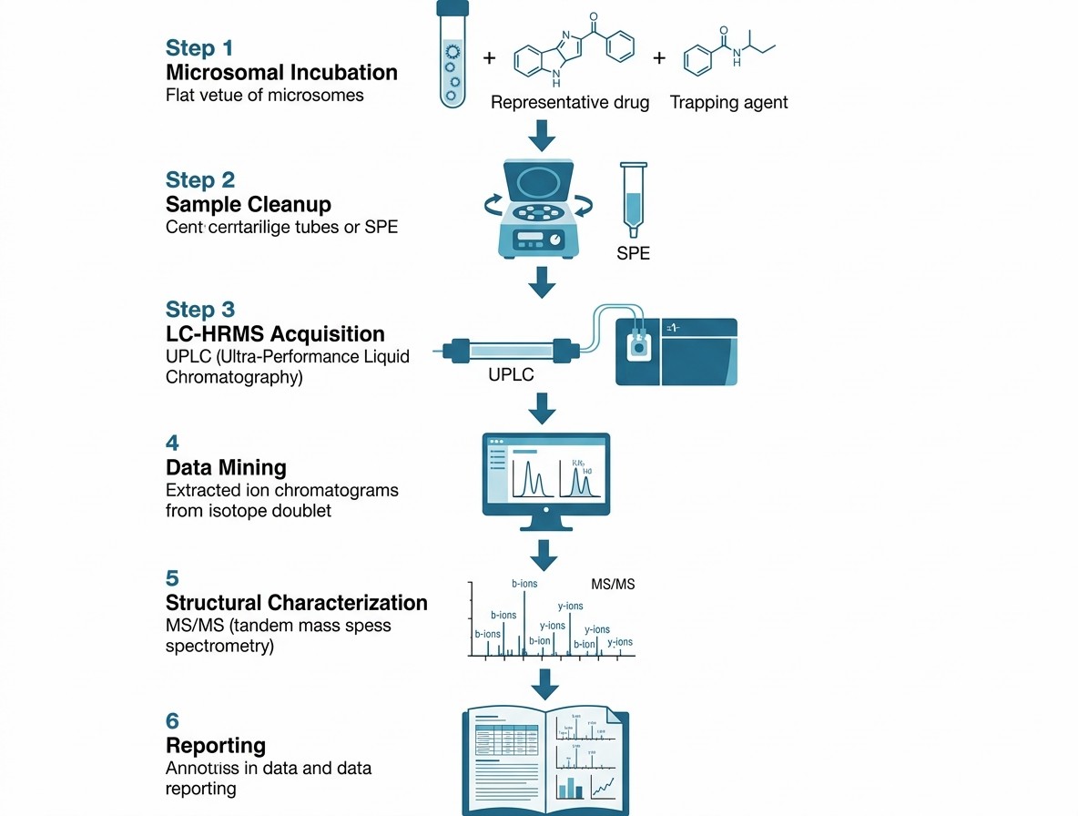
At Creative Proteomics, we offer a comprehensive drug–protein adduct MS service that combines chemical trapping assays (GSH, cyanide, NAC) with direct protein adduct mapping by high-resolution LC-MS/MS. Our integrated workflow enables you to identify reactive metabolite formation, pinpoint the exact proteins and amino acid residues modified, and quantify the extent of covalent binding — all within a single, streamlined service platform.

Key Advantages:

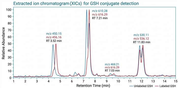
  • GSH, cyanide, and NAC trapping assays with stable isotope labeling for confident adduct identification
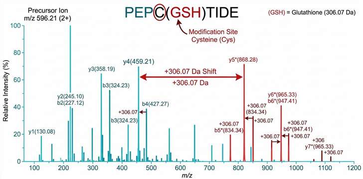
  • Direct protein adduct mapping by tryptic digestion and LC-MS/MS with site-level resolution
  • Kinact/Ki determination for covalent inhibitor characterization
  • High-resolution Orbitrap and Q-TOF mass spectrometry for sensitive and accurate detection
  • Comprehensive data package with annotated spectra, adducted peptide maps, and QC metrics
Drug–protein adduct MS platform featuring LC-MS/MS, GSH trapping, and site-level mapping
Why Adduct Profiling Service Overview Workflow Demo Sample Instrumentation Comparison Deliverables Applications FAQ

Why Drug–Protein Adduct Profiling Matters in Early Drug Discovery

The formation of drug–protein adducts is a well-established mechanism of drug-induced toxicity. Reactive metabolites — electrophilic species generated during Phase I and Phase II metabolism — can covalently modify cellular proteins, leading to protein dysfunction, immune-mediated responses, and organ toxicity. Notable examples include acetaminophen (NAPQI adduction to liver proteins), diclofenac (acyl glucuronide-mediated adduction), and clozapine (reactive nitrenium ion adduction).

Regulatory agencies, including the FDA and EMA, increasingly expect bioactivation assessment as part of preclinical safety packages. The ICH M10 guideline on bioanalytical method validation, while focused on quantification, underscores the broader expectation that drug metabolism data — including reactive metabolite formation — be generated with rigor and reproducibility. Early adduct profiling provides several tangible benefits:

  • Reduced late-stage attrition: Identifying bioactivation liabilities during lead optimization avoids costly failures in development
  • Informed medicinal chemistry: Adduct data guides structural modifications to block or minimize reactive metabolite formation
  • Mechanistic understanding: Knowing which proteins are adducted and at which residues provides insight into toxicity mechanisms
  • Regulatory preparedness: Proactive adduct assessment demonstrates due diligence in candidate selection

For teams developing covalent inhibitors, drug–protein adduct MS serves a dual purpose: confirming target engagement and ruling out off-target adduction that could drive toxicity. For a complete ADME safety assessment, explore our ADME/DMPK research platforms that integrate adduct profiling with metabolic stability, permeability, and bioanalysis services.

Comprehensive Drug–Protein Adduct MS Service Overview

Our service is organized into four integrated modules, each addressing a specific aspect of adduct detection and characterization. We work with you to select the appropriate module(s) based on your compound class, development stage, and specific questions.

ModuleServiceDescriptionApplication
1GSH Trapping AssayIncubate compound with liver microsomes + GSH (unlabeled + stable isotope-labeled 1:1); detect GSH conjugates by LC-HRMS using characteristic isotope doublet patternReactive metabolite screening for lead optimization; bioactivation liability assessment
2Cyanide & NAC TrappingCyanide trapping for iminium ion intermediates; NAC trapping for soft electrophiles; both with stable isotope labelingComprehensive trapping panel for diverse reactive metabolite classes
3Direct Protein Adduct MappingIncubate compound with target protein or microsomes → tryptic digestion → LC-MS/MS → identify adducted peptides by mass shift + MS/MS fragmentation; site localization by diagnostic fragment ionsCovalent inhibitor target engagement; off-target adduction profiling; mechanism-based toxicity investigation
4Kinact/Ki DeterminationTime- and concentration-dependent inactivation assay; intact protein MS or peptide-based readout; fit to kinetic modelCovalent inhibitor potency ranking; SAR support

Each module can be run independently or combined into a full adduct profiling panel. We recommend Module 1 + Module 3 for comprehensive bioactivation assessment, and Module 3 + Module 4 for covalent inhibitor programs.

For related ADME services, explore our metabolite identification (MetID) and toxic metabolite detection capabilities, which complement adduct profiling by providing a complete picture of your compound's metabolic fate.

Our Drug–Protein Adduct MS Workflow: From Sample to Annotated Results

Our integrated workflow covers both trapping-based and direct protein adduct mapping approaches:

1

Microsomal incubation

Test compound incubated with HLMs or recombinant CYPs, NADPH regeneration system, and 1:1 unlabeled/labeled trapping agent

2

Sample cleanup

Protein precipitation or solid-phase extraction to remove matrix components

3

LC-HRMS acquisition

C18 reversed-phase column coupled to Orbitrap or Q-TOF with DDA or DIA acquisition mode

4

Data mining

XICs screened for characteristic isotope doublet pattern confirming adduct identity

5

Structural characterization

MS/MS spectra analyzed to confirm modification site and elucidate adduct structure

6

Reporting

Annotated XICs, MS/MS spectra, and summary table with retention times, m/z values, and relative abundances

Drug–protein adduct MS workflow from incubation to reporting

Representative Data: Adduct Detection and Characterization

GSH conjugate detection by LC-HRMS with isotope doublet pattern

GSH Conjugate Detection by LC-HRMS

Characteristic isotope doublet peaks in extracted ion chromatograms from 1:1 unlabeled and stable isotope-labeled GSH. The doublet pattern provides high-confidence identification of true GSH conjugates, distinguishing them from background signals. Representative data show overlaid unlabeled (blue) and labeled (red) channels with corresponding MS/MS spectrum confirming adduct structure.

Adducted peptide MS/MS spectrum with site localization

Adducted Peptide Identification and Site Localization

Adducted peptides detected by characteristic mass shift in MS1 scan, confirmed through MS/MS fragmentation. Annotated MS/MS spectrum shows b- and y-ion series with modification mass shift on specific fragment ions, enabling precise localization of the adducted residue.

Sample Requirements for Drug–Protein Adduct MS Analysis

Sample TypeRequired AmountConcentrationPurityBuffer ConditionsNotes
Test Compound1–5 mg or 10 mM stock≥1 mM in DMSO≥95%DMSO (≤0.5% final in incubation)Provide molecular weight, structure, and known metabolic pathways
Human Liver Microsomes0.5–2 mg protein0.5–2 mg/mL0.1 M phosphate buffer, pH 7.4Can be supplied by Creative Proteomics
Recombinant CYP50–200 pmol50–100 pmol/mL0.1 M phosphate buffer, pH 7.4Specify isoform(s) of interest
Purified Target Protein50–200 µg1–10 µM≥90%MS-compatible buffer (no glycerol, low detergent)Provide sequence information if available
Trapping Agent (GSH/KCN/NAC)1–5 mM stock≥98%Prepared fresh in incubation bufferSupplied by Creative Proteomics; stable isotope-labeled version available
NADPH Regeneration SystemSupplied by Creative Proteomics

For sample types not listed here, please contact our team to discuss custom preparation protocols.

Instrumentation for High-Resolution Adduct Detection

Module CategoryInstrument / SystemCore CapabilityWhy It Matters
Liquid ChromatographyVanquish Flex UHPLC / nanoLC systemHigh-resolution separation of peptides and small moleculesResolves isomeric adducts; enables detection of low-abundance species
Mass SpectrometryOrbitrap Exploris 480 / Q-Exactive HFHigh-resolution, accurate-mass (HRAM) detection with MS/MS capabilitySub-ppm mass accuracy for confident adduct identification; fast DIA acquisition
Data AnalysisCompound Discoverer / Skyline / Proteome DiscovererAutomated adduct mining, peptide identification, and quantificationStreamlines data processing; supports open-mass search for unexpected adducts
Incubation & Sample PrepHamilton STAR liquid handler / automated SPE systemReproducible sample preparation for high-throughput trapping assaysMinimizes variability; enables batch processing of multiple compounds

Our platform is complemented by LC-MS/MS bioanalysis services for PK curve generation, providing a complete bioanalytical solution for your DMPK program.

Drug–Protein Adduct MS vs. Alternative Methods for Covalent Binding Assessment

TechniqueDetection PrincipleSpecificityThroughputProtein IDSite LocalizationQuantitationRegulatory Context
Drug–Protein Adduct MS (this service)LC-HRMS of trapped conjugates or adducted peptidesHigh — MS/MS confirms adduct structureMedium (10–50 cmpds/week for full panel)Yes — identifies adducted proteinsYes — residue-level by MS/MSSemi-quantitative (relative abundance)ICH M10 relevant
Radiolabeled Covalent Binding AssayScintillation counting of protein-bound radioactivityLow — measures total binding, no structural infoHigh (96/384-well format)NoNoQuantitative (pmol eq/mg protein)Established regulatory precedent
GSH Trapping (LC-UV/FL)UV or fluorescence detection of GSH conjugatesMedium — retention time + UV spectrumHighNoNoSemi-quantitativeScreening only
Intact Protein MSDirect MS of intact protein ± drugHigh — mass shift confirms bindingMediumYes (protein level)NoSemi-quantitativeConfirmatory
Peptide Mapping (Bottom-up MS)Tryptic digestion + LC-MS/MS of adducted peptidesHighest — sequence + siteLow–MediumYesYes (residue-level)Semi-quantitativeGold standard for site mapping

Why choose drug–protein adduct MS? It is the only technique that simultaneously provides (1) detection of adduct formation, (2) identification of the adducted protein(s), and (3) localization of the modification site — all from a single experiment. This makes it the most informative approach for both bioactivation screening and covalent inhibitor characterization.

Deliverables: What You Receive

  • Raw MS data files (.raw or .d format)
  • Processed data: extracted ion chromatograms (XICs) and MS/MS spectra for all identified adducts
  • Adduct identification table: retention time, m/z, mass shift, adduct type, fragment ions
  • Adducted peptide list with site localization (for direct protein mapping)
  • Quantitative estimates: relative abundance of adducts vs. parent compound or unmodified peptide
  • QC summary: replicate reproducibility, blank controls, positive control results
  • Method report: experimental conditions, instrument parameters, data analysis workflow
  • Consultation: expert interpretation of results and recommendations for follow-up studies

Key Applications of Drug–Protein Adduct MS in Drug Discovery

Drug–protein adduct MS addresses critical questions across multiple stages of the drug discovery pipeline. Below are the key application scenarios where this service provides the greatest impact.

Bioactivation Screening for Lead Optimization

Screen lead series for reactive metabolite formation using GSH/cyanide/NAC trapping. Flag compounds that form adducts and guide medicinal chemistry efforts to block bioactivation pathways. Integrate with metabolic soft-spot analysis to identify both sites of metabolism and bioactivation risk simultaneously.

Covalent Inhibitor Target Engagement Confirmation

Confirm that a designed covalent inhibitor forms the intended adduct with its target protein. Map the modification site to verify cysteine (or other residue) selectivity. Determine Kinact/Ki to rank inhibitor potency.

Off-Target Adduction Profiling

Identify unintended protein targets of reactive metabolites or covalent inhibitors using proteome-wide adduct mapping. This is critical for understanding selectivity and predicting toxicity mechanisms.

Mechanistic Investigation of Idiosyncratic Toxicity

When unexpected toxicity arises in preclinical studies, drug–protein adduct MS can identify the proteins modified by reactive metabolites, providing mechanistic hypotheses for the observed toxicity and guiding structure–toxicity relationship (STR) analysis.

Hapten Formation Assessment for Immunogenicity Risk

Drug–protein adducts can act as haptens, triggering immune responses. Identifying adducted proteins and the extent of modification helps assess immunogenicity risk, particularly for drugs that cause hypersensitivity reactions.

FAQ

Frequently Asked Questions

Q: What types of compounds are suitable for drug–protein adduct MS analysis?

Most small-molecule drug candidates are suitable, including those with structural alerts for bioactivation (e.g., anilines, thiophenes, furans, quinones, hydrazines) and designed covalent inhibitors (e.g., acrylamide warheads, Michael acceptors, epoxides). We recommend a preliminary structural assessment to determine the most appropriate trapping agent(s) and workflow.

Q: How does drug–protein adduct MS differ from a standard GSH trapping assay?

A standard GSH trapping assay detects only GSH conjugates formed in microsomal incubations, providing evidence that reactive metabolites are generated. Drug–protein adduct MS goes further — it can identify which specific proteins are adducted and at which amino acid residues, offering mechanistic insight that a simple trapping assay cannot provide.

Q: What is the minimum amount of protein needed for adduct mapping?

For purified target protein adduct mapping, we recommend 50–200 µg per condition. For proteome-wide adduct profiling in microsomes or cell lysates, 200–500 µg of total protein is typically sufficient. Lower amounts may be possible depending on the abundance of the adduct and the sensitivity requirements.

Q: Can you distinguish specific covalent binding from non-specific binding?

Yes. We use multiple control experiments to distinguish specific from non-specific adduction: (1) no-NADPH control (to rule out non-metabolic binding), (2) competition with known inhibitors, (3) time-dependent adduct formation kinetics, and (4) replicate consistency. Specific adducts show time- and concentration-dependence, while non-specific binding appears uniformly across conditions.

Q: How do you report adduct data — what do I receive?

You receive a comprehensive data package including raw MS data, processed chromatograms and spectra, an adduct identification table, site localization data (for direct mapping), quantitative estimates, QC metrics, and a detailed method report. Our team also provides expert interpretation and recommendations for follow-up studies.

References

  1. Mons E, Kim RQ, Mulder MPC. Technologies for Direct Detection of Covalent Protein–Drug Adducts. Pharmaceuticals. 2023;16(4):547. https://doi.org/10.3390/ph16040547
  2. Riffle M, Hoopmann MR, Jaschob D, Zhong G, Moritz RL, MacCoss MJ, Davis TN, Isoherranen N, Zelter A. Discovery and Visualization of Uncharacterized Drug–Protein Adducts Using Mass Spectrometry. Analytical Chemistry. 2022;94(8):3501–3509. https://doi.org/10.1021/acs.analchem.1c04101
  3. Dueñas ME, Peltier-Heap RE, Leveridge M, Annan RS, Büttner FH, Trost M. Advances in High-Throughput Mass Spectrometry in Drug Discovery. EMBO Molecular Medicine. 2023;15:e14850. https://doi.org/10.15252/emmm.202114850

Screen Your Compounds for Bioactivation Liabilities Early

Share your compound structures and we will design a tailored drug–protein adduct profiling strategy for your discovery program.

This service is for research use only and is not intended for diagnostic or clinical applications.

Online Inquiry

Please submit a detailed description of your project. We will provide you with a customized project plan to meet your research requests. You can also send emails directly to for inquiries.

* Email
Phone
* Service & Products of Interest
Services Required and Project Description