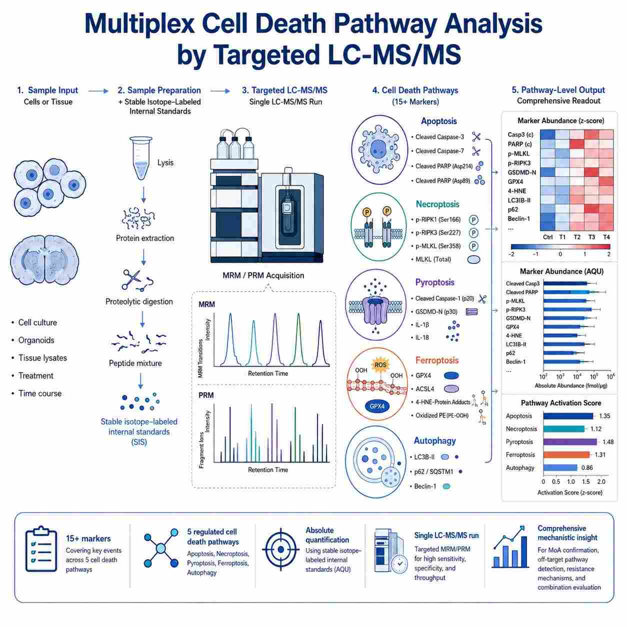
Multiplex Cell Death Pathway Analysis by Targeted LC-MS/MS

Multiplex quantification of protein and metabolite markers across five major regulated cell death pathways — apoptosis, necroptosis, pyroptosis, ferroptosis, and autophagy — from a single sample injection.

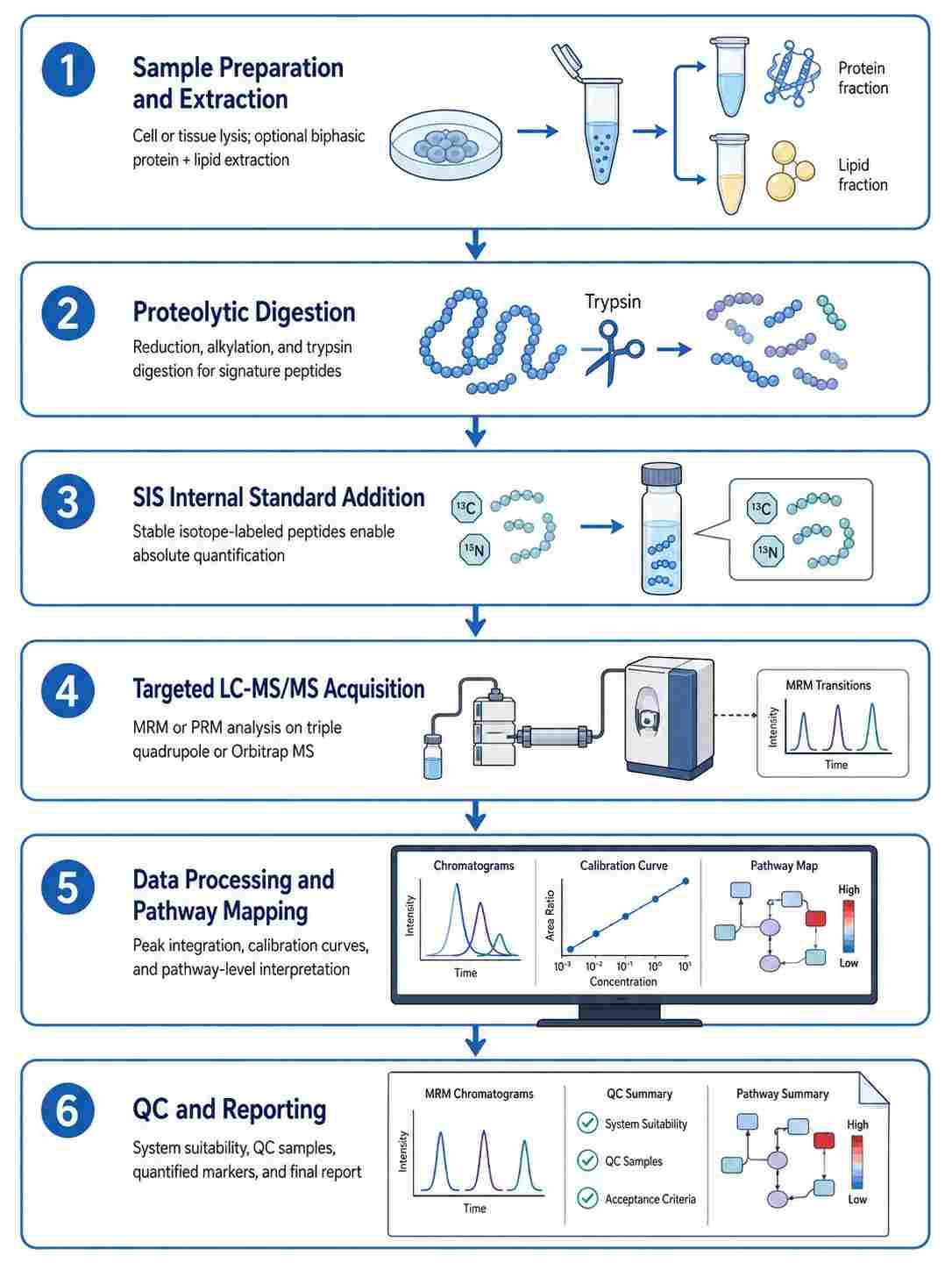
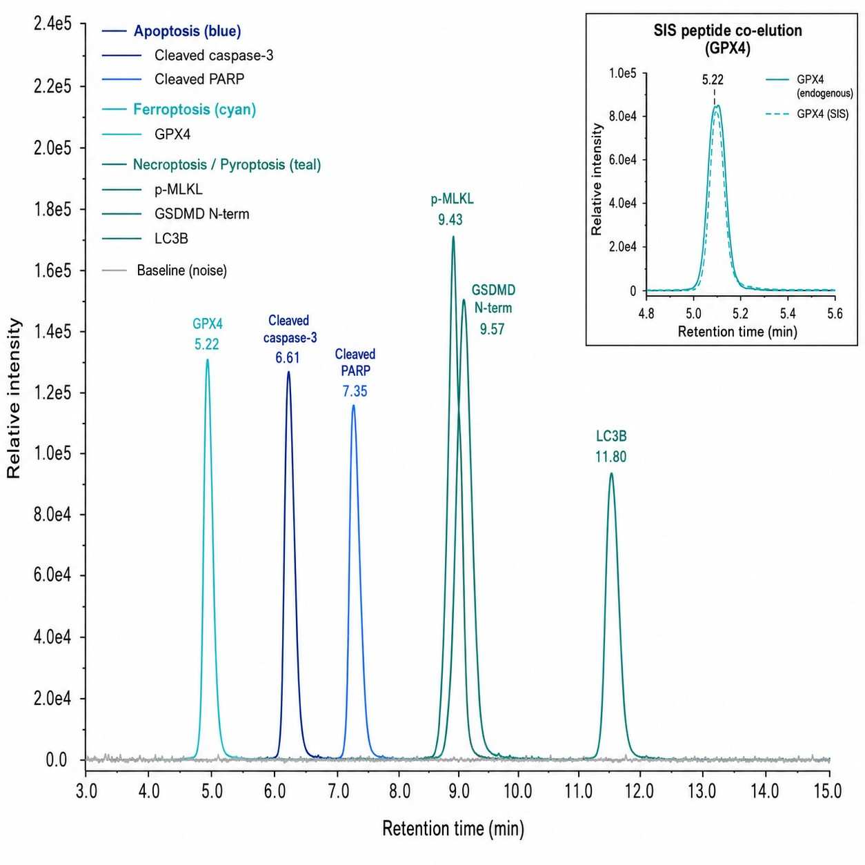
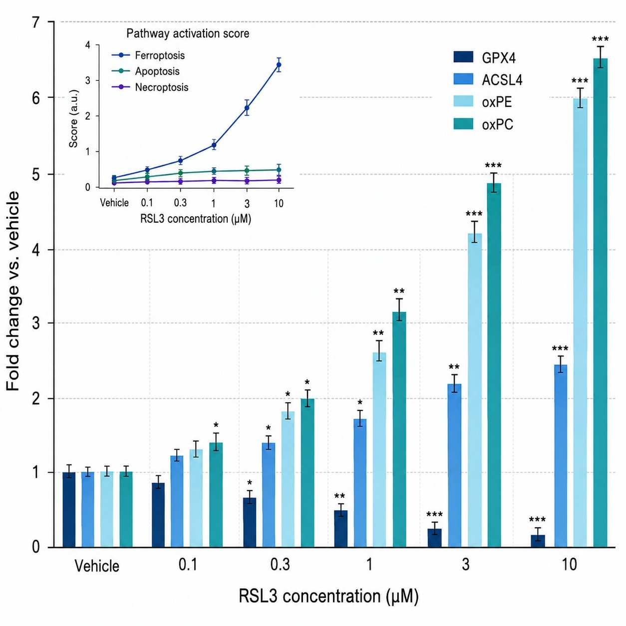
Cell death pathway MS signatures is a targeted mass spectrometry approach that enables simultaneous, absolute quantification of protein and metabolite markers across five major regulated cell death pathways — apoptosis, necroptosis, pyroptosis, ferroptosis, and autophagy — from a single sample injection, providing comprehensive cell death modality profiling with superior specificity and dynamic range compared to conventional antibody-based methods.

Key Advantages:

  • Multiplex quantification of 15+ markers across 5 cell death pathways in a single LC-MS/MS run
  • Absolute quantification with stable isotope-labeled internal standards
  • Isoform-specific detection of cleaved vs. full-length caspases, gasdermins, and MLKL
  • Integrated proteomics + lipidomics for ferroptosis (GPX4, ACSL4, oxidized phospholipids)
  • 50-100 µg total protein sufficient for comprehensive pathway profiling
Cell death pathway MS signatures service overview
What Is Cell Death MS Why Multiplex Matters Pathways Workflow Demo Sample Comparison Applications Case Study FAQ

What Is Cell Death Pathway MS Signatures

Regulated cell death (RCD) encompasses multiple molecularly distinct pathways — including apoptosis, necroptosis, pyroptosis, ferroptosis, and autophagy-associated cell death — each characterized by unique protein markers, lipid mediators, and morphological features. Understanding which cell death modality a drug candidate triggers, and whether off-target cell death pathways are engaged, is critical for mechanism of action (MoA) studies, lead optimization, and safety assessment in drug discovery programs.

Cell death pathway MS signatures leverages liquid chromatography-tandem mass spectrometry (LC-MS/MS) in targeted acquisition modes — multiple reaction monitoring (MRM) or parallel reaction monitoring (PRM) — to achieve precise, multiplexed quantification of cell death-related proteins and lipids. Our targeted MRM-based protein quantification platform has been extensively validated for multiplex biomarker analysis in drug discovery applications. The methodology relies on the selection of proteotypic signature peptides unique to each target protein, combined with stable isotope-labeled (SIS) peptide internal standards for absolute quantification.

Unlike conventional antibody-based methods such as Western blot, ELISA, or activity-based assays (e.g., Caspase-Glo), MS-based analysis offers several fundamental advantages. It provides absolute quantification (not relative fold-change), enables simultaneous detection of 15-20 markers across multiple cell death pathways from a single sample, and can unambiguously distinguish between cleaved (active) and full-length (pro-form) protein isoforms by targeting cleavage-site-spanning signature peptides. This technical capability is particularly valuable for cell death research, where the activation state of caspases, gasdermins, MLKL, and the expression level of GPX4 are the key determinants of cell death modality and commitment.

Our MassTarget™ platform at Creative Proteomics provides a fully integrated cell death pathway MS signature analysis service, from method development through sample processing, data acquisition, and bioinformatics reporting, supporting preclinical drug discovery programs across oncology, neurodegeneration, inflammation, and immuno-oncology.

Why Multiplex Cell Death Pathway Profiling Matters

Drug candidates rarely engage a single cell death pathway in isolation. Growing evidence demonstrates significant cross-talk and co-activation between cell death modalities — a phenomenon increasingly recognized as PANoptosis, where components of apoptosis, necroptosis, and pyroptosis are simultaneously activated. Single-pathway assays (e.g., measuring only caspase-3 activity) can miss this complexity, leading to incomplete or misleading MoA conclusions.

Mechanism of Action Confirmation

Demonstrating that a compound induces cell death through the intended pathway is essential for preclinical validation. For example, a BH3 mimetic should activate the intrinsic apoptosis pathway (caspase-9 → caspase-3 → PARP cleavage), while a ferroptosis inducer should downregulate GPX4 and increase oxidized phospholipids without activating caspases. Multiplex MS profiling provides simultaneous evidence across all relevant pathways from a single sample.

Off-Target Cell Death Detection

Compounds designed to induce one cell death modality may inadvertently trigger alternative pathways, particularly at higher concentrations or in different cell types. Detecting unanticipated necroptosis (p-MLKL) or pyroptosis (GSDMD N-terminal fragment) activation can reveal off-target effects early, guiding medicinal chemistry optimization.

Resistance Mechanism Elucidation

Cancer cells frequently evade therapy by switching between cell death modalities. A tumor initially sensitive to apoptosis induction may acquire resistance through Bcl-2 family upregulation while becoming vulnerable to ferroptosis. Multiplex pathway profiling, combined with our chemoproteomics platform, enables quantitative monitoring of this modality switching and identification of actionable resistance nodes.

Combination Therapy Rationale

The rational design of combination therapies targeting complementary cell death pathways requires quantitative assessment of pathway engagement. For example, combining an apoptosis inducer with a ferroptosis inducer may produce synergistic anti-tumor activity — but only if both pathways are confirmed to be engaged at the molecular level.

Cell Death Pathways Covered

Our cell death pathway MS panel covers five major regulated cell death modalities, each with pathway-specific markers and detection strategies:

APOPTOSIS

Intrinsic and Extrinsic Apoptosis

Key markers: Caspase-3 (cleaved p17), Caspase-8 (cleaved p18), Caspase-9 (cleaved p35), PARP (cleaved 89 kDa fragment), Cytochrome c (cytosolic release), Bcl-2, Bax, Bak

Detection strategy: MRM targeting cleavage-site-spanning peptides for activated caspases; full-length vs. cleaved PARP ratio; Bcl-2 family absolute quantification. Our protease activity MS assays provide complementary caspase substrate cleavage profiling.

Pathway significance: Most established cell death pathway; central to oncology drug development

NECROPTOSIS

Programmed Necrosis

Key markers: MLKL (total and phosphorylated at Ser358), RIPK1, RIPK3

Detection strategy: Phosphosite-specific MRM for p-MLKL; total MLKL quantification for phosphorylation ratio

Pathway significance: Increasingly implicated in inflammatory diseases and neurodegeneration; alternative when apoptosis is blocked

PYROPTOSIS

Inflammatory Cell Death

Key markers: GSDMD (N-terminal fragment), GSDME (N-terminal fragment), Caspase-1 (cleaved p20), NLRP3, ASC, IL-1β, IL-18

Detection strategy: Neo-N-terminus signature peptides for gasdermin cleavage fragments; caspase-1 activation monitoring

Pathway significance: Central to immunogenic cell death (ICD) and innate immune responses; emerging target in immuno-oncology

FERROPTOSIS

Iron-Dependent Lipid Peroxidation

Key markers: GPX4, ACSL4, SLC7A11 (xCT), FTH1, Transferrin Receptor, ALOX family

Detection strategy: Targeted proteomics for protein markers + targeted lipidomics (MRM) for oxidized phospholipids; dual-platform integrated workflow

Pathway significance: Rapidly emerging target in drug-resistant cancers and neurodegeneration; unique iron- and lipid peroxidation-dependent mechanism

AUTOPHAGY

Autophagy-Associated Cell Death

Key markers: LC3B (LC3B-II/LC3B-I ratio), p62/SQSTM1, Beclin-1, ATG5, ATG7

Detection strategy: MRM for LC3B lipidation ratio; absolute quantification of autophagy core proteins

Pathway significance: Context-dependent role in cell survival vs. death; important for combination therapy strategies

Workflow of Cell Death Pathway MS Analysis

Our service follows a systematic workflow designed to deliver robust, reproducible quantification data across multiple cell death pathways.

1

Sample Preparation and Extraction

Cells or tissues are lysed using optimized buffer conditions that preserve protein integrity and prevent post-lysis proteolysis. For integrated proteomics + lipidomics workflows, a biphasic extraction (e.g., MTBE/methanol/water) enables simultaneous recovery of proteins and lipids from the same sample.

2

Proteolytic Digestion

Protein samples are reduced (DTT or TCEP), alkylated (iodoacetamide), and digested with trypsin or alternative proteases (Lys-C, Glu-C) as needed for optimal signature peptide generation. For phosphosite-specific markers (p-MLKL), phosphatase inhibitors are included throughout sample processing.

3

Stable Isotope Internal Standard Addition

SIS peptides for each target marker — synthesized with heavy isotope-labeled amino acids (¹³C/¹⁵N-Arg or -Lys) — are spiked into each sample at known concentrations. These internal standards co-elute with the endogenous peptides and provide the reference for absolute quantification.

4

LC-MS/MS Acquisition

Peptide mixtures are separated by nanoLC or microLC using reversed-phase C18 columns with optimized gradients (60-120 min). The LC system is coupled to a triple quadrupole (for MRM) or high-resolution Orbitrap (for PRM) mass spectrometer operating in targeted acquisition mode.

5

Data Processing and Pathway Mapping

Raw data are processed using Skyline or equivalent software for peak integration, transition ratio verification, and signal-to-noise assessment. Calibration curves constructed from SIS peptide dilution series enable absolute quantification. Results are mapped to individual cell death pathways.

6

Quality Control and Reporting

Each batch includes system suitability standards, blank injections, QC samples at three concentration levels, and replicate analysis. A comprehensive report includes MRM chromatograms, calibration curves, quantified values with statistical analysis, pathway-level heatmaps, and cross-pathway activation assessment.

Six-step workflow of cell death pathway MS analysis

Demo Data: Multiplex Cell Death Marker Quantification

The following representative data demonstrate the analytical performance of our cell death pathway MS signature platform.

MRM chromatogram overlay of cell death markers

MRM chromatogram overlay of cell death markers across three pathways

Dose-response bar chart of cell death marker induction

Dose-dependent cell death marker induction after RSL3 treatment

Cell Death MarkerPathwaySignature PeptideLinear Range (fmol/µg)LLOQ (fmol/µg)Inter-day CV (%)
Cleaved caspase-3 (p17)ApoptosisIETD*SGIGTDDDD0.5 – 5000.9970.58.2
Cleaved PARP (89 kDa)ApoptosisDEVD*GVDEVAK1.0 – 10000.9951.09.5
p-MLKL (Ser358)NecroptosisSIVT(pS)QEPR1.0 – 5000.9941.010.1
GSDMD N-termPyroptosisLLDTGLEESGAGVQK0.5 – 5000.9960.58.8
GPX4FerroptosisFLVGPEGVPR0.5 – 5000.9970.57.5
LC3BAutophagyEDGFLYVYSGENTFGF1.0 – 5000.9931.09.8

Sample Requirements for Cell Death Pathway MS Analysis

Sample TypeRecommended AmountMinimum AmountRecommended ReplicatesStorage Condition
Cell lysate (adherent cells)100-200 µg total protein50 µg total protein3 biological × 3 technical-80°C, avoid freeze-thaw
Cell lysate (suspension cells)1-5 × 10⁶ cells5 × 10⁵ cells3 biological × 3 technical-80°C, avoid freeze-thaw
Tissue biopsy (fresh frozen)5-10 mg wet weight2 mg wet weight3 biological replicatesLiquid N₂ or -80°C
FFPE tissue sections3-5 sections (10 µm)2 sections (10 µm)3 biological replicatesRoom temperature, desiccated
Plasma / serum50-100 µL25 µL3 technical replicates-80°C, single-use aliquots
Mitochondrial / cytosolic fractions50-100 µg per fraction25 µg per fraction3 biological × 2 technical-80°C, avoid freeze-thaw

Notes: (1) For time-course cell death studies, we recommend collecting samples at multiple time points (0, 2, 4, 8, 16, 24 h) to capture the temporal dynamics of pathway activation. (2) For phosphosite-specific markers (p-MLKL), phosphatase inhibitor cocktails must be added to lysis buffers. (3) For integrated proteomics + lipidomics workflows, biphasic extraction is performed to maximize data recovery from limited samples. (4) For ferroptosis studies, antioxidants should be avoided in culture media during the treatment period to preserve lipid peroxidation signals.

Technology Comparison: MS vs. Conventional Cell Death Assays

ParameterMS-Based Quantification (MRM/PRM)Western BlotELISACaspase-Glo Activity Assay
Multiplexing capacity15-20 markers per run1-2 markers per blot1 marker per assay1-2 markers per assay
Quantification typeAbsolute (fmol/µg)Semi-quantitative (relative)Quantitative (concentration)Relative (luminescence units)
Dynamic range3-4 orders of magnitude1-2 orders of magnitude2-3 orders of magnitude1-2 orders of magnitude
Isoform specificityYes (cleaved vs. full-length)Yes (if antibody is specific)Limited (depends on antibody pair)No (measures total activity)
Cross-pathway coverageYes (5 pathways simultaneously)No (pathway-specific)No (pathway-specific)No (pathway-specific)
Lipid marker capabilityYes (integrated lipidomics)NoNoNo
Sample volume required50-200 µg total protein20-50 µg per target50-100 µL per assay10-50 µL per assay
Inter-assay CV5-15%20-40%10-20%10-25%
Throughput20-40 samples/week10-20 samples/week40-80 samples/week40-80 samples/week
Cost per marker (15-marker panel)LowMediumHighMedium
Antibody dependencyNone (peptide-based)HighHighMedium

Selection Guidance. MS-based cell death pathway profiling is the optimal choice when: (1) three or more cell death pathways require simultaneous assessment; (2) absolute quantification is needed for PK/PD modeling; (3) discrimination between cleaved and full-length isoforms is critical for mechanistic interpretation; (4) integrated protein + lipid marker analysis is required (e.g., ferroptosis); (5) sample volume is limited and cannot support multiple individual assays; (6) high inter-study reproducibility is essential for multi-cohort or longitudinal studies. For single-marker, high-throughput screening applications where relative activity data suffices, Caspase-Glo or ELISA may be more practical.

Applications in Drug Discovery

Mechanism of Action Studies

Confirming that a drug candidate induces cell death through the intended pathway is a critical step in preclinical development. MS-based cell death pathway profiling provides direct molecular evidence of pathway engagement by quantifying the activation state of pathway-specific markers across all five major RCD modalities simultaneously, enabling researchers to distinguish between primary and secondary cell death effects. For broader target engagement profiling, our enzyme activity and mechanism studies offer complementary functional readouts.

Lead Optimization

During hit-to-lead and lead optimization phases, medicinal chemists need to compare cell death induction profiles across compound series to guide SAR studies. MS-based multiplex quantification provides quantitative dose-response and time-course data for multiple cell death markers simultaneously, facilitating more informed SAR decisions than single-marker activity assays.

Resistance Mechanism Studies

Acquired resistance to cell death-inducing therapeutics is a major challenge in oncology drug development. Our MS-based panel enables quantitative monitoring of resistance-associated proteins (Bcl-2 family, MLKL, GPX4) across all pathways, providing actionable insights for combination therapy strategies.

Combination Therapy Evaluation

The rational design of combination therapies targeting complementary cell death pathways requires quantitative assessment of synergistic, additive, or antagonistic effects on cell death induction. MS-based multiplex quantification provides the comprehensive dataset needed for combination index calculations across multiple cell death markers. For rapid compound screening in combination studies, our high-throughput MS screening platform enables efficient dose-response matrix analysis.

In Vivo PD Biomarker Analysis

Translating cell death biomarker quantification to in vivo models presents unique challenges, including limited tissue availability. Our MS-based approach is compatible with small tissue biopsies (2-5 mg) and FFPE samples, enabling PD biomarker analysis from xenograft tumors, PDX models, and clinical biopsy specimens.

Case Study: LC-MS/MS Analysis of Ferroptosis Lipid Peroxidation

Background

Ferroptosis is a regulated form of cell death driven by iron-dependent lipid peroxidation, distinct from apoptosis and necroptosis. Understanding the molecular mechanisms of ferroptosis propagation is essential for developing therapeutic strategies targeting this pathway in cancer and neurodegenerative diseases. However, detecting and quantifying the lipid peroxidation signature of ferroptosis requires specialized mass spectrometry-based lipidomics approaches.

Methods

Roeck et al. (2025) developed an optogenetic tool (Opto-GPX4Deg) for light-controlled, single-cell induction of ferroptosis through targeted degradation of GPX4, the central anti-ferroptotic enzyme. To characterize the ferroptosis phenotype at the molecular level, the authors performed LC-ESI-MS/MS-based targeted lipidomics using MRM in negative ion mode. Lipids were extracted using a chloroform/methanol/water biphasic system, separated by C18 reversed-phase chromatography, and analyzed for oxidized PC and oxidized PE species.

Results

LC-MS/MS analysis revealed a significant increase in oxidized PC species in Opto-GPX4Deg-induced ferroptotic cells compared to controls (Fig. 1g), confirming that GPX4 degradation leads to the accumulation of lipid peroxidation products — the hallmark biochemical signature of ferroptosis. Intracellular fatty acid levels showed a marked decrease (Fig. 1h), consistent with metabolic remodeling associated with ferroptotic cell death. These lipidomic changes were observed within hours of GPX4 degradation and preceded loss of plasma membrane integrity.

Conclusion

This study demonstrated that targeted LC-MS/MS lipidomics provides a robust, quantitative tool for detecting the molecular signature of ferroptosis, enabling precise measurement of oxidized phospholipids that are the direct products of ferroptotic lipid peroxidation. The approach offers superior specificity and quantitative accuracy compared to surrogate assays (e.g., C11-BODIPY fluorescence), making it well-suited for preclinical evaluation of ferroptosis-inducing therapeutics.

Source: Roeck BF, Lotfipour Nasudivar S, Vorndran MRH, et al. Ferroptosis spreads to neighboring cells via plasma membrane contacts. Nature Communications. 2025;16:2951. DOI: 10.1038/s41467-025-58175-w

LC-MS/MS lipidomics analysis of ferroptosis lipid peroxidation

Schematic representation of optogenetic GPX4 degradation and LC-MS/MS lipidomics workflow for ferroptosis characterization.

FAQ

Frequently Asked Questions

Q: What cell death pathways are covered in the MS panel?

Our standard cell death pathway panel covers five major regulated cell death modalities: apoptosis (caspase-3/8/9, PARP, cytochrome c, Bcl-2 family), necroptosis (MLKL, p-MLKL, RIPK1, RIPK3), pyroptosis (GSDMD, GSDME, caspase-1, IL-1β), ferroptosis (GPX4, ACSL4, SLC7A11, oxidized phospholipids), and autophagy-associated cell death (LC3B, p62, Beclin-1, ATG5/7). Custom panels can be developed for additional markers or pathways.

Q: Can MS distinguish between different cell death modalities?

Yes. By simultaneously quantifying pathway-specific markers — cleaved caspase-3 for apoptosis, p-MLKL for necroptosis, GSDMD N-terminal fragment for pyroptosis, GPX4 downregulation and oxidized phospholipids for ferroptosis, and LC3B lipidation for autophagy — our MS-based approach provides definitive molecular evidence of which cell death modality(s) are engaged. This is a key advantage over single-marker assays that cannot capture cross-pathway activation.

Q: What is the minimum sample amount for a 15-marker cell death panel?

For cell lysate samples, we recommend 100-200 µg total protein for a standard 15-marker panel. The minimum amount is 50 µg total protein, which is approximately 5-10× less than what would be required for running 15 individual Western blots. For tissue biopsies, 5-10 mg wet weight is typically sufficient.

Q: How many markers can be multiplexed in a single LC-MS/MS run?

Our standard cell death pathway panel includes up to 20 markers covering all five pathways. The panel can be customized to focus on specific pathways or to include additional markers of interest. Each additional marker requires method development and validation.

Q: Can ferroptosis lipid peroxidation be measured alongside protein markers?

Yes. Our integrated proteomics + lipidomics workflow uses a biphasic extraction (MTBE/methanol/water) that simultaneously recovers proteins and lipids from the same sample. This enables quantification of ferroptosis protein markers (GPX4, ACSL4) and oxidized phospholipids (oxPE, oxPC) from a single extraction — maximizing data recovery from limited samples.

Q: Is the cell death pathway MS data suitable for publication?

Absolutely. Our targeted proteomics data meets the highest standards for publication-quality quantitative proteomics, including MIAPE-compliant reporting, SIS internal standard methodology, full transition lists, and inter-assay precision metrics. The absolute quantification format (fmol/µg) is increasingly expected by reviewers in high-impact journals.

Q: How does MS-based cell death profiling compare to Caspase-Glo or ELISA?

MS-based profiling offers several key advantages: (1) 15-20 markers vs. 1-2 markers per assay; (2) absolute quantification vs. relative luminescence units; (3) isoform-specific detection (cleaved vs. full-length); (4) cross-pathway coverage in a single run; (5) integrated lipidomics capability. For single-marker, high-throughput screening, activity-based assays remain practical. For comprehensive pathway characterization, MS is the superior choice.

Q: Can you work with FFPE tissues or small biopsies?

Yes. Our MS-based approach is compatible with FFPE tissue sections (3-5 sections at 10 µm thickness) and small needle biopsies (2-5 mg). We have validated workflows for protein extraction from FFPE samples, including deparaffinization, antigen retrieval, and proteolytic digestion optimized for cross-linked proteins.

References

  1. Roeck BF, Lotfipour Nasudivar S, Vorndran MRH, et al. Ferroptosis spreads to neighboring cells via plasma membrane contacts. Nature Communications. 2025;16:2951.
  2. Albrecht S, Kaisermayer C, Reinhart D, et al. Multiple reaction monitoring targeted LC-MS analysis of potential cell death marker proteins for increased bioprocess control. Analytical and Bioanalytical Chemistry. 2018;410:3197-3207.
  3. Yu Q, Liu X, Keller MP, et al. Sample multiplexing-based targeted pathway proteomics with real-time analytics reveals the impact of genetic variation on protein expression. Nature Communications. 2023;14:555.
  4. Leytens A, Benítez-Fernández R, Jiménez-García C, et al. Targeted proteomics addresses selectivity and complexity of protein degradation by autophagy. Autophagy. 2024;21(2):460-475.

Start Your Cell Death Pathway Profiling Project

Contact our scientific team to discuss your cell death research needs. We will design a customized panel targeting the pathways and markers most relevant to your drug discovery program.

Online Inquiry

Please submit a detailed description of your project. We will provide you with a customized project plan to meet your research requests. You can also send emails directly to for inquiries.

* Email
Phone
* Service & Products of Interest
Services Required and Project Description