SPROX-MS Services: Thermodynamic Target Discovery

Overcome the limitations of thermal stability assays with our SPROX-MS services. By utilizing chemical denaturation and precise methionine oxidation, we provide exact thermodynamic binding data and peptide-level target deconvolution, making it the ideal label-free solution for challenging membrane proteins and complex biological mixtures where traditional heating methods fail.

  • Precise thermodynamic quantification (ΔΔG) in native lysates
  • Ideal for membrane proteins and targets prone to thermal aggregation
  • Peptide-level resolution for mapping allosteric binding pockets
SPROX-MS Services: Thermodynamic Target Discovery
What is SPROX-MS?The Ultimate PatchService CapabilitiesTechnology ComparisonWorkflowDemo ResultsSample RequirementsBioinformaticsCase StudyFAQ

What is SPROX-MS? (Stability from Rates of Oxidation)

Stability of Proteins from Rates of Oxidation (SPROX) coupled with Mass Spectrometry (MS) is an advanced, label-free chemoproteomic technology designed to measure the thermodynamic stability of proteins and detect precise ligand-binding interactions. Unlike traditional assays that rely on physical heating, SPROX utilizes chemical denaturants and a highly specific oxidation reaction to probe protein structures directly within complex biological mixtures.

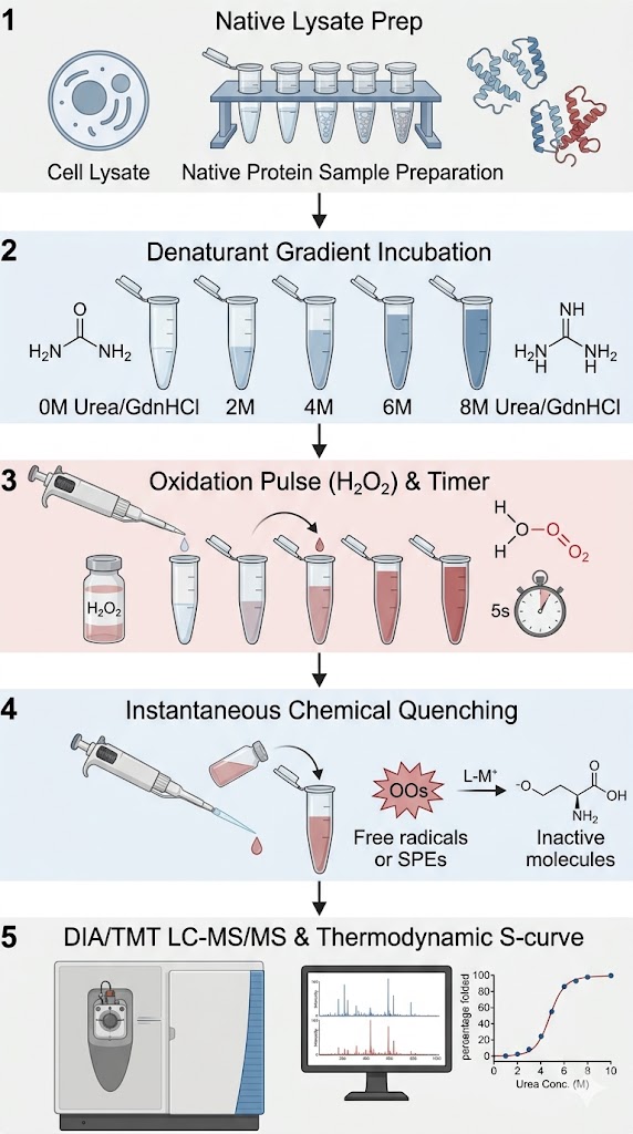
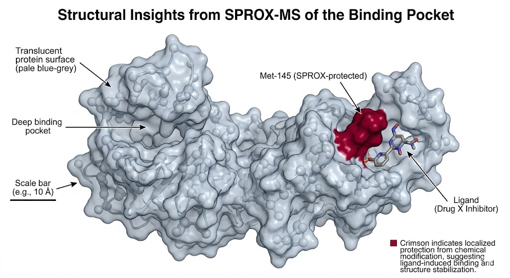
The biophysical principle behind SPROX-MS is elegantly straightforward yet incredibly powerful. In a folded, native protein, hydrophobic amino acids—particularly Methionine residues—are typically buried deep within the structural core or shielded at the protein-protein interaction interfaces. Because they are hidden, they are protected from reacting with the surrounding solvent. However, when we introduce a chemical denaturant like urea or guanidine hydrochloride (GdnHCl), the protein begins to unfold. As the protein unfolds, these previously hidden Methionine residues become exposed to the solvent.

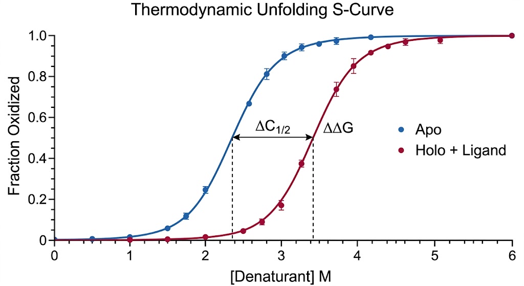
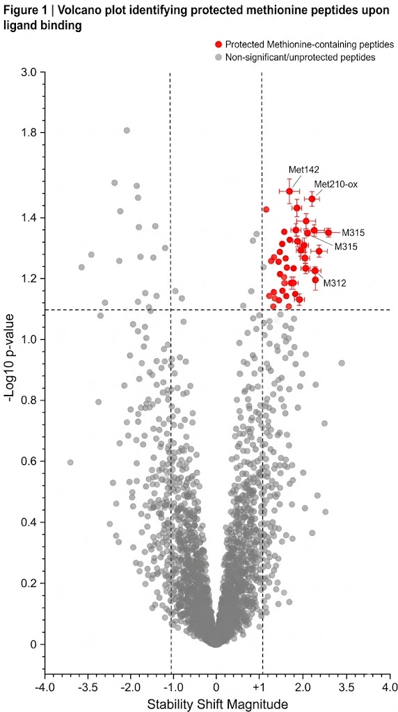
During the SPROX assay, we introduce a brief, highly controlled pulse of hydrogen peroxide (H2O2). The exposed Methionine residues rapidly react with the hydrogen peroxide, undergoing a covalent oxidation event that adds exactly one oxygen atom (+16 Da mass shift) to the amino acid side chain, creating methionine sulfoxide. By utilizing high-resolution mass spectrometry, we can quantify the exact ratio of oxidized versus unoxidized methionine for thousands of peptides simultaneously. When a small molecule drug binds to a protein, it inherently stabilizes the folded state, making it much harder for the chemical denaturant to unfold the protein. This stabilization delays the exposure of the Methionine residue, resulting in a measurable shift in the oxidation rate. By tracking this shift, we identify exactly which proteins your drug bound to, and precisely how tightly it held on.

The "Ultimate Patch" for Difficult Targets (Membrane Proteins)

Target deconvolution is notoriously difficult, but it becomes exponentially harder when dealing with complex, highly hydrophobic targets like multi-pass membrane proteins, ion channels, and large multi-subunit protein complexes. While Thermal Proteome Profiling (TPP) is an excellent tool for soluble, well-behaved globular proteins, it often fails catastrophically when applied to membrane-bound targets.

When you apply heat to a membrane protein complex in a cell lysate, the delicate lipid micelles or detergent structures stabilizing the protein break down. Instead of undergoing a smooth, measurable unfolding transition, the highly hydrophobic transmembrane domains instantly clump together, causing the protein to irreversibly aggregate and crash out of solution. This thermal precipitation completely ruins the assay, yielding chaotic data and massive false negatives.

SPROX-MS is the ultimate patch for this biophysical roadblock. Because SPROX operates entirely at room temperature (isothermally), we completely avoid heat-induced aggregation. Chemical denaturants like urea and GdnHCl are chaotropic agents; they do not just unfold the protein, they actively keep the unfolded hydrophobic chains dissolved in the aqueous solution. Furthermore, our structural biology team can finely tune the chemical denaturant gradient to work in perfect harmony with the specific detergents (such as DDM or LMNG) or lipid nanodiscs used to stabilize your membrane protein. This ensures that the protein unfolds smoothly and predictably, allowing us to capture pristine thermodynamic data on targets that other CROs simply cannot analyze.

Service Capabilities & Orthogonal Validation

Our mass spectrometry platform is engineered to extract maximum biophysical insight from your drug discovery projects. We apply SPROX-MS not just as a screening tool, but as a high-resolution structural probe to answer your most challenging medicinal chemistry questions.

Our Core SPROX Capabilities:

Thermodynamic Lead Optimization

Knowing that a drug binds is only half the battle; you need to know exactly how strongly it binds to drive Structure-Activity Relationship (SAR) optimization. SPROX-MS uniquely allows us to calculate the actual thermodynamic binding free energy change (ΔΔG) directly in a complex lysate, providing a highly accurate, biologically relevant ranking of your lead compounds.

Allosteric Pocket Mapping

Because SPROX analyzes proteins at the peptide level, we do not just identify the target protein; we identify the specific Methionine-containing peptide that was protected by the drug. This allows us to map novel, hidden allosteric binding pockets that sit far away from the primary active site, opening new avenues for drug design.

PPI Disruption

SPROX is exceptionally sensitive to macro-molecular complex stability. We can monitor how a small molecule or biologic drug destabilizes a massive protein complex, proving that your therapeutic is successfully disrupting a pathogenic protein-protein interface.

Overcoming the Methionine Blind Spot (Orthogonal Validation): The primary limitation of SPROX is its reliance on Methionine, an amino acid that makes up only about 2% of the human proteome. If your drug's binding pocket does not contain a Methionine residue nearby, SPROX might miss the interaction. To guarantee 100% proteome coverage and absolute confidence in your results, we offer powerful Orthogonal Validation strategies. We seamlessly combine SPROX with Limited Proteolysis–MS (LiP-MS), which probes global structural changes using enzymatic cleavage, or Hydroxyl Radical Footprinting (HRF-MS), which labels a much broader range of amino acid side chains. This multi-omics approach ensures no target remains hidden.

Technology Comparison: SPROX vs. TPP vs. LiP-MS

To design a successful target discovery campaign, you must align the biophysical perturbation method with the specific nature of your protein target. Here is how SPROX-MS stands apart from other label-free stability techniques.

FeatureSPROX-MSTPP (Thermal Shift)LiP-MS (Limited Proteolysis)
Perturbation MethodChemical Denaturation (Urea/GdnHCl) at Room TempHeat Gradient (Thermal Denaturation)Broad-specificity Enzymatic Digestion
Primary Metric DeliveredThermodynamic Free Energy (ΔΔG)Apparent Melting Temperature (Tm) ShiftProteolytic Protection Score
Membrane Protein SuitabilityExcellent. Prevents irreversible heat aggregation.Poor to Moderate. High risk of precipitation.Good. Depends on detergent compatibility.
Structural ResolutionPeptide-Level (Specific to Methionine residues)Protein-Level (Global stability integration)Peptide-Level (Specific to cleavage sites)
Target RequirementMust possess solvent-accessible MethionineNone. Applicable to most globular proteinsMust possess accessible protease cleavage sites

Our Solution Selection Strategy:

  • Choose TPP for broad, fast, first-pass proteome-wide screening when dealing with standard, soluble globular proteins that behave predictably under heat stress.
  • Choose LiP-MS when you require high-resolution structural footprinting across the entire protein sequence and cannot be limited to mapping only specific amino acid residues.
  • Choose SPROX-MS when you are investigating complex membrane proteins that fail thermal assays, when you are mapping allosteric sites rich in Methionine, or when you explicitly require rigorous, quantitative thermodynamic binding free energy (ΔΔG) values to drive compound optimization.

Optimized SPROX Workflow & QC Checkpoints

The success of a SPROX-MS experiment relies entirely on masterful laboratory execution. Methionine oxidation is a highly dynamic kinetic process. We have optimized every millisecond of our workflow to ensure the oxidation is perfectly controlled and the resulting mass spectrometry data is absolutely pristine.

1

Native Lysate Preparation & Ligand Incubation

We gently extract proteins from your specific cell line or tissue, carefully optimizing the buffer to maintain native folding and complex assembly. The lysate is divided and incubated with your drug to reach binding equilibrium.

2

Chemical Denaturant Gradient

The samples are distributed across a finely tuned 12-to-16 point concentration gradient of a chemical denaturant (typically ultrapure Urea ranging from 0 M to 8 M). The proteins are allowed to equilibrate, reaching their specific unfolded states based on the denaturant severity.

3

Precision Oxidation Pulse

This is our core operational expertise. We introduce a precise concentration of hydrogen peroxide (H2O2) to all samples simultaneously. The oxidation reaction is allowed to proceed for a strictly controlled duration (typically exactly 3 to 5 minutes) to ensure only the exposed Methionines react.

4

Instantaneous Quenching

To prevent over-oxidation and ensure batch-to-batch reproducibility, the reaction is instantaneously halted. We utilize a massive excess of free L-methionine alongside specialized catalase enzymes to instantly neutralize the remaining hydrogen peroxide, freezing the structural snapshot in time.

5

Digestion & High-Resolution LC-MS/MS

The samples are fully denatured, digested into peptides, and often enriched for Methionine-containing sequences. The complex mixture is then analyzed on our state-of-the-art Orbitrap mass spectrometers, utilizing advanced fragmentation techniques to pinpoint exactly which Methionine residues received the +16 Da oxygen tag.

Optimized SPROX-MS workflow detailing chemical denaturation and oxidation pulse.

Demo Results: Unfolding Curves & 3D Mapping

We transform complex, multi-dimensional mass spectrometry data into highly visual, biophysically meaningful deliverables that your medicinal chemistry team can immediately use to guide drug design.

Thermodynamic Unfolding S-Curve showing ΔΔG shift

Thermodynamic Unfolding S-Curves

This is the hallmark of a successful SPROX experiment. We plot the "Fraction of Oxidized Methionine" on the vertical axis against the "Denaturant Concentration" on the horizontal axis. This generates a perfect, sigmoidal S-curve. In our reports, you will see the Apo (drug-free) protein curve in blue, and the Holo (drug-bound) protein curve in red. A clear shift of the red curve to the right definitively proves target engagement and stabilization.

Peptide-Level Volcano Plot for Methionine protection

Peptide-Level Volcano Plots

To highlight your specific hits out of a whole-proteome lysate, we provide rigorous volcano plots. These charts plot the magnitude of the stability shift against the statistical p-value, making the specific Methionine-containing peptides that were protected by your drug immediately obvious against the background noise.

Integrated 3D Structural Mapping of protected Methionine

Integrated 3D Structural Mapping

We do not stop at 2D charts. We take the specific Methionine residues that showed significant protection in the S-curve and map them directly onto a 3D PDB crystal structure or an AlphaFold model of your target. By highlighting these protected residues in bright red on the 3D model, we provide a direct, visual confirmation of the binding pocket's location.

Sample Requirements & Technical Guidelines

Because SPROX relies on precise oxidation chemistry and carefully balanced denaturant baselines, the input samples must be rigorously controlled. Please adhere to the following critical guidelines to prevent assay failure.

Sample TypeMinimum AmountStrict Buffer RestrictionsPreparation Notes
Cell Lysates / Tissues> 5 x 10^7 cells per conditionNO strong reducing agents. High concentrations of DTT, TCEP, or 2-Mercaptoethanol will instantly neutralize the H2O2 probe.Lysis must be performed natively without boiling. Provide any known compound solubility data upfront.
Purified Membrane Proteins> 1 mg total proteinDeclare all detergents. We must know the exact type (e.g., DDM, CHAPS) and critical micelle concentration (CMC) used.Detergent levels must be optimized to prevent premature baseline denaturation of the target before the urea gradient is applied.

Note: Please ship all biological samples overnight on ample dry ice to preserve native protein folding. If your standard purification buffer relies heavily on DTT, please contact our team so we can perform a rapid, native buffer exchange prior to initiating the SPROX workflow.

Bioinformatics: From Oxidation Rates to ΔΔG

Extracting accurate thermodynamic data from thousands of mass spectra requires an advanced, physics-based bioinformatics pipeline. The raw data from the mass spectrometer simply tells us the ratio of the oxidized peptide peak intensity versus the unoxidized peptide peak intensity.

Our proprietary software automatically calculates this "Fraction Oxidized" for every detected Methionine peptide across the entire 16-point urea gradient. We then apply rigorous non-linear regression algorithms to fit this data to a classic two-state thermodynamic unfolding model.

From this mathematical curve fitting, we extract the critical value: the Transition Midpoint (C1/2). This is the exact concentration of denaturant required to unfold 50% of the protein domain. Using established biophysical equations (ΔG = -RT ln Keq), we calculate the folding free energy of the protein. Finally, by subtracting the free energy of the free protein from the free energy of the drug-bound protein, we deliver the highly coveted ΔΔG value. This single number quantifies the exact thermodynamic stabilization energy provided by your drug, giving your team a definitive metric to rank pipeline candidates.

Case Study: Stability Proteomics in OnePot 2D Format

A Comparison of Two Stability Proteomics Methods for Drug Target Identification in OnePot 2D Format. https://www.sciencedirect.com/org/science/article/pii/S1554893721000856

Background

Target identification in complex cellular mixtures requires highly robust biophysical methods. While thermal shift assays are common, they frequently fail for hydrophobic complexes or targets that precipitate easily. Chemical denaturation approaches, such as SPROX, offer unique advantages for keeping proteins in solution and extracting true thermodynamic binding parameters. However, executing chemical denaturation across a whole proteome can be challenging regarding throughput. Researchers required a modernized approach to make SPROX highly multiplexed for efficient drug screening.

Methods

An advanced study outlined a highly optimized "OnePot 2D Format" that modernized the SPROX methodology. Cell lysates were subjected to a precise chemical denaturant gradient (urea) combined with an oxidation pulse in a single, highly multiplexed workflow utilizing isobaric mass tags. The study directly compared the target identification capabilities and the resulting thermodynamic unfolding curves generated by this high-throughput chemical denaturation approach against traditional orthogonal stability methods.

Results

As demonstrated in the data analysis workflows and experimental outcomes of the study (such as the unfolding visualizations detailed in Figure 2 of the referenced publication), the advanced SPROX methodology successfully generated clear, mathematically perfect S-shaped thermodynamic unfolding curves for methionine-containing peptides directly from the complex lysate. The technique successfully identified ligand-induced stability shifts, extracting precise biophysical evidence of target engagement. Most importantly, it demonstrated the immense utility of chemical denaturation in a highly multiplexed format, processing complex mixtures rapidly without the aggregation issues associated with heating.

Conclusion

The application of SPROX, particularly in advanced multiplexed formats, provides a robust, label-free biophysical platform that excels in complex biological environments. It demonstrates an unparalleled ability to extract precise thermodynamic data (ΔΔG) directly from lysates, proving itself as a critical, high-resolution tool for target deconvolution when traditional thermal methods are biophysically unsuitable.

SPROX methodology generating thermodynamic unfolding curves in OnePot 2D format

Advanced SPROX multiplexing yields clean thermodynamic S-curves directly from whole-cell lysates.

FAQ

Frequently Asked Questions

Q: What happens if my drug's binding pocket doesn't contain a Methionine residue?

If a binding event occurs far away from any Methionine residues, SPROX may not detect a localized protection shift, resulting in a false negative. This is why we heavily advocate for an orthogonal screening approach. If we suspect a Methionine blind spot based on the target's sequence, we will seamlessly run your samples through our LiP-MS (Limited Proteolysis) or HRF-MS (Hydroxyl Radical Footprinting) platforms in parallel. These methods probe the protein backbone and other amino acid side chains, ensuring that no binding event escapes detection.

Q: Can SPROX really handle membrane proteins stabilized in detergents?

Yes, and it is often vastly superior to thermal methods for this exact application. When you heat a detergent-solubilized membrane protein, the detergent micelle often falls apart unpredictably, causing immediate precipitation. Because SPROX utilizes chemical denaturants at room temperature, we avoid thermal shock. We can carefully titrate the urea or guanidine gradient to slowly and predictably unfold the protein while it remains soluble within the detergent environment, yielding clean, actionable stability curves.

Q: How do you calculate the binding affinity from a mass spectrometry signal?

We do not measure affinity directly; we measure the shift in structural stability. By fitting the mass spec oxidation ratios across the denaturant gradient, we plot a thermodynamic unfolding curve. The presence of your drug shifts this curve, changing the concentration of urea required to unfold the protein (the C1/2 shift). Using fundamental biophysical equations related to the linear extrapolation method of protein folding, we convert this concentration shift directly into ΔΔG (the change in folding free energy), which directly correlates to binding affinity.

Extract true thermodynamic binding metrics

Submit your target sequence and compound structure for an expert assessment on SPROX applicability and Methionine coverage.

Online Inquiry

Please submit a detailed description of your project. We will provide you with a customized project plan to meet your research requests. You can also send emails directly to for inquiries.

* Email
Phone
* Service & Products of Interest
Services Required and Project Description