BioID2 Mass Spectrometry Service for Target Discovery

Pinpoint novel protein interactions for your toughest targets.

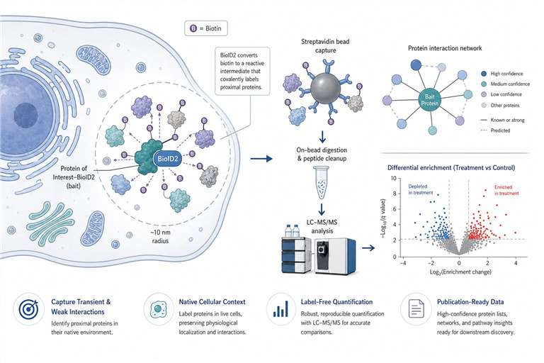
Our BioID2 proximity labeling proteomics service captures transient, weak, and membrane-associated PPIs in their native context.

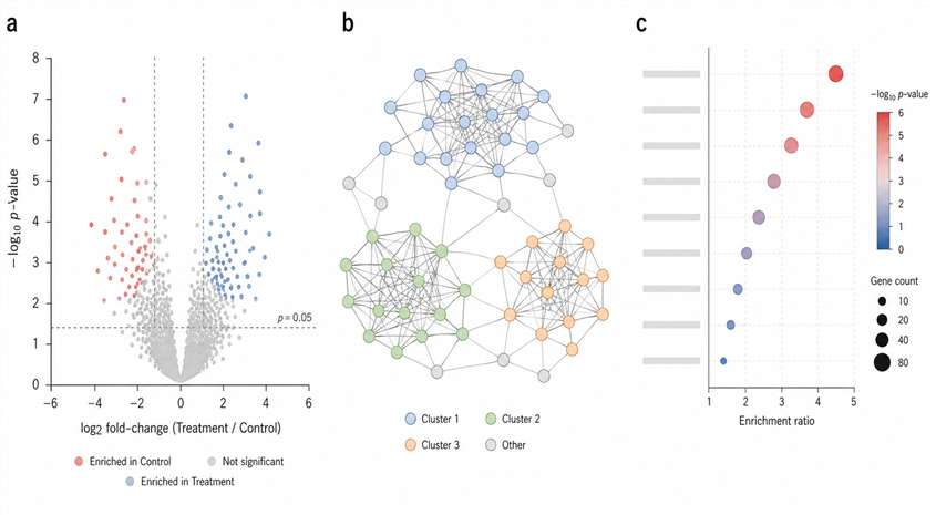
From construct design to MS analysis, we deliver clean, publication-ready interactome maps to accelerate your drug discovery pipeline.

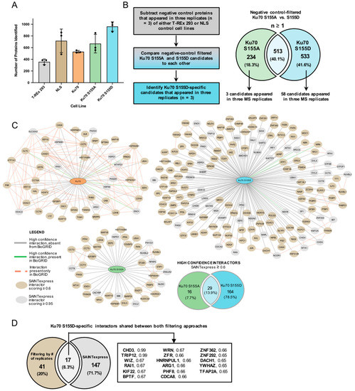
If you've ever struggled to pull down a real protein interactor—getting back nothing but the same high‑abundance contaminants, or losing that one weak, transient binding event that actually matters—you're not alone. Traditional co‑immunoprecipitation works beautifully for rock‑solid complexes. It's the faint, fleeting, and membrane‑buried interactions that slip through. That's where proximity‑dependent biotinylation changes the game, and BioID2 is one of the most forgiving ways to do it.

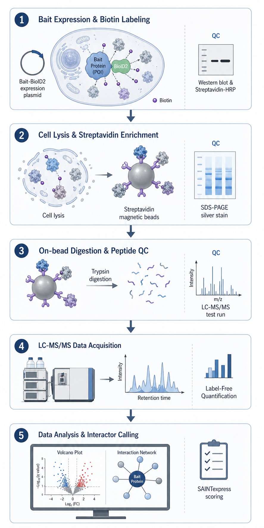
Here, we walk you through how we run BioID2 proximity labeling proteomics, what the data look like, and what you can expect from the service. The short version: we handle everything from construct strategy to the final interaction network, with clear QC gates along the way so you can trust the list of candidates you get back.

Key Advantages:

  • Capture elusive protein interactions.
  • End-to-end service from construct to data.
  • Rigorous QC and negative control design.
  • Deliverables tailored for publication.
BioID2 proximity labeling proteomics service workflow
What Is BioID2 Service Advantage Workflow & QC Demo Results Sample Requirements Bioinformatics Strategy Comparison FAQ Case Study

What Is BioID2 Proximity Labeling and How It Powers Target Discovery

BioID2 is a proximity‑dependent biotin ligase. You fuse it to your protein of interest, add biotin, and the enzyme tags everything within roughly a 10‑nanometer radius. That cloud of biotinylated neighbors—transient binders, membrane‑adjacent partners, even proteins that only touch yours for a few seconds—can then be pulled down on streptavidin beads and identified by high‑resolution mass spectrometry.

Because the labeling happens in living cells, the interactions are captured in their native environment. There is no crosslinking step that can lock in artifacts, and the covalent biotin tag is tough enough that you can use stringent wash conditions to get rid of non‑specific binders. Later methods like TurboID and APEX give you faster labeling kinetics, and we work with those too. But BioID2 remains the go‑to when you need a smaller, less toxic enzyme that performs well in whole‑organism models.

The outcome that matters for target discovery is a shortlist of context‑specific interactors that you can take straight into functional validation—whether that's a co‑IP, a PLA, or a phenotypic readout.

Our BioID2 Service Advantage: From Constructs to Candidates

Some groups just need a mass spectrometry readout; others need help from the very beginning. We are comfortable jumping in at any point.

When you work with us, you get:

  • Full‑workflow coverage. We can design the BioID2 fusion construct, generate stable cell lines or work with transient expression, optimize biotin labeling, perform the enrichment, run the LC‑MS/MS, and deliver the analyzed data. You can hand off one step or the full pipeline.
  • Rigorous negative control design. Every project includes a matched control strategy—empty vector, non‑targeting bait, or a catalytically dead enzyme—so that we can distinguish real neighbors from endogenous biotinylated background.
  • QC checkpoints you can see. At the protein level, we verify biotinylation efficiency by streptavidin‑HRP western blot. At the peptide level, we monitor enrichment quality and digestion completeness before committing samples to deep mass spectrometry runs.
  • Complex samples are welcome. We routinely handle adherent and suspension cell lines, primary cells, and tissue‑derived material. If your model is difficult, we will figure out the right lysis and enrichment conditions during a small‑scale pilot before scaling up.
  • A single scientific point of contact. From method development through final data interpretation, you talk to the same PhD‑level scientist who understands the biology, not a triage desk.

BioID2 Experimental Workflow & QC Checkpoints

The path from a sample to an interaction network has several steps. Here is how it flows in our lab—and where we check progress.

1

Bait expression and biotin labeling

Your BioID2‑tagged protein of interest is expressed in the cell model. We add biotin and incubate for the labeling window you choose (typically 6–24 hours for BioID2). At this point, we confirm expression by western blot and biotinylation by streptavidin‑HRP. If the labeling signal is weak, we adjust biotin concentration or incubation time before proceeding.

2

Cell lysis and streptavidin enrichment

Cells are lysed under denaturing or semi‑native conditions (depending on the bait). Biotinylated proteins are captured on streptavidin‑conjugated beads and washed stringently. We check a small aliquot on SDS‑PAGE with streptavidin‑HRP or silver stain to confirm that the enrichment has worked and that the pattern differs visibly from the negative control.

3

On‑bead digestion and peptide QC

Proteins are reduced, alkylated, and digested directly on the beads with trypsin. Peptide concentration is measured, and a quick LC‑MS/MS test run confirms that the sample complexity and chromatographic quality meet our internal thresholds.

4

LC‑MS/MS data acquisition

Peptides are separated on a nano‑flow UPLC system coupled to a high‑resolution mass spectrometer operating in data‑dependent acquisition (DDA) mode. We use label‑free quantification (LFQ) to compare enrichment across conditions.

5

Data analysis and interactor calling

Raw files are searched against the appropriate protein database. Significantly enriched proteins are identified by comparing the bait condition to the negative control using statistical scoring such as SAINTexpress. The output is a ranked list of high‑confidence proximity interactors.

BioID2 proximity labeling workflow diagram with QC checkpoints

Representative BioID2 Proteomics Results

To give you a sense of what you receive, here are the three types of data views we deliver for a typical BioID2 experiment. The example reflects a comparison between a wild‑type bait and a mutant bait, but the same formats apply regardless of your experimental design.

Demo results of BioID2 proteomics showing volcano plot, network, and enrichment charts

Differential enrichment volcano plot, interaction network, and GO enrichment

1. Differential enrichment volcano plot. Each point is a protein. The x‑axis shows the log₂ fold‑change between conditions, and the y‑axis shows the statistical significance (−log₁₀ p‑value). Proteins that pass the significance and enrichment thresholds are color‑coded. The plot gives you a single‑glance view of which candidates are worth following up.

2. Protein–protein interaction network. High‑confidence interactors are visualized as nodes, with edges representing known or predicted associations. Clusters often resolve into functional modules—a transcriptional complex here, a trafficking machinery there—which helps you prioritize hits that fall into pathways relevant to your biology.

3. GO enrichment and pathway analysis. Enriched gene ontology terms (biological process, molecular function, cellular component) are shown as a bar or bubble chart. This annotation answers the question: "What processes are over‑represented among my hits?" It is particularly useful when you are exploring an uncharacterized bait and need a compass for functional follow‑up.

Sample Submission Requirements

Because BioID2 projects often start with a cell model you have already built, the most common submission is live cells or snap‑frozen cell pellets. The table below covers the typical cases.

Sample TypeRecommended InputContainerShipping ConditionsQC CheckpointsNotes
Adherent or suspension cells (live)≥1×10⁷ cellsT-25 flask or cell pellet, snap-frozenDry ice (−80°C)Cell viability ≥90%; Mycoplasma‑freeInclude empty vector or parental control cells
Transfected cells (transient)≥1×10⁷ cellsSnap-frozen pelletDry ice (−80°C)Confirm expression by western blot before shippingProvide plasmid map and sequence
Cell lysate≥500 µg total protein1.5 mL tube, snap-frozenDry ice (−80°C)Pre‑test biotinylation by streptavidin‑HRPProvide lysis buffer composition
Tissue (mouse, rat)20–50 mg wet weightSnap-frozen, wrapped in foilDry ice (−80°C)Perfusion recommended to reduce blood backgroundContact us before large tissue submissions
Purified plasmid or cDNA1–2 µgTE buffer or driedIce packs (4°C) or ambientConcentration verified by NanoDropShipped separately from cells if needed

For samples not listed here, reach out through the inquiry form. We regularly work with primary neurons, organoids, and xenograft material; the preparation protocol just needs a quick feasibility check first.

Bioinformatics Analysis & Data Deliverables

Every BioID2 project includes a core set of deliverables. We also offer optional add‑ons depending on how far along your analysis pipeline is.

Minimum deliverables — included with every project:

  • Raw MS files (.raw and .mzML formats)
  • Protein identification and label‑free quantification matrix (.csv or .xlsx)
  • List of proteins significantly enriched over the negative control, with fold‑change, p‑value, and confidence score
  • QC summary: biotinylation blot image, enrichment gel image, chromatographic quality metrics

Optional add‑ons — available on request:

  • Cytoscape‑ready interaction network files and publication‑quality figures
  • GO, KEGG, and Reactome pathway enrichment analysis with downloadable result tables
  • Custom volcano plots, heatmaps, and Venn diagrams
  • Extended statistical analysis, including SAINTexpress scoring and permutation‑based FDR estimation
  • A written methods section that you can drop directly into your manuscript

All deliverables are transferred through a secure file‑sharing portal, and we keep your raw data archived for at least 12 months after project close.

Choosing the Right Proximity Labeling Strategy

Not every proximity labeling enzyme is right for every experiment. The table below pulls together the key differences so you can align the method to your model system and biological question.

FeatureBioIDBioID2TurboIDAPEX
OriginBirA* (E. coli)Smaller, humanized BirA*Directed evolution of BirAEngineered ascorbate peroxidase
Labeling time18–24 hours6–24 hours10–60 minutes≤1 minute
Biotin supplementationRequired (µM)Required (µM)Required (µM)Not required (biotin‑phenol probe)
ToxicityLowLowestModerate (high biotin demand)H₂O₂ required (oxidative stress)
Key advantageProven in many modelsCompact size; good in vivo performanceFast labeling; suits kinetic studiesSub‑organelle resolution with EM
Typical applicationGeneral proximity mappingIn vivo models, low‑toxicity needsCell lines, acute stimuliSub‑minute events, synaptic labeling

How to choose:

  • In vivo models and low‑toxicity requirement? Start with BioID2. Its smaller size and lower biotin demand make it the gentlest option for whole‑organism work.
  • Fast signaling events in cell culture? TurboID gives you a snapshot in minutes. That speed is worth the extra biotin if your question is time‑sensitive.
  • Sub‑minute resolution or EM‑level localization? APEX is the answer. The H₂O₂ pulse is harsh but ultra‑precise.
  • Still unsure? Describe your model in the inquiry form. We routinely help teams pick the right enzyme during the feasibility review.

Frequently Asked Questions

Q: How does BioID2 compare to traditional co‑immunoprecipitation?

Co‑IP captures proteins that remain bound through lysis and washing, which strongly favors stable, high‑affinity complexes. BioID2 labels proximal proteins covalently in the living cell before lysis, so transient, weak, and membrane‑embedded interactors are captured. The two methods are complementary; many groups use BioID2 or TurboID to discover candidates and then validate the strongest ones with co‑IP or PLA.

Q: What negative controls should I include?

The gold standard is a BirA*‑dead enzyme fused to the same targeting sequence as your bait. If that is not available, an empty vector or an unrelated bait targeted to the same subcellular compartment works well. We discuss the optimal control during experimental design and can help generate the necessary plasmid if needed.

Q: Can you work with primary cells or in vivo models?

Yes. We have experience with primary neuronal cultures, hematopoietic cells, and tissue‑derived material from mouse models. For in vivo labeling, biotin dosing and timing need pilot testing, which we can do as part of a small‑scale feasibility study before committing to a full project.

Q: What do you need from me to get started?

The essentials: the gene or protein sequence of your bait, the cell line or model you intend to use, and a brief description of your biological question. If you already have a BioID2 construct, we can start from there. If you are starting from scratch, we will design and clone the fusion construct for you.

Q: What bioinformatics analysis is included?

Every project includes protein identification, label‑free quantification, and a statistical comparison of bait versus control to generate a ranked interactor list. Additional analyses—interaction network construction, pathway enrichment, and custom visualization—are available as add‑ons. More details are in the Bioinformatics Analysis section above.

Case Study: BioID2 Resolves Phosphorylation-Dependent DNA Repair Interactions

Swayze, E., et al. (2023) Int. J. Mol. Sci. 24(8), 7041

Background

The Ku70/Ku80 heterodimer sits at the apex of the non‑homologous end joining (NHEJ) pathway, binding double‑strand breaks and recruiting repair factors. A single phosphorylation event—Ku70 Serine‑155 (S155)—had been implicated in shifting the DNA damage response, but the specific protein–protein interactions that explain that shift were unknown. The challenge is a classic one for interaction proteomics: how do you isolate the binding partners of a single phospho‑isoform in a way that conventional immunoprecipitation cannot resolve?

Methods

The authors generated Flp‑In T‑REx 293 cell lines stably expressing BioID2 fusions to wild‑type Ku70, phosphomimetic Ku70 S155D, or phosphoablative Ku70 S155A. Biotin was added for 18 hours, and biotinylated proteins were enriched on streptavidin beads, digested on‑bead, and analyzed by LC‑MS/MS. High‑confidence proximity interactors were called using SAINTexpress (probability score ≥0.6) across three biological replicates. Candidates were then validated by proximity ligation assay (PLA) and co‑immunoprecipitation in doxycycline‑inducible HeLa lines.

Results

The BioID2 screen identified 193 high‑confidence SAINTexpress candidates for the phosphomimetic Ku70 S155D bait. Among these, TRIP12—an E3 ubiquitin ligase previously linked to the DNA damage response—stood out with a near‑perfect SAINTexpress score of 0.99 and was detected exclusively in the S155D condition across all three replicates (see Figure 2 of the original paper). PLA confirmed that Ku70 S155D–TRIP12 proximity was significantly elevated relative to wild‑type Ku70, and endogenous Ku70–TRIP12 association was further detected upon ionizing radiation, confirming physiological relevance.

Conclusion

This study exemplifies the strategic advantage of BioID2 proximity labeling for resolving post‑translational modification‑specific interaction networks. By combining isoform‑specific bait design with rigorous statistical filtering and orthogonal validation, the authors identified a previously unappreciated Ku70–TRIP12 interaction triggered by DNA damage. The work illustrates the full pipeline from hypothesis‑driven bait engineering to mechanistic target deconvolution—precisely the kind of insight our service is designed to support.

BioID2 interactome comparison across Ku70 wild-type, S155D, and S155A conditions identifying TRIP12

Figure 4. Ku70 S155 phospho-specific BioID2 interactome analysis identifies TRIP12 as a DNA damage–responsive interactor. (Adapted from Swayze et al., 2023, Int. J. Mol. Sci., CC BY 4.0)

References

  1. Roux, K. J., Kim, D. I., & Burke, B. (2013). BioID: a screen for protein-protein interactions. Curr Protoc Protein Sci, 74, 19.23.1‑19.23.14. PubMed
  2. Kim, D. I., et al. (2016). An improved smaller biotin ligase for BioID proximity labeling. Mol Biol Cell, 27(8), 1188–1196. PMC
  3. Branon, T. C., et al. (2018). Efficient proximity labeling in living cells and organisms with TurboID. Nat Biotechnol, 36, 880–887. Nature
  4. Swayze, E., et al. (2023). Analysis of Ku70 S155 Phospho-Specific BioID2 Interactome Identifies Ku Association with TRIP12 in Response to DNA Damage. Int. J. Mol. Sci., 24(8), 7041. MDPI

Disclaimer: Creative Proteomics services are for research use only. They are not intended for clinical diagnostic or therapeutic purposes.

Online Inquiry

Please submit a detailed description of your project. We will provide you with a customized project plan to meet your research requests. You can also send emails directly to for inquiries.

* Email
Phone
* Service & Products of Interest
Services Required and Project Description