Drug-Resistance Mechanism Analysis Service — MS-Based Multi-Omics Profiling

Elucidate the molecular mechanisms driving drug resistance with integrated proteomics, metabolomics, and phosphoproteomics — from in vitro models to patient-derived specimens.

Drug resistance remains the single greatest barrier to effective cancer therapy. Whether in targeted therapy, chemotherapy, or immunotherapy, resistant clones inevitably emerge — yet the underlying molecular mechanisms often remain a black box. Genomic approaches alone cannot capture the post-translational signaling rewiring, metabolic reprogramming, and proteome-level adaptations that drive resistance.

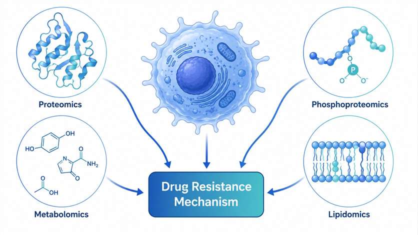
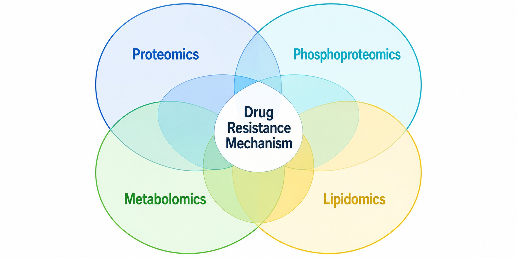
MassTarget™ offers a comprehensive Drug-Resistance Mechanism Analysis Service that integrates mass spectrometry-based proteomics, phosphoproteomics, metabolomics, and lipidomics to systematically map the molecular landscape of drug resistance. Our multi-omics approach covers the full spectrum of resistance mechanisms including drug target alterations, signaling pathway rewiring, metabolic reprogramming, efflux pump upregulation, epithelial-mesenchymal transition, apoptosis evasion, and DNA damage repair activation.

By combining multiple MS-based analytical modalities under one roof, we provide an integrated view of resistance mechanisms that no single-omics approach can deliver — enabling you to identify actionable targets for combination therapy and biomarker strategies.

Drug resistance mechanism analysis multi-omics workflow diagram
Overview Advantages Modes Workflow Platforms Sample Case Study FAQ

Overview of Drug-Resistance Mechanism Analysis

Drug resistance is a multifactorial phenomenon that arises through diverse molecular mechanisms operating at the proteome, signaling, and metabolic levels. Understanding which mechanism(s) drive resistance in a given context is essential for developing effective combination strategies and overcoming treatment failure.

Our service systematically investigates the following resistance mechanisms:

  • Drug target alterations — Mutations, amplifications, or downregulation of drug targets that reduce drug sensitivity
  • Signaling pathway rewiring — Kinase activation, bypass signaling, and compensatory pathway engagement that circumvent target inhibition
  • Metabolic reprogramming — Altered glycolysis, oxidative phosphorylation, nucleotide metabolism, and lipid remodeling that support resistant cell survival
  • Efflux pump upregulation — ABC transporter-mediated drug efflux that reduces intracellular drug accumulation
  • Epithelial-mesenchymal transition (EMT) — Phenotypic switching associated with acquired resistance and metastatic potential
  • Apoptosis evasion — Anti-apoptotic protein upregulation and pro-apoptotic protein downregulation that block cell death
  • DNA damage repair activation — Enhanced repair capacity in response to genotoxic agents

Key Advantages

Multi-Omics Integration

Proteomics, metabolomics, phosphoproteomics, and lipidomics under one roof — no data integration challenges across vendors.

MS-thermal stabilization assay Capability

IMPRINTS-thermal stabilization assay for functional target engagement profiling directly in resistant vs. sensitive cellular contexts.

Broad Sample Compatibility

Cell lines, isogenic resistant/sensitive pairs, PDX tumors, organoids, tissue biopsies, biofluids, and FFPE.

End-to-End Service

From experimental design through sample processing, LC-MS/MS acquisition, bioinformatics analysis, and publication-ready figures.

Published Track Record

Peer-reviewed publications in Nature Communications demonstrating real-world impact of our MS-based resistance profiling.

Rapid Turnaround

Standard 4–6 weeks for proteomics; 3–4 weeks for metabolomics; express options available.

Service Modes

We offer six complementary service modes that can be used individually or in combination to address specific resistance questions.

MODE 1

Resistance Proteome Profiling

Quantitative proteomics (TMTpro, label-free, or DIA) comparing resistant vs. sensitive isogenic cell lines, PDX models, or patient-matched pre/post-treatment biopsies.

Best for: Discovering novel resistance-associated protein targets and pathways.

MODE 2

Phosphoproteomic Kinase Profiling

TiO2-based phosphopeptide enrichment followed by LC-MS/MS to map global phosphorylation changes and activated kinase signaling cascades.

Best for: Understanding signaling pathway rewiring and identifying kinase targets for combination therapy.

MODE 3

Targeted Metabolomics Panel

Quantitative MRM-based measurement of resistance-associated metabolites including nucleotides, amino acids, TCA cycle intermediates, and energy metabolism markers.

Best for: Validating metabolic dependencies and identifying metabolic vulnerabilities.

MODE 4

Untargeted Metabolomics / Lipidomics

Global metabolic and lipid profiling using high-resolution LC-MS to capture the full metabolome and lipidome landscape of resistant cells.

Best for: Comprehensive metabolic and lipid characterization of resistance phenotypes.

MODE 5

MS-thermal stabilization assay Functional Proteomics

IMPRINTS-thermal stabilization assay (Integrated Modulation of Protein Interaction States — thermal stabilization assay) to profile drug-target engagement and resistance-associated protein stability changes across the proteome.

Best for: Functional validation of resistance mechanisms at the proteome-wide level.

MODE 6

Custom Multi-Omics Integration

Tailored combination of any of the above modes with advanced bioinformatics — including correlation analysis, multi-omics network modeling, and machine learning-based biomarker discovery.

Best for: Comprehensive resistance mechanism programs requiring integrated multi-omics interpretation.

Service Workflow

The workflow consists of five essential stages:

1

Experimental Design

Consultation to define the resistant/sensitive model system, select appropriate omics modalities, determine sample size and replicate requirements, and establish the experimental timeline.

2

Sample Preparation

Protein extraction and digestion, phosphopeptide enrichment, metabolite extraction, and lipid extraction with quality control at each step.

3

LC-MS/MS Data Acquisition

High-resolution Orbitrap (Exploris 480, Q Exactive HF-X) and Q-TOF (timsTOF Pro) platforms with optimized acquisition methods for each omics modality.

4

Bioinformatics Analysis

Differential expression analysis, pathway enrichment (KEGG, Reactome, GO), kinase-substrate network mapping, and metabolic pathway mapping.

5

Mechanism Interpretation

Comprehensive report integrating all omics layers, with identification of key resistance drivers, actionable targets for combination therapy, and publication-ready figures.

Drug resistance analysis workflow five steps from design to interpretation

Service Process: Inquiry & Consultation → Project Proposal & Quote → Sample Submission → LC-MS/MS Analysis → Data Analysis & Report Delivery

Technology Platforms

MassTarget's drug-resistance mechanism analysis service is supported by advanced mass spectrometry platforms optimized for multi-omics profiling.

PlatformApplicationKey Specifications
Orbitrap Exploris 480Proteomics, phosphoproteomicsHRAM, 480,000 resolution, FAIMS Pro
Q Exactive HF-XQuantitative proteomics (TMT)120,000 resolution, fast scanning
Triple Quad 6500+Targeted metabolomicsMRM quantitation, high sensitivity
timsTOF Pro4D-proteomics, MS-thermal stabilization assayPASEF, ion mobility separation

Technology Comparison

DimensionProteomicsPhosphoproteomicsMetabolomicsLipidomics
What It MeasuresProtein expression changesKinase signaling activityMetabolite levelsLipid composition
Resistance Mechanism DetectedTarget alteration, EMT, apoptosis evasionPathway rewiring, kinase activationMetabolic reprogrammingMembrane remodeling
Quantification MethodTMTpro / Label-free / DIATMT with TiO2 enrichmentMRM / Untargeted LC-MSLC-MS/MS lipid profiling
Sample Requirement50–200 µg protein1–5 mg protein1×106 cells / 20 mg tissue1×106 cells / 20 mg tissue
Typical Turnaround4–6 weeks4–6 weeks3–4 weeks3–4 weeks

Sample Requirements

Sample TypeRecommended AmountBiological ReplicatesStorageShipping
Cell Pellets≥1×10⁷ cells per condition≥3-80 °CDry ice
Tissue Biopsies20–50 mg per sample≥3Liquid N2 / -80 °CDry ice
PDX Tumor Tissues30–100 mg per sample3–5Liquid N2 / -80 °CDry ice
Organoids≥1×10⁶ cells per condition≥3-80 °CDry ice
Plasma / Serum50–100 µL per sample≥3-80 °CDry ice
CSF / Biofluids100–200 µL per sample≥3-80 °CDry ice
FFPE Sections10 slices (10 µm thickness)≥3Room temperatureAmbient

General Guidelines: Samples should be flash-frozen in liquid nitrogen immediately after collection and stored at -80 °C. Avoid repeated freeze-thaw cycles. For cell-based studies, at least 3 biological replicates are strongly recommended. For metabolomics samples, quenching in liquid nitrogen within seconds of collection is critical to preserve metabolite profiles. For phosphoproteomics, include phosphatase inhibitors during lysis.

Related Services

MassTarget offers a comprehensive portfolio of services that complement drug-resistance mechanism analysis:

Deliverables

  • Comprehensive report with differential expression analysis (volcano plots, heatmaps)
  • Pathway enrichment analysis (KEGG, Reactome, GO)
  • Kinase-substrate network maps (phosphoproteomics projects)
  • Metabolic pathway mapping with metabolite correlation networks
  • Integrated multi-omics correlation analysis
  • Publication-ready figures
  • Raw data files (RAW format) and processed data tables
  • Bioinformatics consultation session to discuss results

Representative Data

Multi-omics integration proteomics phosphoproteomics metabolomics lipidomics drug resistance

Multi-omics integration for drug resistance mechanism elucidation

Case Study: MS-thermal stabilization assay Uncovers DNA Repair Programs Driving Gemcitabine Resistance

Nature Communications (2025) — 10.1038/s41467-025-59505-8

Service Used

Nucleotide quantification by LC-MRM/MS + MS-thermal stabilization assay (IMPRINTS-thermal stabilization assay)

Background

Gemcitabine is a nucleoside analog used as first-line therapy for pancreatic cancer and diffuse large B-cell lymphoma (DLBCL). However, acquired resistance develops rapidly, and the underlying mechanisms at the proteome and metabolome level remain incompletely understood.

Approach

The research team employed IMPRINTS-thermal stabilization assay, a deep functional proteomics implementation of the thermal stabilization assay, to profile biochemical responses to gemcitabine in resistant and sensitive DLBCL cell lines. This was complemented by targeted LC-MRM/MS-based nucleotide quantification to measure intracellular gemcitabine triphosphate levels and nucleotide pool alterations.

Key Findings

  • MS-thermal stabilization assay revealed that gemcitabine-resistant cells activate DNA repair programs, including upregulation of the Fanconi anemia (FA) and homologous recombination (HR) pathways
  • Targeted nucleotide quantification showed altered dNTP pool balance in resistant cells, consistent with enhanced DNA repair capacity
  • Integrated proteomics and metabolomics identified RRM1/RRM2 upregulation as a key metabolic adaptation driving resistance
  • The study demonstrated that combining gemcitabine with ATR or CHK1 inhibitors synergistically overcomes resistance

Impact

This study provided a mechanistic framework for understanding gemcitabine resistance at the proteome and metabolome level, identifying combination therapy strategies (gemcitabine + ATRi/CHK1i) that are now being advanced toward clinical evaluation.

MS-thermal stabilization assay workflow gemcitabine resistance DNA repair activation

IMPRINTS-thermal stabilization assay workflow for profiling gemcitabine resistance mechanisms in DLBCL cells.

FAQ

Frequently Asked Questions

Q: What types of drug resistance mechanisms can your service identify?

Our integrated multi-omics approach can identify a broad range of resistance mechanisms, including drug target alterations (mutations, amplifications, downregulation), signaling pathway rewiring (kinase activation, bypass signaling), metabolic reprogramming (glycolysis, OXPHOS, nucleotide metabolism), efflux pump upregulation, EMT, apoptosis evasion, and DNA damage repair activation.

Q: What sample types are compatible with your drug resistance analysis service?

We accept cell pellets, isogenic resistant/sensitive cell line pairs, PDX tumor tissues, organoids, tissue biopsies (fresh frozen or FFPE), plasma/serum, CSF, and other biofluids. For cell-based studies, we recommend at least 1×10⁷ cells and 3 biological replicates per condition.

Q: How do you integrate proteomics and metabolomics data for resistance mechanism analysis?

Our bioinformatics pipeline performs correlation analysis across omics layers, mapping protein expression changes to metabolite level alterations. We use pathway enrichment (KEGG, Reactome) and network-based approaches to identify converging resistance mechanisms. The integrated analysis is presented in a comprehensive report with cross-omics correlation heatmaps and pathway-level integration.

Q: What is MS-thermal stabilization assay and how does it help study drug resistance?

MS-thermal stabilization assay (thermal stabilization assay coupled with mass spectrometry) is a label-free proteomics method that measures protein thermal stability changes in response to drug treatment or resistance-associated perturbations. It can identify drug-target engagement directly in cellular contexts and reveal resistance-associated protein stability changes across the proteome — providing functional insights that expression-level proteomics alone cannot capture.

Q: How long does a typical drug resistance mechanism analysis project take?

Standard timelines are 4–6 weeks for proteomics/phosphoproteomics and 3–4 weeks for metabolomics/lipidomics. Combined multi-omics projects typically require 6–8 weeks. Express options are available for time-sensitive programs.

Q: Do you provide bioinformatics support and publication-ready figures?

Yes. Our service includes comprehensive bioinformatics analysis and publication-ready figures including volcano plots, heatmaps, pathway enrichment maps, kinase-substrate networks, and integrated multi-omics correlation visualizations. We also provide a results discussion session with our bioinformatics team.

Ready to Uncover the Mechanisms Behind Drug Resistance?

Partner with MassTarget™ to decode resistance pathways and advance your therapeutic pipeline. Our scientific team will work with you to design the optimal multi-omics strategy for your specific resistance question.

Online Inquiry

Please submit a detailed description of your project. We will provide you with a customized project plan to meet your research requests. You can also send emails directly to for inquiries.

* Email
Phone
* Service & Products of Interest
Services Required and Project Description