Proteomics-Based Target Discovery — From Disease Biology to Validated Therapeutic Targets

An integrated mass spectrometry proteomics platform that identifies, validates, and characterizes drug targets — from global proteome profiling to target engagement and mechanism of action.

Target discovery is the critical front end of the drug development pipeline, yet it remains the stage with the highest attrition. The gap between a compelling disease hypothesis and a drug-ready target requires orthogonal lines of proteomic evidence — abundance changes, interaction networks, post-translational modifications, thermal stability shifts, and structural dynamics — that no single proteomics experiment can deliver alone.

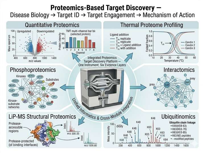
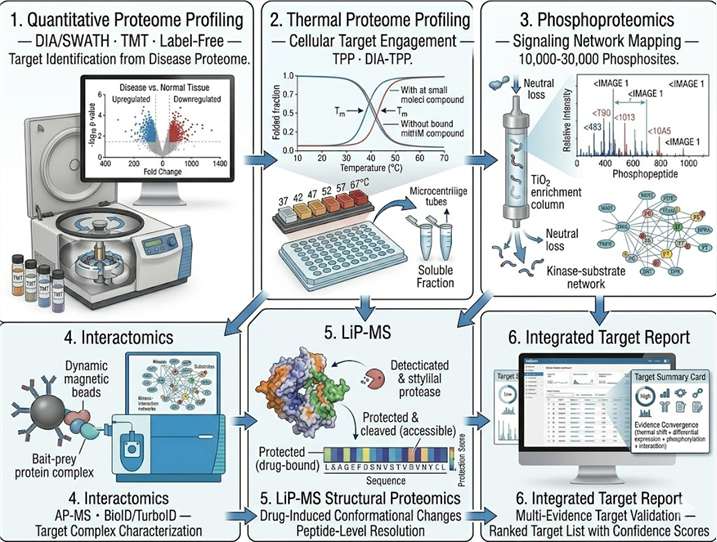
Our Proteomics-Based Target Discovery solution integrates six complementary quantitative and functional proteomics modules into a single, instrument-harmonized workflow. Every experiment runs on the same Orbitrap platform with unified sample preparation and data processing standards, so cross-experiment comparisons are quantitative, not anecdotal. From identifying differentially expressed proteins in disease tissue to confirming target engagement in living cells, our platform delivers the multidimensional proteomic evidence that distinguishes a validated drug target from an interesting observation.

Proteomics-Based Target Discovery integrated platform — six proteomics modules for target identification, validation, and mechanism of action
What Is Proteomics-Based Target Discovery Solution Modules Case Study FAQ

What Is Proteomics-Based Target Discovery?

Proteomics-based target discovery is the systematic application of mass spectrometry-driven proteomic technologies to identify and validate protein targets from disease-relevant biological systems. Unlike genomics or transcriptomics, which measure potential, proteomics measures the actual effector molecules — the proteins that carry out disease processes and that drugs ultimately modulate. A proteomics-based discovery program generates four interdependent layers of evidence that collectively build the case for a therapeutic target:

Abundance Evidence

Quantitative proteomics — DIA/SWATH proteomics or TMT-based quantification — compares protein expression across disease vs. normal, treated vs. untreated, and responder vs. non-responder cohorts. A protein consistently up-regulated in diseased tissue that is also down-regulated upon effective treatment provides a strong initial rationale.

Engagement Evidence

Thermal proteome profiling detects whether a protein engages a small molecule in the cellular environment by measuring ligand-induced thermal stabilization shifts. A target that shows a dose-dependent thermal shift upon compound treatment has passed a functional engagement test that gene expression alone cannot provide.

Mechanistic Evidence

Phosphoproteomics and interactomics map the signaling pathways and protein complexes in which the target operates. A target that sits at a signaling node controlling a disease-relevant pathway, and whose perturbation alters downstream phosphorylation, carries mechanistic credibility.

Structural Evidence

LiP-MS (limited proteolysis–mass spectrometry) detects drug-induced conformational changes across the proteome, confirming that a compound physically alters its target's structure. When combined with thermal profiling (DIA-TPP), this provides orthogonal structural and thermodynamic evidence of target binding.

When all four evidence layers converge on the same protein, the confidence in that target is fundamentally different from what a single differential expression dataset can provide. This is the principle behind our integrated platform: every module feeds into a unified evidence framework that transforms candidate proteins into validated targets.

Proteomics Target Discovery Solution Modules

MODULE 1

Quantitative Proteome Profiling for Target Identification

The foundational module. We compare proteomes across disease and control cohorts using DIA/SWATH proteomics (data-independent acquisition for unbiased, reproducible quantification) and shotgun label-free proteomics (data-dependent acquisition for deep proteome coverage). For studies requiring highest multiplexing capacity, TMTpro 16-plex or 18-plex labeling enables simultaneous quantification across multiple conditions, time points, or patient samples in a single MS run.

  • DIA/SWATH — reproducible quantification of 6,000–9,000 proteins per sample
  • TMT/TMTpro — up to 18-plex isobaric labeling for multi-condition comparisons
  • Label-free — deep proteome coverage with flexible experimental design
  • Volcano plot analysis, pathway enrichment, and protein–protein interaction network mapping

Deliverable: A ranked list of differentially expressed proteins with fold-change, statistical significance, pathway annotation, and disease-relevance scores — the starting point for target nomination.

MODULE 2

Thermal Proteome Profiling for Target Engagement & Validation

Once a candidate target is identified, the next question is whether a compound physically engages it in the cellular environment. Thermal proteome profiling answers this question proteome-wide: cells or lysates are heated across a temperature gradient, soluble proteins are quantified at each temperature by quantitative MS, and proteins whose thermal stability shifts in the presence of a compound are identified as engaged targets.

  • Label-free TPP for target identification — unbiased, no compound modification required
  • Isobaric TPP (TMT-TPP) for dose–response target engagement curves
  • DIA-TPP — data-independent acquisition for highest quantitative precision
  • Simultaneously identifies on-target engagement AND off-targets

Deliverable: Thermal stability curves for the target protein and any off-targets, with melting temperature (Tm) shifts quantified, confirming dose-dependent cellular target engagement.

MODULE 3

Phosphoproteomics for Signaling Pathway Mapping

Disease-relevant targets rarely act in isolation — they sit within signaling networks. Phosphoproteomics activation mapping quantifies thousands of phosphorylation sites across the kinome and beyond, revealing which signaling pathways are activated or suppressed in disease, and how a target or compound alters the signaling landscape.

  • TiO2- or IMAC-based phosphopeptide enrichment from 1–5 mg protein input
  • Quantification of 10,000–30,000 phosphosites per experiment
  • Kinase–substrate relationship inference from phosphorylation motifs and network analysis
  • Integration with quantitative proteomics for phosphorylation stoichiometry estimation

Deliverable: A signaling network map with differentially phosphorylated sites annotated by kinase, pathway, and functional domain, identifying the signaling nodes that the target controls.

MODULE 4

Interactomics for Target Complex & PPI Network Characterization

Most drug targets function as part of multi-protein complexes. Interactomics (AP-MS and proximity-labeling MS) identifies the full complement of proteins that physically associate with the target — its interactome. This reveals which complexes the target participates in, which regulatory subunits control its activity, and whether targeting it might disrupt essential complexes (potential toxicity) or disease-specific ones (therapeutic window).

  • Affinity purification–MS (AP-MS) — for stable, high-affinity interactions
  • Proximity-dependent biotinylation (BioID/TurboID) + MS — for transient and proximal interactions
  • Label-free or TMT-based quantification of specific vs. control interactors
  • SAINT/CRAPome filtering for high-confidence interaction assignment

Deliverable: A high-confidence interactome map identifying all specific protein–protein interactions of the target, with functional annotation of each interacting complex.

MODULE 5

Structural Proteomics (LiP-MS) for Drug-Induced Conformational Changes

LiP-MS (Limited Proteolysis–Mass Spectrometry) detects structural changes in proteins induced by drug binding, metabolite interaction, or post-translational modification — without requiring purified protein or crystallization. By exposing native protein extracts to a non-specific protease under controlled conditions, LiP-MS generates structure-dependent peptide patterns that shift when a ligand alters the target's conformation.

  • Proteome-wide detection of drug-induced conformational changes
  • Identification of binding-affected regions — complementary to HDX-MS
  • Compatible with DIA-TPP for combined structural + thermodynamic target engagement evidence
  • Works with cell lysates, tissue homogenates, and subcellular fractions

Deliverable: A structural perturbation map showing which protein regions undergo conformational change upon drug binding, confirming physical target engagement at peptide-level resolution.

MODULE 6

Ubiquitinomics & Degradation Proteomics

For targets that are degraded via the ubiquitin–proteasome system — whether endogenous turnover or PROTAC-induced degradation — ubiquitinomics provides a quantitative view of ubiquitination dynamics. DiGly remnant profiling (K-ε-GG) after trypsin digestion captures endogenous ubiquitination sites at proteome scale, enabling you to track which proteins are ubiquitinated, at which lysine residues, and how ubiquitination patterns change upon treatment.

  • DiGly peptide enrichment (K-ε-GG antibody) for ubiquitin remnant profiling
  • Quantification of 5,000–15,000 ubiquitination sites per experiment
  • PROTAC selectivity profiling — does degradation extend beyond the intended target?
  • Time-course ubiquitinome analysis for degradation kinetics

Deliverable: A quantitative ubiquitination landscape showing which proteins and lysine sites are ubiquitinated, with fold-changes upon treatment, confirming degradation selectivity for the target of interest.

Why Integrated Proteomics Matters for Target Discovery

Cross-Module Evidence Convergence

When your differential proteomics, thermal profiling, phosphoproteomics, and interactomics data all point to the same protein — and the LiP-MS data confirms drug-induced structural change — the target is validated by four independent physical measurements, not one statistical observation. This is the standard of evidence that preclinical review committees and investment boards require.

Same Platform, Comparable Data

All six modules run on the same Orbitrap platform with shared sample preparation, chromatography, and data processing. When you compare your thermal profiling dataset to your phosphoproteomics dataset, the mass accuracy, resolution, and quantification algorithm are identical — eliminating the systematic offsets that occur when different CROs run different experiments on different instruments.

Multi-Omics Integration Ready

Proteomics data does not exist in a vacuum. Our platform connects with AI-driven multi-omics integration that combines proteomics with transcriptomics, metabolomics, and fluxomics data to build systems-level models of disease biology. Systems pharmacology MS modeling translates these multi-omics signatures into actionable target hypotheses.

Seamless Transition to Lead Discovery

A validated target from our proteomics platform transitions directly into our automated compound-target binding HT-MS screening — the same instrument, the same sample prep, and the same data portal. The transition from "we have a target" to "we have a hit" is measured in days, not months, because the platform is already configured for your target.

Case Study

IP-MS Interactome Profiling Identifies HDAC4/HDAC1/HDAC2 Epigenetic Complex as a DNA Repair Regulator

Di Giorgio E, Dalla E, Tolotto V, D'Este F, Paluvai H, Ranzino L, Brancolini C. (2024) HDAC4 influences the DNA damage response and counteracts senescence by assembling with HDAC1/HDAC2 to control H2BK120 acetylation and homology-directed repair. Nucleic Acids Research, 52(14): 8218–8240.

Study design: Cellular senescence — a state of irreversible cell-cycle arrest — contributes to aging and age-related disease, yet the molecular mechanisms that enforce and maintain senescence remain incompletely understood. The research team at Università degli Studi di Udine investigated the role of HDAC4 in the DNA damage response, hypothesizing that HDAC4 might function in genome maintenance pathways relevant to senescence bypass. They employed Creative Proteomics' immuno-precipitation proteomics (IP-MS) service to characterize the HDAC4 interactome, identifying HDAC1 and HDAC2 as the core components of a novel epigenetic complex that regulates DNA double-strand break repair pathway choice.

Key results: The IP-MS analysis revealed that HDAC4 physically interacts with HDAC1 and HDAC2 to form a deacetylase complex that removes acetyl groups from histone H2B at lysine 120 (H2BK120ac). This HDAC4/HDAC1/HDAC2 complex modulates homology-directed repair (HDR) efficiency through dynamic deacetylation of H2BK120. When HDAC4 is degraded during Ras-induced senescence, this complex disassembles, H2BK120ac accumulates, BRCA1 and CtIP recruitment to DNA damage sites is impaired, and cells accumulate unrepaired DNA damage — which in turn reinforces the senescence transcriptional program. Forced HDAC4 expression reduces γH2AX genomic spreading and partially restores DNA repair capacity. The study establishes HDAC4 as a potential therapeutic target for modulating senescence and DNA repair in cancer and aging, and demonstrates that the HDAC4/HDAC1/HDAC2 complex represents a node for pharmacological intervention in senescence-associated pathologies.

Relevance to our proteomics-based target discovery platform: This study exemplifies the integrated target discovery logic that our platform operationalizes: interactomics (IP-MS identification of HDAC4's binding partners, revealing the HDAC1/HDAC2 complex as the functional unit), mechanistic proteomics (linking interactome data to a specific post-translational modification — H2BK120ac — and a specific biological process — HDR), and target validation (demonstrating that loss of the target complex produces a disease-relevant phenotype — senescence — and that restoration of the target partially rescues normal function). For target discovery programs seeking to move beyond differential expression lists to mechanistically validated targets, our integrated proteomics platform provides the same workflow: AP-MS or proximity-labeling MS to define the target's interactome, phosphoproteomics or modification-specific proteomics to map the signaling context, and thermal profiling or LiP-MS to confirm that the target can be engaged by small molecules — all on a single, harmonized MS platform.

IP-MS HDAC4 interactome identifies HDAC4/HDAC1/HDAC2 epigenetic complex — proteomics-based target discovery case study Figure 1
FAQ

Frequently Asked Questions

Q: How does an integrated proteomics platform reduce target validation risk compared to single-experiment approaches?

Single-experiment target discovery — for example, a differential proteomics comparison of disease vs. normal tissue — identifies hundreds of differentially expressed proteins but provides no information about which ones are druggable, which ones engage a compound, or which ones sit at actionable signaling nodes. An integrated platform adds three additional evidence layers: thermal profiling confirms whether a compound physically engages the candidate target in live cells; phosphoproteomics reveals whether the target controls disease-relevant signaling; and interactomics identifies whether the target participates in complexes that can be pharmacologically modulated. When all modules converge on the same protein, the confidence in that target is fundamentally higher, and the attrition risk in downstream lead discovery is correspondingly lower. Equally important, when modules disagree — for example, a protein is differentially expressed but shows no thermal shift — the platform flags this discrepancy early before resources are committed to an invalid target.

Q: What types of biological samples are compatible with your proteomics discovery platform?

The platform accommodates the full range of sample types encountered in target discovery: cultured cell lines (adherent or suspension, standard or genetically modified), primary cells (immune cells, neurons, patient-derived cells), fresh-frozen tissue biopsies (tumor, brain, liver, muscle, adipose), FFPE tissue sections (for retrospective clinical cohort studies), biofluids (plasma, serum, CSF, synovial fluid, BALF), subcellular fractions (membrane, nuclear, cytosolic, mitochondrial), and immunoprecipitated or affinity-enriched protein complexes. For thermal proteome profiling experiments, we recommend fresh or fresh-frozen material — FFPE-derived proteins have been heat-treated during embedding and are not suitable for thermal stability measurements. Our project consultation team will guide you on sample preparation, recommended input amounts, and MS-compatible lysis buffer conditions for your specific sample type.

Q: Which quantitative proteomics approach — DIA/SWATH, TMT, or label-free — is best for my target discovery study?

The choice depends on your study design. DIA/SWATH provides the highest quantitative reproducibility and is ideal for large cohort studies (20–200 samples) where each sample must be measured once and compared across groups — the data-independent acquisition ensures every peptide is quantified in every run with low missing values. TMT (isobaric labeling) provides the highest multiplexing capacity (up to 18 conditions in one MS run) with excellent quantitative precision, making it ideal for dose–response or time-course designs with a moderate number of conditions. Label-free (DDA shotgun) provides the deepest proteome coverage per sample and is the most flexible for exploratory studies with a small number of samples. In practice, many discovery programs combine DIA for the initial cohort screen and TMT for the follow-up dose–response validation. We discuss these trade-offs during experimental design consultation.

Q: How do thermal proteome profiling and LiP-MS complement each other in target engagement studies?

Thermal proteome profiling (TPP) measures ligand-induced changes in protein thermal stability — a thermodynamic readout of target engagement. A protein that is stabilized upon compound binding melts at a higher temperature. TPP is sensitive and proteome-wide, but it does not reveal WHERE on the protein the compound binds. LiP-MS measures ligand-induced changes in protease accessibility — a structural readout. When a compound binds, it protects (or, less commonly, exposes) specific peptide regions from protease cleavage. The peptide-level resolution of LiP-MS can localize the binding site to a specific domain or region, while TPP provides the thermodynamic confirmation that binding occurred. Running both assays on the same compound-treated samples provides two orthogonal physical measurements — thermodynamic and structural — of target engagement. If both converge on the same protein, the evidence for direct binding is substantially stronger than either assay alone.

Q: Can your platform handle membrane protein targets?

Yes. Membrane proteins — GPCRs, ion channels, transporters, and receptor tyrosine kinases — constitute the largest class of drug targets and require specialized proteomics handling. Our platform uses MS-compatible detergents (e.g., DDM, CHS, LMNG) for membrane protein solubilization, nanodisc or SMALP reconstitution for maintaining native-like lipid environments, and optimized digestion protocols that account for the limited tryptic cleavage sites in transmembrane helices. For thermal profiling of membrane proteins, we use detergent-screened conditions that preserve thermal denaturation behavior in the membrane fraction. For interactomics, rapid proximity-labeling approaches (TurboID) capture transient and hydrophobic interactions that are lost during traditional AP-MS. Our membrane protein proteomics workflows have been validated on GPCRs, ion channels (TRP, Kv, Nav), and single-pass and multi-pass transmembrane receptors.

Q: How do you handle data integration and IP security across the multi-module workflow?

All raw MS data (.raw files), processed quantification matrices, and analysis reports from every module reside on a dedicated, access-controlled server with project-specific encryption. You access results through a secure data portal where all modules are organized under a single project identifier — your differential proteomics results are viewable alongside thermal profiling data and interactomics networks in the same interface. Cross-module analyses (e.g., proteins that are both differentially expressed AND thermally stabilized) can be performed directly in the portal through integrated filtering and visualization tools. Data never transits through third-party cloud services or subcontractors because all six proteomics modules are performed in-house. At program completion, we transfer the complete data package — raw files, processed results, and integrated analysis reports — to your designated repository, with a structured data dictionary documenting all processing parameters.

References

  1. Meissner F, Geddes-McAlister J, Mann M, Bantscheff M. The emerging role of mass spectrometry-based proteomics in drug discovery. Nature Reviews Drug Discovery. 2022;21:637–654.
  2. Mateus A, Kurzawa N, Becher I, Sridharan S, Helm D, Stein F, Typas A, Savitski MM. Thermal proteome profiling for interrogating protein interactions. Molecular Systems Biology. 2020;16(3):e9232.

Start Your Target Discovery Program

From disease proteome profiling to validated target — six integrated modules, one platform, one partner.

Online Inquiry

Please submit a detailed description of your project. We will provide you with a customized project plan to meet your research requests. You can also send emails directly to for inquiries.

* Email
Phone
* Service & Products of Interest
Services Required and Project Description