Tissue Binding MS — Multi-Tissue fu Determination by RED & LC-MS/MS

Reliable tissue binding data for PBPK modeling, Vss prediction, and DDI assessment. We measure fraction unbound (fu) across 7 tissue types (brain, liver, kidney, lung, heart, muscle, tumor) using RED paired with high-sensitivity LC-MS/MS. Multi-species coverage. Systematic NSB correction. Integrated with our ADME platform.

  • 7 tissue types: brain, liver, kidney, lung, heart, muscle, tumor
  • Gold-standard RED methodology
  • High-sensitivity LC-MS/MS for fu < 0.5%
  • Multi-species: mouse, rat, dog, monkey, human
  • Systematic NSB assessment and correction
  • Flexible method selection: RED, UF, or UC
  • Integrated ADME platform compatibility
Tissue binding MS service overview with multi-tissue fu analysis by RED and LC-MS/MS
Why Multi-Tissue Tissue Types Advantages When to Use Workflow Comparison Sample Demo Case Study FAQ

Why Multi-Tissue Binding Data Matters for Your PBPK Model

Your PBPK model is only as good as its tissue-specific inputs. Without measured fraction unbound (fu) for each compartment, models default to predicted values that introduce significant uncertainty into Vss and clearance projections. We measure fu across seven tissue types — brain, liver, kidney, lung, heart, muscle, and tumor — using rapid equilibrium dialysis (RED) paired with high-sensitivity LC-MS/MS. Multi-species coverage (mouse, rat, dog, monkey, human) enables cross-species scaling. Systematic NSB correction ensures your fu values reflect true tissue binding, not assay artifacts. For a complete overview of our ADME capabilities, visit our ADME/DMPK/PK-PD Research Platforms page.

Tissue Types We Cover

Most CROs limit tissue binding to brain only. We don't. Here's the full panel and why each tissue matters for your PK model:

Brain

Role: CNS target exposure, Kp,uu calculation

Determines whether CNS-active compounds reach therapeutic concentrations.

Liver

Role: Metabolic clearance, hepatic uptake

Primary clearance organ; fu,liver directly impacts CLint scaling.

Kidney

Role: Renal clearance, transporter-mediated disposition

Critical for renally cleared compounds and OAT/OCT substrate assessment.

Lung

Role: Inhalation drugs, pulmonary target engagement

Essential for respiratory drug programs.

Heart

Role: Cardiac safety, tissue distribution

Relevant for cardioactive compounds and cardiac toxicity assessment.

Muscle

Role: Vss calculation, peripheral distribution

Largest tissue mass in the body; major contributor to Vss.

Tumor

Role: Oncology PK/PD, tumor penetration

Determines free drug exposure at the tumor site for efficacy prediction.

All tissues are available in rat, mouse, dog, monkey, and human (where ethically sourced). Custom tissue requests are handled on a project-by-project basis.

Key Advantages of Our Tissue Binding MS Service

Gold-Standard RED Methodology

Rapid equilibrium dialysis (RED) is the most widely accepted method for tissue homogenate binding, recommended by FDA and EMA guidance for ADME studies.

Multi-Tissue Panel

7 tissue types — far broader than the industry standard of brain-only.

Multi-Species Coverage

Rat, mouse, dog, monkey, and human tissue available for cross-species scaling.

Systematic NSB Correction

We assess and correct for nonspecific binding using recovery controls at every assay run.

Flexible Method Selection

Choose between RED (gold standard), ultrafiltration (rapid screening), or ultracentrifugation (fallback for membrane-incompatible compounds).

Integrated ADME Platform

Combine tissue binding with PPB, metabolic stability, and permeability in a single study workflow.

When to Use Tissue Binding MS

Tissue binding isn't a one-size-fits-all assay. Here are the scenarios where it adds direct value to your drug program.

PBPK Model Building

Tissue-specific fu values are required for each compartment in a PBPK model. Without measured tissue binding, models fall back on predicted values that introduce significant uncertainty into Vss and clearance projections.

Volume of Distribution (Vss) Prediction

The Oie-Tozer equation and other Vss prediction methods take fu,tissue as a direct input. Accurate Vss predictions are essential for determining appropriate dosing regimens in first-in-human studies.

DDI Risk Assessment with Tissue-Specific Transporters

For compounds that are substrates of OATP1B1/1B3 (liver), OAT1/3 (kidney), or P-gp/BCRP (brain), combining tissue binding data with transporter expression data enables more accurate DDI predictions than plasma binding alone.

Oncology — Tumor Penetration Studies

Free drug concentration in tumor tissue drives antitumor efficacy. Tumor binding data, combined with tumor PK, determines the unbound tumor-to-plasma ratio (Kp,uu,tumor).

CNS Drug Development

Brain tissue binding is a mandatory parameter for calculating Kp,uu,brain — the gold-standard metric for CNS target exposure. Without fu,brain, you can't distinguish between poor brain penetration and high brain tissue binding.

Special Populations — Pediatric and Disease-State Modeling

Tissue composition changes with age and disease (e.g., hepatic fibrosis, renal impairment). Measured tissue binding in disease-state tissue homogenates improves model accuracy for special populations.

For a broader view of how tissue binding fits into your ADME workflow, see our plasma protein binding MS and metabolic stability assay services. Also explore our drug permeability MS service for a complete ADME panel.

Our Tissue Binding MS Workflow

A standardized, six-step process that delivers reproducible fu data across every tissue type and species we handle.

1

Tissue Homogenate Preparation

Fresh or flash-frozen tissue is weighed, homogenized in PBS (1:4 w/v), and centrifuged to remove debris. Homogenate protein concentration is measured for QC.

2

RED Setup

The test compound is spiked into tissue homogenate at the desired concentration (typically 1–10 µM). Homogenate goes into the RED device sample chamber; PBS buffer goes into the adjacent chamber.

3

Equilibrium Dialysis

The RED device incubates at 37°C with orbital shaking for 4–6 hours (or until equilibrium is confirmed by time-course sampling). For compounds with MW < 1,000 Da, equilibrium is typically reached within 4 hours.

4

LC-MS/MS Analysis

Post-dialysis samples from both chambers are matrix-matched, protein-precipitated, and analyzed by LC-MS/MS using MRM transitions optimized for each compound. Our instrumentation achieves LLOQ down to 0.1 ng/mL, enabling accurate fu determination even for fu < 0.5%.

5

fu Calculation and NSB Correction

Fraction unbound is calculated as the ratio of buffer-side concentration to homogenate-side concentration. Recovery controls are run to assess NSB; fu values are corrected when recovery falls below 70%.

6

QC and Report Generation

Every assay includes positive controls (warfarin, antipyrine), negative controls (blank matrix), and duplicate/triplicate determinations. The final report includes raw data, QC metrics, and corrected fu values.

Tissue binding MS workflow 6-step process from homogenate preparation to QC report

Our LC-MS/MS bioanalysis service provides the analytical backbone for this workflow, ensuring high-sensitivity detection across all tissue matrices.

RED vs Ultrafiltration vs Ultracentrifugation for Tissue Homogenate

DimensionRED (Gold Standard)UltrafiltrationUltracentrifugation
PrinciplePassive diffusion across semipermeable membranePressure-driven filtration through membraneSedimentation of protein-bound drug by centrifugal force
ThroughputMedium (96-well RED devices available)High (96-well filter plates)Low (batch mode, limited tube capacity)
NSB RiskLow — minimal membrane surface areaModerate — NSB to filter membrane and deviceLow — no membrane contact
fu Range0.1% – 99%0.5% – 99% (limited by NSB at low fu)1% – 99% (limited by incomplete sedimentation)
Cost per CompoundModerateLowHigh (requires ultracentrifuge)
Regulatory AcceptanceFDA/EMA preferred methodAccepted with validationAccepted with validation

Selection Strategy: RED is our default for most compounds — it offers the best balance of regulatory acceptance and low NSB risk. Ultrafiltration works well for rapid screening of unstable compounds or when RED equilibrium time is prohibitive. Ultracentrifugation is the fallback for compounds that exhibit high NSB on dialysis membranes (e.g., highly lipophilic compounds with LogP > 4).

Sample Requirements

Tissue TypeMinimum WeightBuffer RatioReplicatesStorage Stability
Brain100 mg1:4 w/v PBS3−80°C ≥ 90 days
Liver200 mg1:4 w/v PBS3−80°C ≥ 90 days
Kidney150 mg1:4 w/v PBS3−80°C ≥ 90 days
Lung150 mg1:4 w/v PBS3−80°C ≥ 90 days
Heart200 mg1:4 w/v PBS3−80°C ≥ 90 days
Muscle250 mg1:4 w/v PBS3−80°C ≥ 90 days
Tumor100 mg (or ≥ 50 mg for micro-dialysis)1:4 w/v PBS3−80°C ≥ 90 days

Compound Requirements: 1–10 μM final concentration in homogenate. Minimum 50 μL of 10 mM DMSO stock solution. Compound purity ≥ 95% preferred. For highly protein-bound compounds (fu < 1%), a higher spiking concentration may be recommended to ensure accurate detection.

Deliverables

  • Fraction unbound (fu) for each tissue, calculated from triplicate determinations
  • Recovery (%) for each compound-tissue combination
  • NSB-corrected fu (when recovery < 70%)
  • Positive control data (warfarin and antipyrine fu values for assay validation)
  • Raw MRM chromatograms for all samples
  • QC summary — equilibrium confirmation, matrix effect assessment, carryover check
  • Method details — RED/UF/UC parameters, LC-MS/MS conditions, data analysis protocol
  • Optional: Kp and Kp,uu calculation using matched plasma binding data (available through our plasma protein binding MS service)

Representative Tissue Binding Data

Tissue binding MS demo fu data across 3 tissues in rat and human

Fraction Unbound Across Three Tissues in Rat and Human

The chart shows fu values for warfarin (high binding, fu ~1%) and antipyrine (low binding, fu ~90%) measured across brain, liver, and kidney in both rat and human. Recovery for all compounds exceeded 85%, confirming minimal NSB under standard assay conditions. These values are consistent with published literature, confirming the reproducibility of our assay across species and tissue types.

Case Study: Multi-Tissue Brain Binding in Alzheimer's Disease Research

Gustafsson S, Sehlin D, Lampa E, et al. "Heterogeneous drug tissue binding in brain regions of rats, Alzheimer's patients and controls: impact on translational drug development." Sci Rep. 2019;9:5308. https://doi.org/10.1038/s41598-019-41828-4

Background

Understanding regional drug tissue binding in the brain is critical for CNS drug development. Gustafsson et al. (2019) asked a straightforward question: is drug tissue binding uniform across different brain regions in rats, Alzheimer's disease (AD) patients, and age-matched controls?

Methods

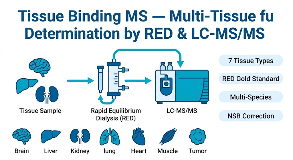
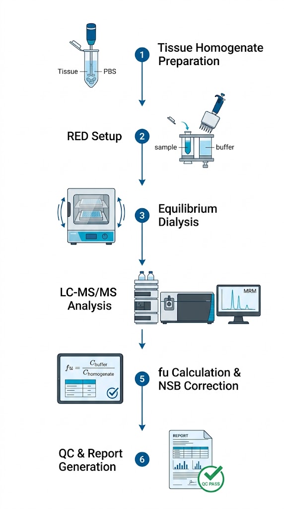
The team used equilibrium dialysis combined with LC-MS/MS to measure fu,brain,ROI for three CNS drugs — citalopram, donepezil, and risperidone — across five brain regions (frontal cortex, temporal cortex, hippocampus, thalamus, and striatum). Brain tissue homogenates were prepared at a 1:4 dilution in PBS, dialyzed against buffer for 5 hours at 37°C, and analyzed by LC-MS/MS.

Results

The answer was no — regional binding is far from uniform. Donepezil showed a 2.5-fold difference in fu between the hippocampus (fu = 0.038) and the thalamus (fu = 0.095) in human AD brain tissue. Regional binding patterns also differed between AD patients and controls, suggesting that disease state alters tissue composition and binding characteristics. In rats, the variability was less pronounced but still present, with the lowest fu values consistently in the hippocampus.

Conclusions

Single-region brain tissue binding measurements may not adequately represent the entire brain for CNS drug development. Multi-region assessment — an approach we routinely offer — provides more accurate Kp,uu,brain values and improves PBPK model predictions for CNS-active compounds.

Case study multi-region brain tissue binding in Alzheimer's disease research

Heterogeneous drug tissue binding across brain regions in rats, AD patients, and controls. Adapted from Gustafsson et al. (2019), Sci Rep.

FAQ

Frequently Asked Questions

Q: Which tissues can you measure for binding studies?

We cover seven tissue types as standard: brain, liver, kidney, lung, heart, muscle, and tumor. If you need something outside that list — spleen, pancreas, skin, bone marrow — just ask. We can usually accommodate custom tissues.

Q: What's the difference between RED, ultrafiltration, and ultracentrifugation?

RED relies on passive diffusion across a semipermeable membrane — it's the gold standard with the lowest NSB risk. Ultrafiltration uses pressure-driven filtration and offers higher throughput, but NSB risk is higher. Ultracentrifugation separates bound from free drug by sedimentation, avoiding membrane-related NSB entirely, but throughput is lower and equipment cost is higher. We'll help you pick the right method based on your compound's properties.

Q: How do you handle NSB in tissue homogenate assays?

We run recovery controls for every compound-tissue combination. Recovery equals total drug measured post-dialysis divided by the spiked concentration. When recovery drops below 70%, we apply a correction factor and flag it in the report. For compounds that consistently show low recovery, we recommend switching to ultracentrifugation.

Q: What species tissue do you offer?

Rat, mouse, dog, monkey, and human — for all seven tissue types. Additional species (minipig, rabbit, guinea pig) are available by special request.

Q: What's the typical turnaround time?

A standard 7-tissue × 1-species study takes 3–4 weeks from tissue receipt. Larger panels (multiple species × multiple tissues) typically run 4–6 weeks. Rush timelines can be arranged for critical milestones.

Q: Can I combine tissue binding with other ADME assays?

Yes — and we encourage it. Our integrated ADME platform lets you run tissue binding alongside plasma protein binding, metabolic stability, CYP inhibition, and permeability assays using the same compound batch. That means less compound consumption, fewer freeze-thaw cycles, and consistent data across assays. Contact us to design a panel that fits your program.

Plan Your Tissue Binding Study with the MassTarget™ Team

Tell us which tissues and species you need, and our scientists will design a customized tissue binding study for your drug discovery program.

For research use only. Not for use in diagnostic procedures.

Online Inquiry

Please submit a detailed description of your project. We will provide you with a customized project plan to meet your research requests. You can also send emails directly to for inquiries.

* Email
Phone
* Service & Products of Interest
Services Required and Project Description