HDAC Activity MS

Label-free mass spectrometry-based histone deacetylase activity assays for direct, quantitative measurement of HDAC inhibition and selectivity.

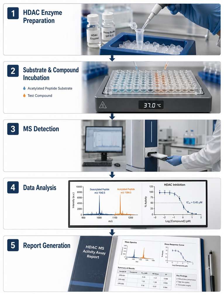
At Creative Proteomics, our HDAC MS activity assay service is designed to address the challenges researchers face with conventional HDAC screening: compound interference in optical readouts, limited substrate flexibility, and the need for detailed kinetic characterisation alongside routine IC₅₀ determination.

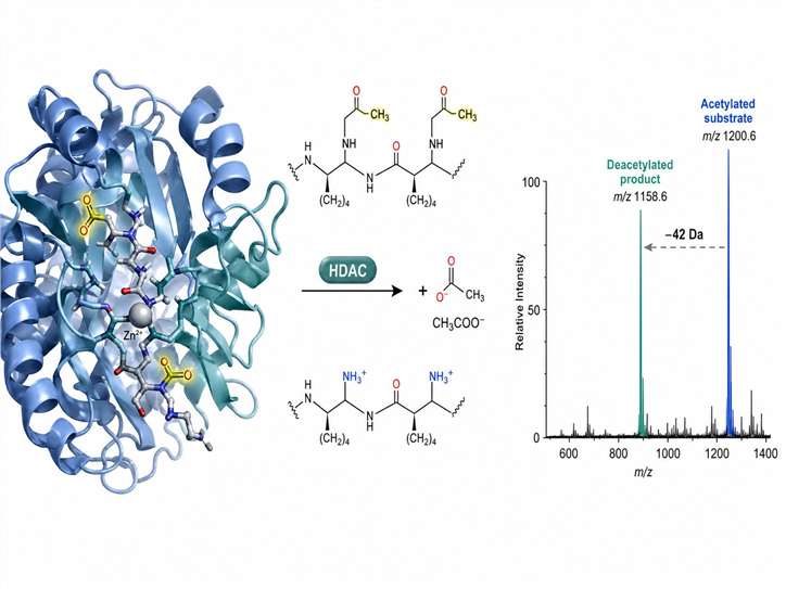
Our platform detects HDAC-catalysed deacetylation directly by MS, measuring the characteristic mass shift (−42 Da) as acetylated substrates convert to deacetylated products. No fluorescent labels, no colorimetric reagents, no coupled enzyme systems — just clean, interference-free data for your HDAC inhibitor screening programmes.

Key Advantages:

  • Label-free detection — no compound autofluorescence or quenching interference
  • Direct deacetylation readout — measure deacetylated product formation without coupled enzymes
  • IC₅₀, Kₘ, and mechanism-of-action from a single MS workflow
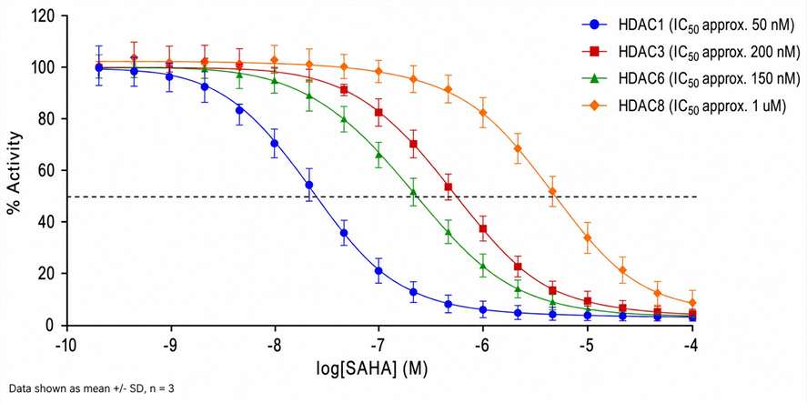
  • Class I/II HDAC isoforms (HDAC1–11) supported
  • Peptide and full-length protein substrates, including non-histone targets
  • DMSO-tolerant at standard screening concentrations
HDAC MS activity assay principle diagram with enzyme, substrate, deacetylation reaction, and mass spectrum
What Is HDAC MS Key Advantages Service Overview Workflow Tech Comparison Sample Case Study FAQ

What Is HDAC MS Activity Assay?

An HDAC MS activity assay uses mass spectrometry to measure histone deacetylase catalytic activity in vitro. Here is how it works: a purified HDAC enzyme is incubated with an acetylated peptide or protein substrate, and mass spectrometry directly detects and quantifies the deacetylated product alongside the remaining acetylated substrate. The result is a precise deacetylation ratio that reports on HDAC activity.

The key difference from conventional HDAC activity assays — whether fluorogenic (e.g., Boc-Lys(Ac)-AMC), colorimetric, or radiometric (³H-acetate release) — is that MS measures the actual mass difference between the acetylated and deacetylated forms of the substrate. When HDAC removes an acetyl group (42 Da) from the ε-amino group of a lysine residue, the mass spectrometer detects that shift directly. This eliminates several common sources of assay interference: compound autofluorescence in fluorogenic HDAC assays, signal quenching in colorimetric formats, and the hazards and disposal costs associated with radiometric assays.

The approach works with both MALDI-TOF and LC-MS/MS platforms. In MALDI-TOF mode, quenched reaction mixtures are spotted onto a target plate with matrix and acquired at rates of seconds per sample — throughput suitable for moderate-to-high screening campaigns. LC-MS/MS mode adds chromatographic separation as an extra dimension of specificity, particularly useful for complex substrate mixtures, full-length histone substrates with multiple acetylation sites, or reactions where isobaric interferences may be present.

This makes HDAC MS activity assays a strong fit for drug discovery programmes where compound interference with optical readouts is a known problem, where direct deacetylation measurement is preferred over surrogate signals, and where detailed kinetic characterisation (Kₘ, Vₘₐₓ, mechanism of inhibition) is required alongside routine IC₅₀ determination.

Key Advantages of MS-Based HDAC Activity Detection

Label-Free Detection Eliminates Compound Interference

Fluorescence-based HDAC assays using fluorogenic acetylated lysine substrates are vulnerable to compound autofluorescence, fluorescence quenching, and inner-filter effects — all well-documented sources of false positives and false negatives in high-throughput screening. Because our MS readout is based on mass, not light, these optical interferences simply do not apply. Compounds that would be flagged as "hits" or "non-hits" due to optical artifacts in fluorescence assays can be evaluated cleanly by MS.

Direct Deacetylation Readout — No Coupled Enzyme Systems

Many popular HDAC assay formats detect deacetylase activity indirectly. Fluorogenic HDAC substrates require enzymatic development with trypsin or similar proteases to release the fluorophore from the deacetylated lysine — an additional enzymatic step that introduces variability and potential interference. Our MS-based HDAC assays measure the deacetylated product directly, giving a more faithful representation of HDAC catalytic activity without surrogate signals or coupled enzyme steps.

Broad Substrate Flexibility — Including Non-Histone Targets

MS-based detection imposes no constraints on substrate length or sequence beyond the requirements of mass spectrometric detection. We work with both short synthetic peptides (8–20 amino acids) containing a single acetylated lysine and full-length histone proteins (H3, H4) with defined acetylation patterns. Importantly, we also support non-histone HDAC substrates — proteins such as p53, α-tubulin, HSP90, and others regulated by reversible acetylation.

Kinetic Characterisation from a Single Workflow

Because MS simultaneously detects both acetylated substrate and deacetylated product ions, a single reaction yields a direct deacetylation ratio. This makes the assay inherently ratiometric, reducing well-to-well variability from pipetting errors and enabling robust kinetic measurements. Kₘ determinations for acetylated peptide substrates, Vₘₐₓ measurements, and mechanism-of-inhibition studies can all be performed within the same analytical workflow.

Isoform-Specific Assay Conditions

Each HDAC isoform has distinct catalytic properties, substrate preferences, and inhibitor sensitivity profiles. We individually optimise assay conditions for each HDAC isoform — including buffer composition, metal cofactor requirements (Zn²⁺ for class I/II HDACs), substrate identity and concentration, and reaction time. This isoform-specific optimisation ensures the measured activity faithfully reflects the target enzyme's native catalytic behaviour.

Service Overview — HDAC MS Activity Assay Capabilities

We offer six service modes tailored to different stages of the HDAC drug discovery pipeline:

MODE 1

Single HDAC Isoform IC₅₀ Determination

For individual HDAC targets, we design and execute dose-response inhibition assays using your compounds of interest. Each assay is optimised for substrate identity and concentration (at Kₘ), reaction time (linear range), and enzyme concentration. IC₅₀ values are calculated from 10-point dose-response curves with technical replicates.

Typical output: IC₅₀ values with 95% confidence intervals, dose-response curves, raw MS spectra at each compound concentration.

MODE 2

HDAC Selectivity Panel Profiling

For compounds that need profiling across multiple HDAC isoforms, we offer panel screening using our MS-based platform. Selectivity profiling can be configured as full dose-response across the panel (for detailed selectivity ratios) or as single-concentration percentage inhibition screening (for rapid selectivity fingerprinting). Our panel covers class I (HDAC1, HDAC2, HDAC3, HDAC8) and class II (HDAC4, HDAC5, HDAC6, HDAC7, HDAC9, HDAC10) isoforms.

Typical output: HDAC selectivity heatmap, percentage inhibition at screening concentration, IC₅₀ values for confirmed hits, selectivity ratio table.

MODE 3

Mechanism-of-Action Studies

Understanding whether a compound is competitive, non-competitive, or mixed-type with respect to the acetylated substrate is critical for medicinal chemistry optimisation. Our MS platform supports detailed kinetic mechanism studies including substrate competition assays (IC₅₀ at multiple acetylated peptide concentrations), time-dependence assays, reversibility assessment via dilution-jump experiments, and residence time determination for slow-binding HDAC inhibitors.

Typical output: Mechanism classification, Kᵢ determination, double-reciprocal plots, residence time estimates.

MODE 4

Non-Histone Substrate HDAC Activity Measurement

Many HDACs deacetylate non-histone protein substrates with important biological functions — HDAC6 deacetylates α-tubulin, HDAC1 deacetylates p53, and HDACs regulate HSP90 acetylation status. We offer custom assay development using your non-histone substrate of interest, with MS-based detection providing direct readout of substrate-specific deacetylation.

Typical output: Substrate-specific deacetylation activity data, IC₅₀ values with non-histone substrate, comparison with histone substrate activity.

MODE 5

HDAC Inhibitor Kinetic Characterisation

For compounds that show time-dependent inhibition or slow-binding kinetics, we offer detailed kinetic characterisation including apparent rate constant (kobs) determination at multiple inhibitor concentrations, target residence time (τ = 1/koff) calculation, kinact/KI determination for covalent HDAC inactivators, and binding mechanism classification (one-step vs. two-step binding).

Typical output: Kinetic constants (kobs, koff, kinact/KI), residence time, binding mechanism classification.

MODE 6

Custom Assay Development

For novel HDAC targets, non-standard substrate requirements, or specialised assay conditions, our scientists work with your team to develop and validate a fit-for-purpose MS-based HDAC activity assay. This includes substrate identification and optimisation, assay buffer and condition screening, signal-to-noise optimisation, and full assay validation (Z′ factor, intra-assay and inter-assay precision, linearity, and stability).

Typical output: Validated assay protocol, optimisation data summary, validation report.

For related capabilities, see our high-throughput MS screening platform and transferase (HAT/HMT) MS activity assays services.

HDAC MS Activity Assay Workflow

The workflow consists of five essential stages:

1

HDAC Enzyme Preparation

Purified recombinant HDAC enzyme (class I or class II isoform) is prepared in assay buffer optimised for the specific isoform — typically 50 mM HEPES, pH 7.5, 150 mM NaCl, 0.01% Tween-20, with appropriate metal cofactors (Zn²⁺ for class I/II HDACs). Enzyme concentration is titrated to establish linear reaction conditions.

2

Substrate and Compound Incubation

Purified HDAC enzyme, acetylated peptide or protein substrate (at Kₘ concentration), and test compound (at desired concentration range) are combined in the reaction volume. Reactions are incubated at 37 °C for a predetermined linear-range time point, then quenched with acidified organic solvent or heat denaturation.

3

MS-Based Product Detection

Quenched reaction mixtures are analysed by MALDI-TOF MS (Bruker rapifleX or equivalent) or LC-MS/MS (Xevo G3 QTof or equivalent). For MALDI-TOF, samples are spotted onto a target plate with matrix and acquired at rates of seconds per sample. For LC-MS/MS, samples are injected onto a C18 column with a rapid gradient for separation and detection of acetylated and deacetylated peptide species.

4

Data Analysis

The deacetylation ratio is calculated from the peak intensities of the deacetylated product and the remaining acetylated substrate. IC₅₀ values are determined by fitting percentage inhibition data to a four-parameter logistic model. For kinetic studies, Kₘ and Vₘₐₓ are calculated from substrate-velocity curves, and mechanism-of-inhibition data are analysed using appropriate enzyme kinetic models.

5

Report Generation

A comprehensive report is compiled including raw MS spectra, deacetylation ratios at each compound concentration, dose-response curves with fitted IC₅₀ values, quality control metrics (Z′ factor, S/B ratio, coefficient of variation), and any additional kinetic parameters requested.

HDAC MS activity assay workflow diagram showing five steps from enzyme preparation to report generation

For more information about our MALDI-TOF high-throughput screening capabilities, please visit the dedicated page.

Platform Instrumentation

Our HDAC MS activity assay platform integrates advanced mass spectrometry systems to support sensitive and reproducible HDAC inhibitor screening.

InstrumentManufacturerApplicationKey Specifications
rapifleX MALDI-TOF/TOFBruker DaltonicsHigh-speed MALDI-TOF MS for HDAC activity screening10 kHz laser, 200–500 kDa mass range, TOF/TOF fragmentation
Xevo G3 QTofWaters CorporationLC-MS/MS for HDAC substrate and product quantificationQTof analyser, StepWave ion optics, 20,000 FWHM resolution
RapidFire 365Agilent TechnologiesHigh-throughput solid-phase extraction MS for HDAC assays<10 sec per sample cycle, online SPE-MS/MS
Q Exactive HFThermo Fisher ScientificHigh-resolution LC-MS/MS for complex HDAC substrate analysisOrbitrap analyser, 240,000 resolution at m/z 200
TripleTOF 6600+SciexHigh-resolution LC-MS/MS for HDAC kinetic studies30,000 resolution, 100–150 SWATH windows per cycle

Technology Comparison — MS vs. Conventional HDAC Assay Formats

DimensionFluorescence (Fluorogenic HDAC Substrate)Colorimetric (HDAC Activity Kit)Radiometric (³H-Acetate Release)FRET-Based (HDAC Assay)ADP-Glo (Indirect HDAC Readout)MS-Based (Label-Free HDAC Assay)
Detection PrincipleFluorophore release after tryptic cleavage of deacetylated substrateChromophore development upon deacetylationScintillation counting of released ³H-acetateFluorescence resonance energy transfer between donor/acceptorLuciferase-based ATP detection (coupled enzyme)Direct mass measurement of substrate and product
Label RequirementFluorogenic cap on lysine side chainChromogenic substrate modification³H-acetyl group on substrateFluorophore pair on substrateRequires ATP consumptionNone — label-free
Optical Interference SusceptibilityHigh — compound autofluorescence at common excitation/emission wavelengthsModerate — compound colour can interfere with absorbance readoutNone — radiometric detection is optically blindHigh — compound fluorescence can interfere with FRET signalLow — luminescence readout, but compound quenching possibleNone — mass-based readout is optically blind
Substrate FlexibilityLimited to short fluorogenic peptidesLimited to kit-specific substratesLimited to ³H-acetylated peptidesLimited to peptide substrates with fluorophore pairCompatible with any ATP-utilising enzymeFull flexibility — peptides, full-length proteins, non-histone substrates
ThroughputHigh (384/1536-well plates)Moderate (96/384-well plates)Low (scintillation counting)High (384/1536-well plates)High (384-well plates)Moderate-to-high (MALDI-TOF: seconds/sample; RapidFire: <10 sec/sample)
Kinetic Information DepthLimited (single end-point or few time points)Limited (end-point only)Low (end-point, single time course)Moderate (real-time possible)Limited (end-point ATP depletion)High (direct product/substrate ratio, multi-time point)
Z′ Factor Range0.5–0.8 (dependent on compound library optical properties)0.4–0.70.6–0.80.5–0.70.5–0.70.6–0.8 (consistent across compound types)

Selection Strategy: For HDAC inhibitor screening programmes where compound libraries contain fluorescent or coloured compounds — which applies to most modern drug discovery collections — MS-based detection offers a decisive advantage. The label-free, direct readout eliminates the primary source of false positives in fluorescence-based HDAC assays while providing richer kinetic information than any end-point format. For projects requiring non-histone substrate measurement or full-length protein substrates, MS is the only viable option among the formats compared.

For related enzyme assay services, see our Kinase MS Activity Assays and DNMT activity MS assays.

Sample Requirements

Sample TypeRecommended QuantityConcentrationPurityFormat
Test compound (small molecule)≥50 µL of 10 mM stock10 mM in DMSO≥95% purity96-well plate or microcentrifuge tube
HDAC enzyme (if client-provided)≥100 µL at 1 µM≥1 µM≥80% purity by SDS-PAGELow-bind tube on dry ice
Acetylated peptide substrate≥200 µL of 1 mM stock1 mM in H₂O or DMSO≥95% purity by HPLCMicrocentrifuge tube
Full-length histone/protein substrate≥50 µg≥0.5 mg/mL≥90% purityLow-bind tube on dry ice
Assay buffer (if special requirements)≥5 mL of 2× stock2× final concentrationN/ASterile tube at 4 °C

Notes: All samples should be shipped on dry ice unless otherwise specified. Compounds with known solubility issues should be accompanied by solubility data and recommended solvent. For full-length protein substrates, please provide acetylation status information if available.

Deliverables

  • Raw MS spectra for all assay conditions
  • Deacetylation ratios (product/substrate peak intensity ratios) for each reaction
  • Dose-response curves with fitted IC₅₀ values and 95% confidence intervals
  • HDAC selectivity heatmap (for panel profiling service)
  • Kinetic parameters (Kₘ, Vₘₐₓ, Kᵢ) where applicable
  • Mechanism-of-inhibition classification (competitive, non-competitive, mixed-type)
  • Quality control metrics (Z′ factor, S/B ratio, intra-assay CV, inter-assay CV)
  • Comprehensive final report with methods, results, and data interpretation

Representative HDAC MS Assay Data

Dose-response curves showing HDAC inhibition by a reference inhibitor across multiple isoforms.

HDAC inhibition dose-response curves for SAHA across HDAC1, HDAC3, HDAC6, and HDAC8 isoforms

Example HDAC inhibition dose-response curves — SAHA (vorinostat) across four HDAC isoforms

Case Study — SAMDI-MS for HDAC8 Activity and Substrate Specificity Profiling

Gurard-Levin Z.A., Mrksich M. "The Activity of HDAC8 Depends on Local and Distal Sequences of Its Peptide Substrates." Biochemistry 47(23):6242-6250 (2008). https://pmc.ncbi.nlm.nih.gov/articles/PMC2605276/

Background

Understanding how HDAC enzymes recognise and deacetylate specific lysine residues on protein substrates is fundamental to both HDAC biology and drug discovery. Traditional HDAC activity assays using fluorogenic substrates provide limited information about substrate sequence preferences because the fluorogenic cap dominates substrate recognition. Gurard-Levin and Mrksich (2008) addressed this limitation by applying SAMDI (Self-Assembled Monolayers for MALDI) mass spectrometry to profile HDAC8 activity across a library of peptide substrates.

Methods

The authors immobilised acetylated peptide substrates on self-assembled monolayers of alkanethiolates on gold. Each peptide contained a single acetylated lysine residue flanked by varying sequence contexts derived from the HDAC8 substrate histone H4 (residues 12–23). HDAC8 was incubated with the peptide arrays, and SAMDI-TOF MS was used to directly measure the mass shift (−42 Da) corresponding to lysine deacetylation on each peptide. The label-free SAMDI readout enabled simultaneous, quantitative measurement of deacetylation kinetics across multiple peptide substrates on a single array.

Results

The SAMDI-MS assay revealed that HDAC8 deacetylase activity is strongly modulated by both local residues immediately surrounding the acetylated lysine and by a distal basic sequence motif (KRHR, corresponding to H4 residues 17–20). Peptides containing the full H4(12–23) sequence showed approximately 3-fold higher deacetylation rates compared to peptides containing only the local residues around the acetylation site. Kinetic analysis demonstrated that the distal KRHR sequence acts through an exosite-binding mechanism — a secondary binding site on the HDAC8 surface distinct from the catalytic centre — to enhance catalytic efficiency.

Conclusion

This study demonstrates the power of MS-based HDAC activity measurement for uncovering substrate recognition mechanisms that are invisible to conventional fluorescence-based assays. The SAMDI-MS approach enabled label-free, multiplexed, quantitative measurement of HDAC8 deacetylation activity across multiple peptide substrates, revealing an exosite-dependent regulatory mechanism that provides new insights for HDAC8 inhibitor design.

SAMDI-MS workflow for HDAC8 activity profiling on peptide arrays

SAMDI-MS workflow for HDAC8 deacetylation activity measurement on self-assembled monolayer peptide arrays.

FAQ

Frequently Asked Questions

Q: What HDAC isoforms can you assay using MS?

We offer MS-based activity assays for class I (HDAC1, HDAC2, HDAC3, HDAC8) and class II (HDAC4, HDAC5, HDAC6, HDAC7, HDAC9, HDAC10) histone deacetylases. Each isoform is assayed under optimised buffer conditions with isoform-specific acetylated peptide substrates.

Q: Can you measure HDAC activity on full-length histone proteins?

Yes. In addition to synthetic acetylated peptides, our MS platform supports full-length histone H3 and H4 substrates with defined acetylation patterns. This is particularly valuable when studying context-dependent deacetylation or non-histone substrate recognition.

Q: What is the throughput of MS-based HDAC screening?

Using MALDI-TOF MS in automated mode, our platform can process up to several hundred samples per hour — throughput comparable to plate-reader-based fluorescence assays while providing the direct mass readout that eliminates optical interference concerns.

Q: How do you ensure data quality and reproducibility?

Every HDAC assay includes positive and negative controls, Z′ factor determination, intra-assay and inter-assay precision assessment, and signal-to-background ratio optimisation. We routinely achieve Z′ factors >0.5 for HDAC MS activity assays.

Q: Can you measure HDAC activity in cell lysates or nuclear extracts?

Our standard service uses purified recombinant HDAC enzymes for biochemical activity measurement. For cellular context, we offer an optional cell-based HDAC activity profiling service using enriched nuclear extracts — please inquire for details.

Q: What is the typical turnaround time for HDAC IC₅₀ determination?

Single-target HDAC IC₅₀ determination is typically completed within 2-3 weeks from sample receipt, including assay optimisation, dose-response screening, data analysis, and final reporting. Multi-isoform selectivity panels require additional time depending on the number of isoforms.

References

  1. Gurard-Levin Z.A., Mrksich M. The Activity of HDAC8 Depends on Local and Distal Sequences of Its Peptide Substrates. Biochemistry. 2008;47(23):6242-6250.
  2. Kuo H.Y., DeLuca T.A., Miller W.M., Mrksich M. Profiling Deacetylase Activities in Cell Lysates with Peptide Arrays and SAMDI Mass Spectrometry. Analytical Chemistry. 2014;86(1):196-203.
  3. Peng L., Yuan Z., Seto E. Histone Deacetylase Activity Assay. Methods in Molecular Biology. 2015;1288:335-349.

Plan an HDAC MS screening campaign with the MassTarget™ team

Share your HDAC target and compound details and our scientists will design a tailored MS-based screening strategy for your drug discovery programme.

For research use only. Not for use in diagnostic procedures.

Online Inquiry

Please submit a detailed description of your project. We will provide you with a customized project plan to meet your research requests. You can also send emails directly to for inquiries.

* Email
Phone
* Service & Products of Interest
Services Required and Project Description