Tissue Slice (PCLS) MS Drug Response

Comprehensive LC-MS drug response profiling in precision-cut tissue slices for translational ADME and toxicity research.

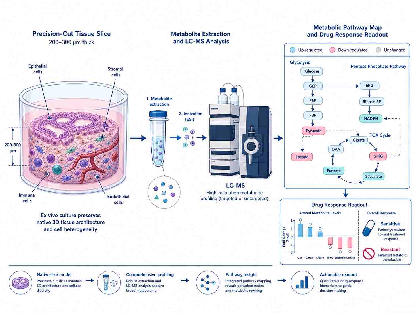
Precision-cut tissue slices (PCTS) — including precision-cut lung slices (PCLS), precision-cut liver slices, and precision-cut intestinal slices (PCIS) — represent the gold-standard ex vivo model for drug metabolism and toxicity research. By combining precision-cut tissue slice technology with LC-MS metabolomics, lipidomics, and targeted drug quantification, our Tissue Slice (PCLS) MS Drug Response service delivers a comprehensive readout of drug-tissue interactions in a physiologically relevant, human-relevant platform.

Unlike 2D cell cultures that rapidly lose CYP enzyme activity or animal models that suffer from species-specific metabolic differences, precision-cut tissue slices retain the native tissue architecture — including all resident cell types (epithelial, stromal, immune, endothelial), intact extracellular matrix, and metabolic zonation — making them the most translationally predictive ex vivo model for ADME, drug-induced toxicity, and pharmacometabolomics studies.

Key Capabilities:

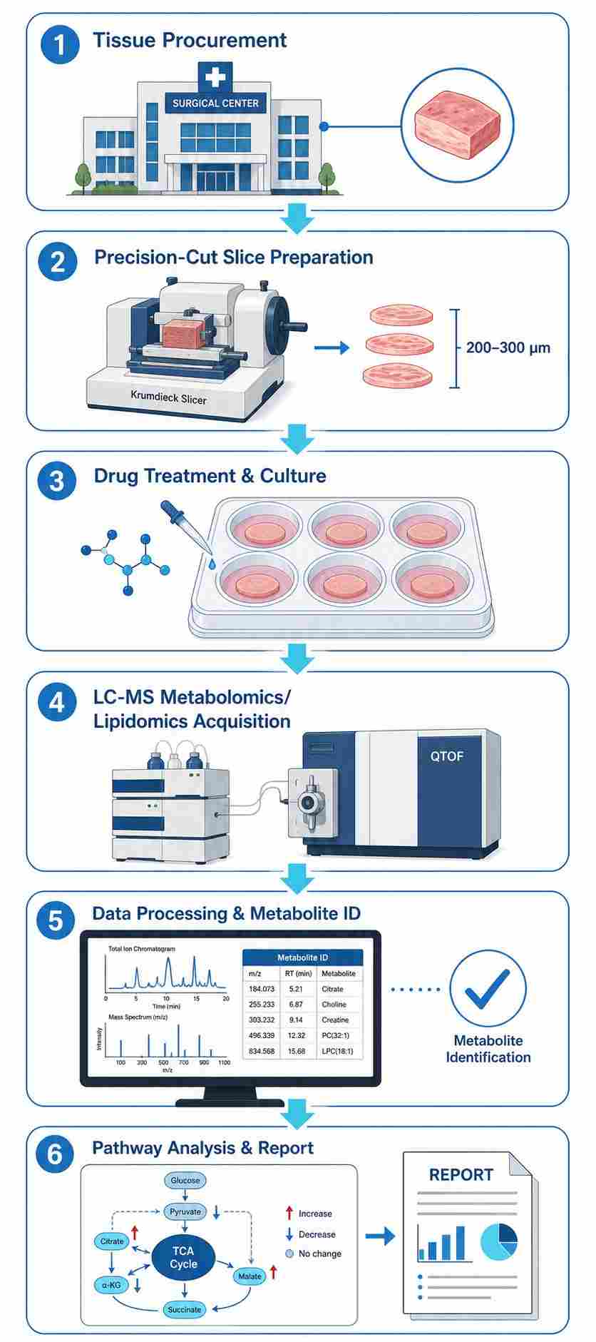
  • Multi-organ PCLS capability: lung (healthy and diseased), liver (healthy, fibrotic, steatotic), intestine (small and colon), and tumor tissue.
  • Integrated LC-QTOF-MS untargeted metabolomics/lipidomics and LC-TQ-MS targeted drug quantification from the same tissue slices.
  • Validated slice preparation protocol (Krumdieck slicer, 200-300 µm) with >80% viability post-cutting.
  • Flexible service model: full-service (tissue-to-report), slice-only, or analysis-only options.
  • Stable isotope tracing (¹³C) for metabolic flux analysis in drug-treated tissue slices.
  • Comprehensive pathway enrichment analysis with KEGG, Reactome, and HMDB mapping.
Precision-cut tissue slice (lung/liver) in culture well with LC-MS analysis and metabolic pathway map showing drug-response readout.
Overview Why PCLS Workflow Capabilities Sample Data Outputs Applications Case Study FAQ

What Is Tissue Slice (PCLS) MS Drug Response?

Precision-cut tissue slices (PCTS) are thin, uniform sections of solid tissue (typically 200-300 µm thick) prepared using a Krumdieck tissue slicer under oxygenated buffer conditions. These slices retain the native three-dimensional tissue architecture — including all resident cell types, intact extracellular matrix, cell-cell interactions, and metabolic zonation — making them the most physiologically relevant ex vivo model for studying drug metabolism, drug-induced toxicity, and pharmacometabolomics.

Our Tissue Slice (PCLS) MS Drug Response service integrates this ex vivo tissue slice platform with high-resolution LC-MS metabolomics, lipidomics, and targeted drug quantification to provide a comprehensive, multi-parameter readout of drug-tissue interactions. Whether you need human-specific metabolite profiles for IND filing, multi-organ ADME data across lung, liver, and intestine, or mechanistic insight into drug-induced toxicity pathways, our service delivers publication-ready data from as few as one tissue slice per condition.

This service is part of our broader cell-based MS drug screening portfolio, which also includes organoid metabolomics by LC-MS for 3D culture models and cellular metabolomics screening for 2D cell systems.

Why PCLS for Drug Metabolism & Toxicity Studies

Selecting the right model system is critical for generating meaningful drug metabolism and toxicity data. Each model offers distinct trade-offs between physiological relevance, throughput, and cost.

Feature2D Cell CultureOrganoidsPrecision-Cut Tissue Slices (PCLS)In Vivo Animal Models
Tissue architectureMonolayer, no 3D structure3D but limited stromal/immune cellsFull native 3D architecture preservedComplete tissue context
Cell type diversitySingle or co-culture (2-3 types)Epithelial-focusedAll resident cell types (epithelial, stromal, immune, endothelial)Complete organism
CYP enzyme activityLost within 4-24 hVariable, often reducedPreserved for ≥72 hNative activity
Metabolic zonationAbsentAbsentPreserved (periportal to pericentral gradients)Native
ECM & cell-matrix interactionsAbsentMatrigel-dependentNative ECM preservedNative
Human relevanceHuman cells possibleHuman tissue-derivedHuman tissue-derivedSpecies differences
ThroughputHighMediumMediumLow
Ethical concernsMinimalMinimalMinimal (3R-compliant)High
Regulatory acceptanceLimitedEmergingGrowing (NAMs initiatives)Gold standard
Cost per data pointLowMediumMediumHigh

When to Choose PCLS

Precision-cut tissue slices are the optimal choice when:

  • You need human-specific metabolite profiles for IND filing or clinical translation — PCLS from human donors captures the full repertoire of human phase I and phase II drug-metabolizing enzymes, transporters, and tissue-specific metabolic pathways that rodent models may miss.
  • Your drug candidate targets the lung, liver, or intestine — PCLS can be prepared from any solid organ, enabling tissue-specific ADME profiling that is not possible with hepatocyte suspensions or organoids.
  • You want to study drug-induced toxicity in a multicellular context — PCLS preserves the immune-stromal-epithelial interactions that drive DILI, pulmonary toxicity, and intestinal toxicity.
  • You need to reduce animal use (3Rs) — PCLS is recognized as a New Approach Methodology (NAM) by regulatory agencies, providing human-relevant data while reducing and replacing animal studies.

For studies focused on 2D cell monolayers, our cellular metabolomics screening service may be more appropriate. For 3D organoid models, we offer dedicated organoid metabolomics by LC-MS.

Service Workflow

Our Tissue Slice (PCLS) MS Drug Response service follows a streamlined six-step workflow designed to maximize tissue viability, data quality, and turnaround efficiency.

1

Tissue Procurement & Ethical Sourcing

We source fresh human and preclinical tissue from accredited tissue banks and surgical centers with full ethical approval and donor consent. Tissue types include lung (healthy and diseased), liver (healthy, fibrotic, steatotic), intestine (small and large), and tumor tissue. Tissue is transported in ice-cold UW solution or HBSS and processed within 2-4 hours of resection to ensure maximal viability.

2

Precision-Cut Slice Preparation

Tissue cores (8-10 mm diameter) are prepared using a Krumdieck tissue slicer under ice-cold, oxygenated buffer conditions. Slices are cut at 200-300 µm thickness — optimal for nutrient and oxygen diffusion while preserving tissue architecture. Viability is assessed by ATP content and LDH release (target: >80% viability post-cutting). Slices are cultured in serum-free William's E medium with insulin, dexamethasone, and antibiotic supplementation at 37 °C in 5% CO₂.

3

Drug Treatment & Culture

Drug candidates are added to the culture medium at specified concentrations and time points. We support dose-response studies (typically 3-6 concentrations), time-course experiments (0-72 h), and multi-compound panels. Vehicle controls (DMSO ≤0.1%), time-zero controls, and blank controls are included in every experiment. For stable isotope tracing studies, we can substitute ¹³C-labeled nutrients in the culture medium.

4

LC-MS Metabolomics & Lipidomics Acquisition

Metabolites and lipids are extracted from individual tissue slices using a biphasic solvent system (acetonitrile/methanol/water or MTBE/methanol/water for lipidomics). Extracts are analyzed on our LC-QTOF-MS platform (Agilent 6546 Q-TOF) for untargeted metabolomics and lipidomics, and LC-TQ-MS (Agilent 6470 Triple Quad) for targeted drug and metabolite quantification. We also offer high-throughput metabolite profiling for larger screening panels.

5

Data Processing & Metabolite Identification

Raw LC-MS data are processed using our MassTarget™ bioinformatics pipeline. Features are detected, aligned, and filtered using blank subtraction and QC-based correction. Metabolite identification follows Metabolomics Standards Initiative (MSI) guidelines: Level 1 (retention time + MS/MS matching against authentic standards), Level 2 (MS/MS library matching), and Level 3 (putative annotation). For targeted drug quantification, we use stable isotope-labeled internal standards where available.

6

Pathway Analysis & Report

Metabolomics data are interpreted through pathway enrichment analysis (KEGG, Reactome, HMDB), metabolic flux analysis (for ¹³C tracing studies), and integration with proteomics or transcriptomics data when available. The final report includes PCA/PLS-DA plots, volcano plots, heatmaps, pathway enrichment maps, and a complete metabolite list with identification confidence levels. For pathway-level interpretation, we also offer metabolic pathway drug-response mapping.

Six-step PCLS MS drug response workflow: Tissue Procurement → Precision-Cut Slice Preparation → Drug Treatment & Culture → LC-MS Acquisition → Data Processing → Pathway Analysis & Report.

Key Capabilities & Platform

LC-MS Platforms

PlatformApplicationMass AccuracyResolutionKey Features
Agilent 6546 LC-QTOF-MSUntargeted metabolomics & lipidomics<1 ppm40,000 (FWHM)All-ion MS/MS, isotope profiling, wide dynamic range
Agilent 6470 LC-TQ-MSTargeted drug & metabolite quantificationUnit resolutionUnit resolutionMRM acquisition, 500 MRM transitions/s, 0.1 pg/mL LLOQ
Agilent 1290 Infinity II UHPLCChromatographic separationN/AN/ABinary pump, 1300 bar, 0.001-5 mL/min flow range

Tissue Type Capabilities

Tissue TypeSpeciesApplicationsTypical Yield (slices per donor)Culture Duration
Lung (healthy)Human, rat, mousePulmonary drug metabolism, inhalation toxicology, fibrosis50-100≥72 h
Lung (COPD/IPF/diseased)HumanDisease-specific drug response, pharmacometabolomics30-60≥48 h
Liver (healthy)Human, rat, mouse, dog, monkeyHepatic clearance, metabolite ID, DILI, CYP induction80-150≥96 h
Liver (fibrotic/steatotic)HumanDILI in diseased liver, NASH drug testing40-80≥72 h
Intestine (small)Human, ratFirst-pass metabolism, oral drug ADME60-120≥48 h
Intestine (colon)Human, ratColonic drug metabolism, microbiome interactions40-80≥48 h
Tumor (various)HumanTumor drug response, resistance mechanisms20-60≥48 h

For complementary enzyme-level analysis, we offer CYP450 inhibition panel MS and continuous-flow MS kinetics services.

Study Design & Sample Requirements

Proper study design is essential for generating robust, publication-quality PCLS drug response data. Below are our recommended sample requirements and study design parameters.

Sample TypeRecommended AmountPreparationStorage & Shipping
Fresh tissue (lung, liver, intestine, tumor)≥1 cm³ per condition; ≥3 biological replicatesTransport in ice-cold UW/ViaSpan or HBSS; process within 2-4 h of resection4 °C transport; overnight courier
Pre-cut tissue slices≥3 slices per condition (200-300 µm thickness)Cut using Krumdieck tissue slicer; culture in William's E medium with supplementsCulture medium transport at 4 °C; overnight courier
Snap-frozen tissue≥20 mg per conditionSnap-freeze in liquid N₂ immediately after collection; store at -80 °C-80 °C storage; dry ice shipping
Conditioned media from slice culture≥200 µL per replicateCentrifuge at 13,000 g, 4 °C, 10 min; collect supernatant-80 °C storage; dry ice shipping
Replicates≥3 biological replicates per group (recommended: 5-6)Randomized block design with vehicle controlsN/A
Turnaround4-8 weeks (standard project)Depending on study size, tissue type, and analytical scopeN/A

Study Design Recommendations:

  • Dose-response studies: Include at least 3 concentrations spanning the predicted therapeutic range plus a toxic concentration.
  • Time-course studies: Include 0 h (baseline), 6 h, 24 h, 48 h, and 72 h time points to capture both early metabolic shifts and late toxicity.
  • Species comparison: Include human and at least one preclinical species (rat or mouse) for translational ADME studies.
  • Vehicle controls: DMSO concentration should not exceed 0.1% and must be matched across all treatment groups.
  • Blank controls: Include tissue-free culture medium controls to distinguish drug-derived signals from endogenous metabolites.

Data Outputs & Interpretation

Our standard deliverables package includes:

DeliverableFormatDescription
Raw LC-MS data.d (Agilent) or .mzMLFull MS1 and MS/MS acquisition files
Processed feature table.xlsxDetected features with m/z, RT, intensity, and QC metrics
Metabolite identification list.xlsxAnnotated metabolites with MSI confidence level, MS/MS match score, and pathway assignment
Targeted quantification report.xlsxAbsolute concentrations (or relative fold changes) for target analytes with calibration curves and QC data
PCA/PLS-DA score plot.pdf/.pngMultivariate analysis showing group separation and outliers
Volcano plot.pdf/.pngFold change vs p-value visualization with significance thresholds
Heatmap.pdf/.pngHierarchical clustering of significantly changed metabolites
Pathway enrichment map.pdf/.pngKEGG/Reactome pathway analysis with enrichment scores
Metabolic flux analysis (if applicable).pdf/.xlsx¹³C labeling patterns, isotopologue distributions, and calculated flux values
Final report.pdfComplete study summary with methods, results, and interpretation

Applications

Our Tissue Slice (PCLS) MS Drug Response service supports a wide range of drug discovery and development applications:

Drug Metabolism & Metabolite Profiling (ADME)

Identify and quantify phase I and phase II metabolites of drug candidates in human-relevant tissue. Determine metabolic clearance pathways, species differences, and potential for drug-drug interactions. PCLS captures both hepatic and extrahepatic metabolism (lung, intestine) that standard hepatocyte assays may miss.

Drug-Induced Toxicity (DIT) Metabolomics

Detect early metabolic signatures of drug-induced liver injury (DILI), pulmonary toxicity, and intestinal toxicity before histopathological changes appear. Untargeted metabolomics can identify novel toxicity biomarkers and mechanism-based safety signals.

Pharmacometabolomics in Human Tissue

Characterize the metabolic response to drug treatment in human tissue, revealing pathway-level effects that complement traditional PK/PD endpoints. Particularly valuable for drugs targeting metabolic pathways (e.g., metabolic disease, oncology).

Species Comparison of Drug Metabolism

Compare metabolite profiles across human, rat, mouse, dog, and monkey tissue slices to select the most translationally relevant preclinical species for toxicology studies. Critical for IND-enabling studies where human-specific metabolites must be identified early.

Drug Efficacy in Diseased Tissue

Evaluate drug response in diseased human tissue (fibrotic lung, steatotic liver, tumor) to assess efficacy in a disease-relevant metabolic context. Provides more translatable efficacy data than healthy tissue or cell line models.

Multi-Organ ADME Profiling

Profile drug metabolism across lung, liver, and intestine from the same donor to build a complete picture of first-pass metabolism, hepatic clearance, and pulmonary metabolism in a single integrated study.

Metabolic Flux Analysis with Stable Isotope Tracing

Use ¹³C-labeled nutrients to trace metabolic pathway activity in drug-treated tissue slices. Reveals the metabolic mechanism of action, identifies pathway bottlenecks, and quantifies flux changes invisible to steady-state metabolomics alone. For pathway-level analysis, see our metabolic pathway drug-response mapping service.

Case Study

Global ¹³C Tracing and Metabolic Flux Analysis of Intact Human Liver Tissue Ex Vivo

Source: Grankvist N, Jönsson C, et al. Nature Metabolism. 2024;6:1963-1975. DOI: 10.1038/s42255-024-01119-3 (CC BY 4.0)

Background

Understanding human liver metabolism is fundamental to drug development, yet most knowledge comes from animal models or cell cultures that do not fully recapitulate human physiology. The authors aimed to establish a method for measuring metabolism in intact human liver tissue with sufficient depth and resolution to detect unexpected metabolic activities and pharmacologic responses.

Methods

Human precision-cut liver slices (200-250 µm thick) were prepared from surgical resections of 10 donors using a Krumdieck tissue slicer. Slices were cultured in medium where all 20 amino acids and glucose were uniformly labeled with ¹³C (U-¹³C). After 24 h of culture, metabolites were extracted and analyzed by non-targeted LC-MS. Metabolic flux analysis (MFA) was performed using a custom metabolic network model encompassing 144 intracellular fluxes.

Results

The study produced several key findings:

  • Preserved metabolic function: Cultured liver slices maintained ATP levels, albumin secretion, VLDL-triglyceride secretion, and urea synthesis at near-physiological levels for at least 24 h.
  • Unexpected metabolic activities: The authors discovered that human liver tissue performs de novo creatine synthesis and direct BCAA transamination — activities previously thought to be absent in human liver based on rodent studies.
  • Individual metabolic phenotypes preserved: The rate of glucose release from liver slices correlated with donor fasting plasma glucose levels (R² = 0.86), demonstrating that donor-specific metabolic traits are retained ex vivo.
  • Pharmacologic inhibition: Treatment with a glycogen phosphorylase inhibitor (CP-91149, 10 µM) significantly reduced glucose release from liver slices, confirming that the system can detect drug-induced metabolic effects (Figure 4d).
  • Metabolic flux quantification: MFA revealed that under fasted-like conditions, 89% of released glucose originated from glycogenolysis rather than gluconeogenesis, providing quantitative pathway-level insight.

Conclusions

This study demonstrates that precision-cut human liver slices combined with global ¹³C tracing and non-targeted LC-MS provide an unprecedented window into human liver metabolism and drug response. The platform is suitable for drug metabolism studies, pharmacometabolomics, and metabolic flux analysis in human-relevant tissue.

Figure 4 from Grankvist et al. 2024 — metabolic flux analysis showing substrate uptake/release rates, glucose production correlation with donor blood glucose, and pharmacologic inhibition of glycogen phosphorylase in human liver tissue slices.

Figure 4: Metabolic flux analysis of human precision-cut liver slices. Adapted from Grankvist et al. (2024), Nature Metabolism, CC BY 4.0.

FAQ

Frequently Asked Questions

Q: What is PCLS MS drug response and how is it different from standard cell-based metabolomics?

PCLS (precision-cut tissue slices) MS drug response combines ex vivo tissue slice technology with LC-MS metabolomics to study drug effects in intact human or animal tissue. Unlike cell-based metabolomics, PCLS preserves the native 3D tissue architecture, all resident cell types, and metabolic zonation, providing more physiologically relevant data for ADME and toxicity studies.

Q: What tissue types can be used?

We work with lung (healthy and diseased), liver (healthy, fibrotic, steatotic), intestine (small and colon), and tumor tissue from human and preclinical species (rat, mouse, dog, monkey). Each tissue type requires optimized slice preparation and culture protocols.

Q: How do you maintain tissue slice viability during culture?

Slices are cultured in serum-free William's E medium supplemented with insulin, dexamethasone, and antibiotics at 37 °C in 5% CO₂. Viability is monitored by ATP content, LDH release, and histological assessment at each time point. Under optimized conditions, slices maintain >80% viability for 72-96 h depending on tissue type.

Q: What is the minimum tissue amount needed for LC-MS analysis?

For untargeted metabolomics, a single tissue slice (5-15 mg wet weight, 200-300 µm thickness) is sufficient per condition. For targeted drug quantification, we recommend pooling 2-3 slices per condition. For ¹³C tracing studies, 3-5 slices per condition are recommended to ensure sufficient material for isotopologue analysis.

Q: Can you analyze tissue slices that we prepare ourselves?

Yes. We offer a flexible service model: (1) full-service — we procure tissue, prepare slices, treat with drugs, and perform LC-MS analysis; (2) slice-only — you prepare slices and send them to us for drug treatment and LC-MS analysis; (3) analysis-only — you perform drug treatment and send us conditioned slices or extracts for LC-MS analysis.

Q: What metabolites and lipids can you detect from tissue slice samples?

Our untargeted LC-QTOF-MS platform detects 500-2,000 metabolite features and 300-1,000 lipid species per tissue slice, covering amino acids, organic acids, nucleotides, carbohydrates, bile acids, eicosanoids, phospholipids, glycerolipids, sphingolipids, and sterols. Targeted LC-TQ-MS can quantify specific drugs and metabolites with LLOQ as low as 0.1 pg/mL.

Q: How many biological replicates do you recommend for tissue slice studies?

We recommend a minimum of 3 biological replicates (slices from different donors or different tissue blocks) per condition, with 5-6 replicates for studies requiring high statistical power (e.g., biomarker discovery, regulatory submission). Technical replicates (duplicate injections) are included for each sample.

Q: How do you ensure data quality and slice-to-slice reproducibility?

Our QC protocol includes: (1) pre-cut viability assessment (ATP >80%), (2) pooled QC injections every 10 samples, (3) blank subtraction and batch correction, (4) internal standards (isotope-labeled metabolites), (5) slice-to-slice CV monitoring (target<25%), and (6) randomized block design to minimize batch effects.

Q: Can metabolomics and targeted drug quantification be performed from the same slices?

Yes. We use a split-extract approach: a portion of the tissue extract is used for untargeted metabolomics and the remaining portion is used for targeted drug quantification. This enables direct correlation between drug exposure and metabolic response within the same biological sample.

Q: How long does a typical PCLS drug response project take?

Standard projects (1-5 compounds, 3 doses, 3 time points, single tissue type) are completed within 4-6 weeks. Larger studies (multi-compound panels, multi-organ, ¹³C tracing) typically require 6-8 weeks. Expedited timelines are available for urgent projects.

References

  1. Grankvist N, Jönsson C, et al.Global ¹³C tracing and metabolic flux analysis of intact human liver tissue ex vivo. Nature Metabolism. 2024;6:1963-1975.
  2. Dewyse L, De Smet V, et al.Improved Precision-Cut Liver Slice Cultures for Testing Drug-Induced Liver Fibrosis. Frontiers in Medicine. 2022;9:862185.
  3. Kollareth DJM, Sharma AK.Precision cut lung slices: an innovative tool for lung transplant research. Frontiers in Immunology. 2024;15:1504421.
  4. de Graaf IAM, Olinga P, et al.Preparation and incubation of precision-cut liver and intestinal slices for application in pharmaceutical and toxicological research. Nature Protocols. 2010;5:1540-1551.

Plan a PCLS Drug Response Study with the MassTarget™ Team

Share your tissue type, drug candidates, and study goals — our scientists will design a tailored PCLS drug response strategy for your drug discovery program.

Online Inquiry

Please submit a detailed description of your project. We will provide you with a customized project plan to meet your research requests. You can also send emails directly to for inquiries.

* Email
Phone
* Service & Products of Interest
Services Required and Project Description