Target-Responsive Accessibility Profiling (TRAP) Services

Bridge the gap between target identification and structural mapping with our Target-Responsive Accessibility Profiling (TRAP) services. TRAP-MS provides residue-level resolution directly in complex cell lysates, identifying precise binding pockets and hidden allosteric sites without the need for protein purification or ligand tagging.

  • Residue-level resolution (mapping reactive lysines)
  • 100% Label-free and Lysate-based structural probing
  • Direct 3D structural mapping of binding interfaces
Target-Responsive Accessibility Profiling (TRAP) Services
What is TRAP-MS?Why TRAP?Service CapabilitiesTechnology ComparisonWorkflowDemo ResultsSample RequirementsBioinformaticsCase StudyFAQ

What is TRAP-MS? (Defining High-Resolution Accessibility Mapping)

Target-Responsive Accessibility Profiling (TRAP) coupled with high-resolution mass spectrometry is an advanced structural proteomics technique. It is designed to map the exact locations where small molecules, peptides, or proteins interact with their targets. Instead of simply confirming that a drug binds to a protein, TRAP-MS reveals the specific amino acid residues that make up the binding pocket.

The technology relies on covalent labelling mass spectrometry. Proteins naturally possess reactive amino acids on their surfaces, most notably lysines. In a TRAP assay, we introduce a gentle, pan-specific chemical probe that covalently attaches to these solvent-accessible lysines.

When a drug binds to its target protein, it physically occupies a specific pocket on the protein's surface. This binding event physically shields the underlying lysines, preventing the chemical probe from attaching to them. We use high-resolution mass spectrometry to measure the labelling efficiency across the entire proteome. By comparing the drug-free sample to the drug-bound sample, we can pinpoint the exact lysine residues that exhibit a significant decrease in labelling. This "ligand-induced protection" acts as a highly precise footprint, revealing the exact location of the binding pocket directly on the protein's surface.

Why TRAP? The Bridge Between Target ID and Structural Biology

For decades, drug discovery has faced a frustrating gap. Traditional target deconvolution methods can tell you which protein your drug binds to, but they leave medicinal chemists blind as to where it binds. Without knowing the 3D structure of the binding pocket, optimizing the drug's affinity and selectivity is incredibly difficult.

Historically, researchers turned to X-ray crystallography or Cryo-EM to get this structural data, but these methods require heavily purified proteins and fail frequently. Another alternative is HDX-MS / HDX-driven Epitope Mapping, which offers fantastic peptide-level resolution. However, HDX-MS also strictly requires highly purified, recombinant protein. If your target is a complex membrane protein, an intrinsically disordered protein (IDP), or a target that loses its natural conformation outside of the cell, HDX-MS simply cannot be performed.

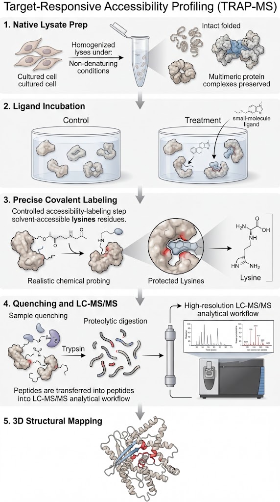
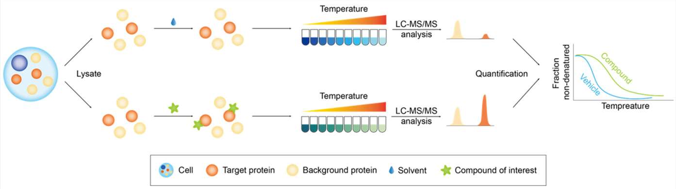
This is exactly why we utilize TRAP-MS. TRAP operates perfectly in native cell lysates and tissue homogenates. It allows us to probe the structural accessibility of proteins while they remain in their natural, physiological environment, surrounded by their native interacting partners. It bridges the gap by providing high-resolution structural mapping without the impossible prerequisite of protein purification.

Service Capabilities: From Phenotypic Hits to Allosteric Sites

Our TRAP-MS platform is specifically designed to support medicinal chemistry and structural biology teams who need actionable, spatial data to drive their drug development pipelines.

Phenotypic Hit Deconvolution with 3D Pockets

When you discover a promising compound through a phenotypic screen, identifying the target is only the first step. We take your unmodified hit compound, incubate it in a disease-relevant lysate, and use TRAP-MS to not only identify the target protein but to immediately map the exact 3D pocket the drug occupies. This transitions your project from a biological observation directly into structure-based drug design.

Allosteric Site Discovery

Standard active-site assays and competitive probes are entirely blind to allosteric modulators. Because TRAP-MS measures solvent accessibility across the entire surface of the protein simultaneously, it is exquisitely sensitive to allosteric binding. We can detect ligands that bind to cryptic pockets and map the resulting global conformational shifts that occur across the protein structure.

Protein-Protein Interaction (PPI) Mapping

TRAP is not limited to small molecules. We routinely use this platform to map the interaction interfaces between large protein complexes. By monitoring which lysines are shielded when two proteins assemble, we provide the high-resolution interface data required to design targeted PPI inhibitors or molecular glues.

Technology Comparison: TRAP vs. TPP vs. HDX-MS

Choosing the right biophysical tool is critical. Here is how TRAP-MS compares to other established label-free technologies in our portfolio.

FeatureTRAP-MSThermal Proteome Profiling (TPP)HDX-MSLimited Proteolysis–MS (LiP-MS)
Spatial ResolutionHigh (Residue-Level, specific to Lysines)Low (Global Protein-Level)Very High (Peptide-Level)High (Peptide-Level)
Sample EnvironmentNative Cell Lysates & TissuesNative Cell Lysates & TissuesPurified Proteins OnlyNative Cell Lysates & Tissues
Ligand Tag Required?NoNoNoNo
Primary Output3D Binding Pocket MapsTarget Identification (Tm shift)High-Res Conformational DynamicsStructural Footprinting

Our Solution Selection Strategy:

  • Choose TPP when you need a broad, unbiased first-pass screen to identify the primary target of a drug, and you do not immediately need to know the exact binding pocket.
  • Choose HDX-MS when you already possess highly purified recombinant protein and need the highest possible resolution to track real-time conformational dynamics.
  • Choose TRAP-MS when your target cannot be purified, or when you need to transition a phenotypic hit directly into a 3D binding model by mapping the exact binding pocket directly within a complex cell lysate.

End-to-End TRAP Workflow: Lysate to 3D Insights

Executing a TRAP assay requires meticulous control over chemical kinetics to ensure we capture native structures without causing artificial denaturation. Our workflow is rigorously optimized at every step.

1

Native Lysate Preparation

We gently extract proteins from your requested cell line or tissue. We use highly optimized, non-denaturing buffers to ensure that all proteins and endogenous complexes remain in their native folded states.

2

Ligand Incubation

The native lysate is divided into control and treatment aliquots. We incubate the sample with your unmodified drug candidate across a concentration gradient to establish binding equilibrium.

3

Precise Covalent Labeling

This is our most critical QC checkpoint. We introduce a specifically titrated concentration of our covalent probe (e.g., formaldehyde or DEPC). The concentration and reaction time are strictly controlled to ensure only highly accessible lysines are labeled, preventing over-labeling which could cause the protein to artificially unfold.

4

Quenching & LC-MS/MS

The labeling reaction is instantaneously quenched. The proteins are denatured, digested into peptides, and subjected to deep, high-resolution LC-MS/MS. We use advanced acquisition methods to maximize the coverage of lysine-containing peptides across the proteome.

5

3D Structural Mapping

Raw mass spectrometry data is processed through our bioinformatics pipeline to calculate accessibility scores. The significantly protected residues are then directly mapped onto existing PDB crystal structures or AlphaFold models for your review.

End-to-End TRAP Workflow diagram showing Native Lysate to 3D Insights

Demo Results: 3D Hotmaps & Residue-Level Volcano Plots

We do not just hand you a spreadsheet of peptide masses. We deliver visually intuitive, structural data that your chemistry team can use immediately.

Residue-Level Volcano Plots displaying significant lysine protection

Residue-Level Volcano Plots

We provide rigorous statistical plots demonstrating binding confidence. The x-axis displays the fold-change in solvent accessibility, while the y-axis displays the statistical p-value. Lysines that are significantly protected by your drug appear as distinct, highly significant hits in the upper quadrants of the plot, separating true binding events from background noise.

3D Accessibility Hotmaps visualizing protected residues

3D Accessibility Hotmaps

This is the ultimate deliverable. We take the statistically significant lysines from the volcano plot and highlight them directly on a 3D structural model of your target protein. Residues showing high protection are rendered as vibrant red spheres directly in the binding pocket, while unaffected residues remain blue. This gives you a clear, undeniable visual model of exactly how your drug engages the target.

Dose-Response Accessibility Curves for specific target engagement

Dose-Response Accessibility Curves

To definitively prove that the protection is driven by specific target engagement, we plot the accessibility of the key lysine residues across multiple drug concentrations. A smooth, dose-dependent decrease in accessibility confirms that the shielding is directly tied to the drug binding.

Sample Requirements & Project Guidelines

The chemical nature of the TRAP assay means that sample buffer composition is absolutely critical. Contaminating chemicals will scavenge our probes and ruin the profiling data.

Sample TypeMinimum AmountStrict Buffer RestrictionsPreparation Notes
Cell Lysates / Pellets5 - 10 mg total protein (or >5x10^7 cells)Strictly NO Primary Amines. Buffers containing Tris, Glycine, or Ammonium salts will scavenge the probe and cause assay failure.Use gentle, native lysis methods (e.g., HEPES, PBS, NP-40). Do not boil or use SDS.
Tissue Homogenates> 50 mg (wet weight)NO exogenous Lysine additives.Tissues must be flash-frozen immediately upon collection. Avoid multiple freeze-thaw cycles.
Small Molecule Ligands2 - 5 mg dry powderDMSO must be kept < 1% final concentration in the assay.Compound must be >95% pure. Please provide maximum solubility data upfront.

Bioinformatics: Automated 3D Accessibility Scoring

The challenge in chemical proteomics is separating true ligand-induced protection from the natural variance of a complex lysate. Our dedicated bioinformatics pipeline manages this complexity.

First, our software quantifies the exact ratio of labeled versus unlabeled peptide peaks for every detected lysine across the proteome. We then calculate a precise ΔAccessibility Score, comparing the drug-treated state to the vehicle control. We apply strict False Discovery Rate (FDR) corrections to eliminate random biological noise.

The true power of our pipeline is in the translation step. Our proprietary algorithms automatically map the identified peptide sequences back to the full-length protein sequence, locate the specific reactive lysine residue, and align it with known PDB structures or AlphaFold predictions. This seamless translation from raw MS spectra to spatial 3D coordinates ensures you receive an actionable structural model, rather than just a list of raw data points.

Case Study: Therapeutic Target ID via TRAP-MS

Therapeutic Target Identification and Drug Discovery Driven by Chemical Proteomics. https://www.mdpi.com/2079-7737/13/8/555

Background

Traditional target identification methods often leave researchers with a list of proteins, but no actionable structural data. For drug discovery, identifying the target is insufficient; researchers must know exactly where the drug binds to guide chemical optimization. This case demonstrates the power of TRAP-MS to transition a project from simple target identification to high-resolution structural mapping.

Methods

Researchers utilized a chemical proteomics approach highly analogous to TRAP. Native cancer cell lysates were incubated with therapeutic lead compounds. The lysates were then subjected to covalent labeling targeting reactive lysines. This was followed by comprehensive LC-MS/MS analysis to monitor ligand-induced solvent accessibility changes across the entire cellular proteome simultaneously.

Results

As detailed in the methodological workflow (conceptualized in Figure 4 of the referenced study), the profiling approach successfully identified the specific targetome of the tested compounds. More importantly, the data revealed that specific reactive lysines located exactly at the ligand-binding interfaces showed significant, dose-dependent protection (a sharp decrease in solvent accessibility) in the presence of the drug. The data cleanly separated the true binding interfaces from the thousands of unaffected background lysines in the lysate.

Conclusion

TRAP-MS provides the crucial spatial resolution necessary to confirm binding pockets directly within complex, native biological environments. By monitoring changes in surface accessibility, the technology effectively turns a standard "Target ID" screening hit into a validated, actionable "3D Binding Model" to drive therapeutic development.

Conceptual workflow of TRAP-MS for high-resolution target identification and binding site mapping

Conceptual workflow demonstrating target identification mapping via chemical proteomics.

FAQ

Frequently Asked Questions

Q: Why does TRAP focus primarily on reactive lysines?

Lysines are highly abundant on the solvent-exposed surfaces of almost all proteins, making them excellent global reporters for structural changes. Furthermore, the primary amine group on the lysine side chain is highly nucleophilic, allowing us to use gentle, highly specific covalent probes that react rapidly under physiological conditions without requiring harsh, denaturing environments.

Q: Can TRAP be used to map binding sites on membrane proteins?

Yes. While membrane proteins are notoriously difficult to purify for crystallography or HDX-MS, TRAP works beautifully in native lysates and membrane preparations. As long as the drug binding pocket involves or is located near lysines on the cytosolic or extracellular domains of the membrane protein, TRAP will detect the ligand-induced protection.

Q: Is TRAP compatible with covalent inhibitors?

Absolutely. TRAP is uniquely suited for mapping irreversible covalent inhibitors. If your drug covalently binds to a specific residue in the active site, it will permanently block our chemical probes from accessing that region. This results in a massive, unmistakable drop in accessibility at that specific site, providing definitive proof of the exact covalent attachment point.

Ready to map your exact binding pockets?

Submit your target sequence and compound structure for an expert assessment on TRAP applicability and lysine coverage.

Online Inquiry

Please submit a detailed description of your project. We will provide you with a customized project plan to meet your research requests. You can also send emails directly to for inquiries.

* Email
Phone
* Service & Products of Interest
Services Required and Project Description