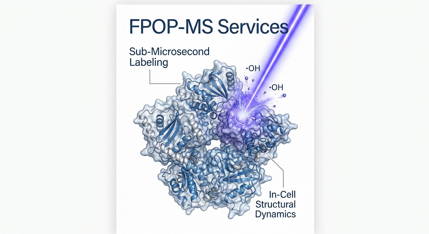
Fast Photochemical Oxidation of Proteins (FPOP-MS) Services

Accelerate your structural biology discovery with our Fast Photochemical Oxidation of Proteins (FPOP-MS) services. By utilizing sub-microsecond hydroxyl radical labeling, we capture transient conformational dynamics and in-cell target engagements that traditional methods miss. We deliver residue-level structural evidence without altering your protein's native folded state.

  • Sub-microsecond labeling speed for transient states
  • In-cell and complex lysate compatibility
  • Residue-level 3D structural mapping
Fast Photochemical Oxidation of Proteins (FPOP-MS) Services
What is FPOP-MS?Service CapabilitiesTechnology ComparisonWorkflow & QCDemo ResultsSample RequirementsCase StudyBioinformaticsFAQ

What is FPOP-MS? (Sub-Microsecond Protein Footprinting)

Fast Photochemical Oxidation of Proteins (FPOP-MS) is an advanced hydroxyl radical footprinting technique that maps protein solvent accessibility. Utilizing a pulsed UV excimer laser, FPOP photolyzes hydrogen peroxide to generate a microsecond burst of hydroxyl radicals. These radicals irreversibly label exposed amino acid side chains before the protein can unfold.

As a highly specialized extension of Hydroxyl Radical Footprinting (HRF-MS), FPOP relies on extreme speed. Traditional analytical techniques are often too slow to study highly dynamic proteins. If a protein changes its shape in milliseconds, a technique that takes minutes to label the protein will only capture a blurred, time-averaged picture. Because the entire FPOP radical reaction happens in less than one microsecond, the labeling is completely finished before the protein even has a chance to change its shape in response to the oxidation. This sub-microsecond snapshot capability allows us to capture true, native structural dynamics, fleeting intermediate states, and deeply hidden allosteric binding pockets.

Service Capabilities & Boundaries (Our Expertise & Suitable Projects)

Our FPOP-MS platform is specifically engineered to address the most challenging structural biology bottlenecks in modern drug discovery. We specialize in mapping therapeutic targets that refuse to crystallize, characterizing protein interactions that happen too quickly for standard mass spectrometry, and evaluating drug binding in complex biological matrices.

Projects We Excel At:

Transient Conformational States & Enzyme Kinetics

Many enzymes and receptors transition through crucial, ultra-fast intermediate states during catalysis or activation. We capture these fleeting structural intermediates, providing a high-resolution window into the mechanistic action of your target that equilibrium-based methods simply cannot see.

Targeted Protein Degradation (PROTACs & Molecular Glues)

The formation of ternary complexes (Target-PROTAC-Ligase) is highly dynamic. We utilize FPOP to map the induced-proximity interfaces and confirm the structural cooperativity of these complexes, ensuring your degrader molecule is stabilizing the correct conformational orientation for ubiquitination.

Intrinsically Disordered Proteins (IDPs)

Because IDPs completely lack a fixed 3D structure, traditional methods like X-ray crystallography are useless. Our ultra-fast labeling takes instant snapshots of these highly flexible ensembles in their natural solution phase, allowing us to track how specific ligands or post-translational modifications shift the conformational equilibrium.

Membrane Proteins & In-Cell Structural Proteomics

Integral membrane proteins are notoriously difficult to analyze outside of a lipid bilayer. FPOP easily penetrates lipid nanodiscs or detergent micelles. Furthermore, unlike HDX-MS, the irreversible chemistry and extreme speed of FPOP allow us to perform labeling directly inside living cells. We can map target engagement and drug binding in the complex, authentic physiological environment of the cell, perfectly complementing our Live-cell ABPP screening services.

We easily identify remote conformational changes triggered by small molecules, pinpointing allosteric pockets with residue-level precision, even when the orthosteric active site remains completely unmodified.

Technology Comparison: FPOP-MS vs. HDX-MS vs. XL-MS

Choosing the right structural mass spectrometry tool is a critical strategic decision that heavily impacts your discovery timeline. While FPOP-MS is exceptionally powerful, it operates alongside other sophisticated techniques to form a comprehensive structural toolkit.

FeatureFPOP-MSHDX-MSXL-MS
Labeling SpeedSub-microsecond (ultra-fast)Seconds to hours (slow kinetics)Minutes to hours
ReversibilityIrreversible (covalent side-chain modification)Reversible (back-exchange is a constant risk)Irreversible (covalent distance constraints)
In-cell CompatibilityExcellent (radical chemistry survives cellular environments)Poor (intracellular water causes total back-exchange)Moderate to Good (cross-linkers can permeate cells)
Structural ResolutionResidue level (specific amino acid side chains)Peptide level (regional segments of the backbone)Distance constraints between specific reactive residues

Our Solution Selection Strategy:

  • Choose HDX-MS if you need routine, comprehensive tracking of slow backbone conformational dynamics, standard epitope mapping for therapeutic antibodies, or structural comparability analysis for biosimilars in highly purified, simple buffer systems.
  • Choose XL-MS when your primary goal is to map the direct protein-protein interaction interfaces and measure exact atomic distance constraints. This is ideal for building topological 3D models of large, multi-subunit protein complexes that are difficult to analyze natively.
  • Choose FPOP-MS when your project demands the capture of ultra-fast transient states, involves highly flexible Intrinsically Disordered Proteins (IDPs), requires strict residue-level resolution, or necessitates performing native structural analyses directly within living cells or crude cellular lysates.

End-to-End FPOP Workflow & Strict Dosimetry QC

The biggest technical challenge in FPOP-MS is balancing the radical yield: ensuring that the protein is adequately labeled to produce a strong signal, without generating so many radicals that the protein is destroyed or forced to unfold by the oxidation itself. We have engineered a highly controlled technical workflow that guarantees pristine data through strict dosimetry (dose control).

1

Sample Preparation & Reagent Mixing

Once we receive your samples, we carefully mix them with a precise, low concentration of hydrogen peroxide and a specific radical scavenger (typically glutamine or histidine). The scavenger acts as an essential buffer, strictly limiting the lifespan of the hydroxyl radicals to the microsecond timescale.

2

Laser Irradiation (The Technical Core)

The sample is pumped continuously through a fused-silica microcapillary. A KrF excimer laser (operating at 248 nm) fires precisely timed, nanosecond-duration pulses. The liquid flow rate is mathematically matched to the laser repetition frequency so that each target segment of your sample receives exactly one laser pulse, preventing double-exposure.

3

In-Line Dosimetry QC

This is our critical quality checkpoint. We measure the UV absorbance of an internal standard (a dosimeter peptide, such as adenine) in real-time. This proves that the exact correct "dose" of hydroxyl radicals was generated in every single run, ensuring lot-to-lot reproducibility and protecting against over-oxidation.

4

Quenching & Digestion

Immediately after exiting the laser path, the sample flows into a quench buffer containing catalase and free methionine to instantly destroy any unreacted peroxide and halt all downstream secondary oxidations. We then digest the irreversibly labeled protein using specialized, high-efficiency proteases.

5

LC-MS/MS & Bioinformatics

The digested peptides are analyzed on our high-resolution Orbitrap or TOF mass spectrometers. Our algorithms scan the spectra to locate the exact amino acid residues that received stable oxygen additions (+16 Da, +32 Da mass shifts).

FPOP-MS experimental workflow featuring precision excimer laser irradiation and strict dosimetry control

Demo Results: Visualizing Oxidation at the Residue Level

We do not just hand you a raw list of fragmented mass spectra or a spreadsheet of mass-to-charge ratios. Our dedicated bioinformatics team transforms this highly complex oxidation data into visual, publication-ready models that immediately answer your structural questions for your internal milestone reviews or IND filings.

Differential plot highlighting statistically significant differences in oxidation levels

Residue-Level Differential Plot (Volcano/Bubble)

We provide comprehensive differential plots that highlight the statistically significant differences in oxidation levels for specific amino acid side chains. For example, if a specific tyrosine residue is highly oxidized in the free apo-protein, but heavily protected when your small molecule drug is added, it will appear as a distinct, significant data point, instantly pinpointing the active binding site or allosteric pocket.

3D Structural Oxidation Heatmap on PDB model

3D Structural Oxidation Heatmap

We take the quantitative oxidation differences and map them directly onto your target's PDB model, Cryo-EM map, or high-confidence AlphaFold structure. Using a high-contrast scientific color scale, we highlight shielded binding pockets in deep blue (indicating protection) and exposed, highly accessible structural loops in red. This provides a clear, three-dimensional topographical map of your protein's dynamic surface.

In-Cell vs. In-Vitro Topography comparisons

In-Cell vs. In-Vitro Topography

For our cutting-edge in-cell FPOP projects, we provide comparative analyses demonstrating how your target's conformation inside a living, crowded cell differs from its purified state in a test tube. This unique data set reveals the true physiological context of your drug's mechanism of action, confirming whether your molecule behaves the same way in vivo as it does in your biochemical assays.

Sample Requirements & Preparation Guidelines

Because FPOP-MS relies entirely on the precise photochemical generation and reactivity of hydroxyl radicals, the sample buffer environment must be strictly and carefully controlled. Many common laboratory buffers actively scavenge (destroy) radicals and will cause the experiment to fail completely.

Sample TypeRecommended Amount / ConcentrationBuffer Matrix RestrictionsPurity
Purified Protein Complex> 10 µM (Minimum volume: 50 µL)Strictly avoid radical scavengers (e.g., HEPES, Tris, glycerol, DTT, DMSO). PBS, sodium phosphate, or pure water is highly preferred.> 90%
Live Cells (for In-Cell FPOP)10^6 to 10^7 cells per conditionCultured in standard media; we will perform specialized rapid-washing steps immediately prior to laser exposure.N/A

Note: Please ship all purified protein samples overnight on ample dry ice to preserve structural integrity and prevent degradation prior to laser irradiation.

Case Study: Structural Proteomics via FPOP

Fast photochemical oxidation of proteins (FPOP): A powerful mass spectrometry–based structural proteomics tool. https://www.jbc.org/article/S0021-9258%2820%2930149-6/fulltext

Background

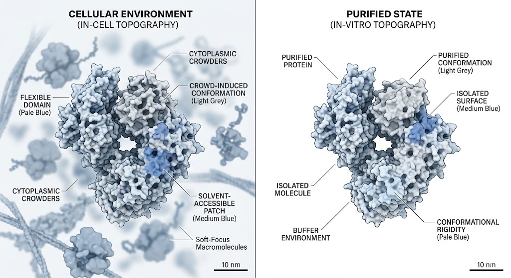
Traditional structural techniques often fail to capture highly dynamic or transient protein conformations. This is largely due to slow labeling speeds in footprinting assays or the requirement for rigid, unnatural crystallization in X-ray methodologies. Researchers required a powerful analytical method to map the true, solvent-accessible surfaces of proteins as they exist in a dynamic, liquid environment, without altering their natural folding pathways or capturing only a time-averaged structural blur.

Methods

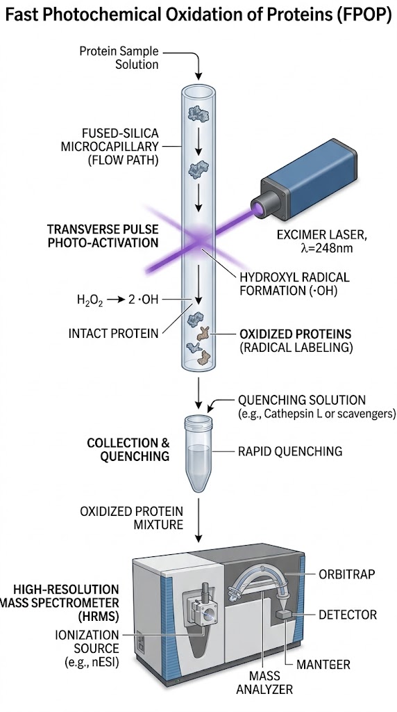
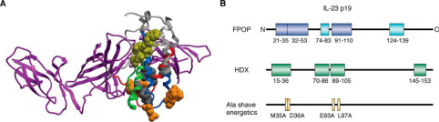
Researchers utilized FPOP-MS to overcome these fundamental biophysical limitations. The target proteins were exposed to a precisely controlled, pulsed UV laser system that photolyzed an optimized concentration of hydrogen peroxide, generating a brief, intense burst of hydroxyl radicals. These highly reactive radicals covalently modified the solvent-exposed amino acid side chains within a sub-microsecond timeframe. The radical lifetime was strictly limited by a scavenger molecule. The samples were then rapidly quenched, enzymatically digested, and analyzed using high-resolution liquid chromatography-tandem mass spectrometry (LC-MS/MS).

Results

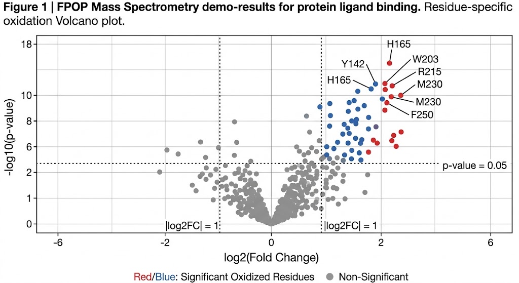
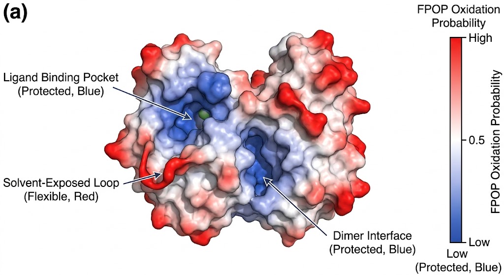
As detailed in the structural mapping data (specifically highlighted in the mass spectra and topographical mapping figures of the study), FPOP successfully labeled the target proteins faster than any unfolding events could physically occur. The high-resolution mass spectrometry analysis accurately identified specific oxidized residues (such as prominent modifications on exposed methionines, tyrosines, and tryptophans). By calculating the oxidation fractions, the researchers created a high-fidelity, residue-level topographical map of the protein's native conformation, successfully pinpointing previously hidden interaction interfaces and solvent channels.

Conclusion

FPOP-MS provides incredibly robust, residue-level structural proteomics data. It is an exceptionally powerful tool for investigating higher-order protein structures and fast-kinetic dynamics that easily evade classical analytical methodologies, supporting critical mechanism-of-action decisions in early-stage drug design and discovery.

Structural proteomics insights derived from FPOP mass spectrometry mapping

FPOP mapping data plots demonstrating residue-level structural topography and solvent accessibility.

Bioinformatics & Data Deliverables

Our dedicated data analysis pipeline converts massive arrays of complex MS/MS spectra into actionable structural insights. We employ strict statistical thresholds to eliminate background noise, delivering robust evidence for your internal milestone decisions, IP filings, or peer-reviewed publications.

Minimum Deliverables:

  • Precise residue-level identification of all valid oxidation sites across the entire protein sequence.
  • Quantitative differential modification analysis (comparing apo-protein vs. ligand-bound states, or wild-type vs. mutant).
  • Normalization data charting the internal dosimeter peptide response, ensuring absolute quantitative confidence.
  • Fully annotated high-resolution MS/MS spectra for confident modification tracking.
  • A detailed methodology report including all dosimetry QC metrics, laser parameters, and quench protocols.

Optional Add-ons:

  • High-Resolution 3D Mapping: We mathematically project the differential oxidation data directly onto PyMOL or Chimera 3D models, delivering publication-ready high-resolution rendering files.
  • In-Cell Target Engagement Analysis: For complex cellular lysates, we map not just your primary target, but the broader protein-protein interaction network affected by your compound, identifying critical off-target binding events.
FAQ

Frequently Asked Questions

Q: Will the intense UV laser destroy or unfold my protein?

No. The KrF excimer laser pulse lasts only a few nanoseconds, and the hydroxyl radical lifetime is limited to roughly one microsecond by the scavenger in the buffer. This entire labeling process is orders of magnitude faster than the time it takes for a protein to physically unfold or alter its tertiary conformation. We effectively capture the structure long before the protein "realizes" it has been oxidized, leaving the native structure completely intact at the moment of measurement.

Q: Why must I avoid HEPES or Tris buffers in FPOP experiments?

Chemicals like HEPES, Tris, glycerol, and DTT are highly effective at scavenging (absorbing) hydroxyl radicals. If these chemicals are present in your buffer, they will consume the radicals instantly before the radicals have a chance to interact with and label your target protein, resulting in a failed experiment with zero signal. We highly recommend using simple, non-reactive phosphate buffers (like PBS) or ultrapure water.

Q: Can you perform FPOP-MS on intact live cells?

Yes. Because FPOP utilizes a laser that can penetrate biological membranes and hydroxyl radicals that are generated instantly within the intracellular environment, we can successfully perform in-cell footprinting. This advanced technique allows us to map target engagement and protein conformations in their true physiological state, accounting for molecular crowding and intracellular binding partners.

Capture ultra-fast structural dynamics with the MassTarget™ team

Share your target details and our scientists will design a custom FPOP-MS strategy to map transient conformations and in-cell engagements.

Online Inquiry

Please submit a detailed description of your project. We will provide you with a customized project plan to meet your research requests. You can also send emails directly to for inquiries.

* Email
Phone
* Service & Products of Interest
Services Required and Project Description