LiP-Quant Target Deconvolution Services

Unlock the "black box" of phenotypic screening with our LiP-Quant target deconvolution services. Utilizing label-free limited proteolysis and advanced machine learning scoring, we identify direct binding targets, calculate proteome-wide apparent Kd values, and map binding pockets at peptide-level resolution—directly in complex physiological lysates.

  • 100% label-free target identification
  • Apparent Kd and dose-response quantification
  • Peptide-level 3D binding site mapping
LiP-Quant Target Deconvolution Services
What Is LiP-Quant?Service CapabilitiesTechnology ComparisonWorkflow & ML ScoringDemo ResultsSample RequirementsCase StudyBioinformaticsFAQ

What Is LiP-Quant? (Label-Free Target Identification)

LiP-Quant (Quantitative Limited Proteolysis) is an advanced label-free chemoproteomic technology used to identify direct protein targets of bioactive compounds. By combining limited Proteinase K digestion with high-resolution mass spectrometry and machine learning, LiP-Quant detects ligand-induced structural changes to reveal precise binding pockets and apparent Kd values directly within complex cell lysates.

When you discover a promising compound through a phenotypic screen, the next big hurdle is figuring out exactly which protein it binds to. Traditional target deconvolution methods often require you to attach a chemical tag (like biotin or a photo-crosslinker) to your compound to physically "fish" for the target. The problem? Adding these bulky tags often destroys the compound's natural biological activity, alters its cell permeability, or completely blocks its ability to fit into the binding pocket, leading to dead ends. LiP-Quant elegantly solves this by identifying targets based purely on native structural changes, requiring absolutely zero chemical modification to your valuable lead compounds.

Service Capabilities & Boundaries (Our Expertise & Suitable Projects)

Our mass spectrometry team specializes in turning mysterious screening hits into clear, actionable mechanism-of-action data. We built our LiP-Quant platform to handle the complex, real-world biological samples that discovery teams struggle with, offering a clear pathway from an initial hit to an optimized lead compound.

Projects We Excel At:

Phenotypic Screen Deconvolution

Finding the direct binding targets for highly active molecules when you have no prior mechanism data. If your compound works beautifully in a cell-based assay but you do not know why, LiP-Quant can scan the entire proteome to identify the primary driver of the phenotype.

Off-Target Toxicity Profiling

Scanning the entire proteome to see what else your lead candidate binds to. Many clinical trials fail due to unforeseen off-target toxicities. By running LiP-Quant on human cell lysates, we provide a comprehensive off-target liability profile, helping you predict and avoid side effects before entering costly animal studies.

Natural Product Discovery

Identifying the therapeutic targets of complex natural extracts or highly complex macrocycles. Natural products are notoriously difficult to modify with chemical tags due to their complex stereochemistry and multiple reactive groups. LiP-Quant allows us to test these compounds exactly as nature synthesized them.

Binding Site & Allosteric Mapping

Pinpointing the exact pocket where your drug binds. Because LiP-Quant operates at the peptide level, it does not just tell you which protein is bound; it tells you where the drug is sitting. This provides your medicinal chemists with the precise structural data they need to optimize the molecule's potency and selectivity.

Our Service Boundaries: To generate high-confidence, quantitative dose-response data, LiP-Quant requires a highly pure compound and a carefully constructed concentration gradient. We need a sufficient amount of your pure compound (typically >2 mg) to run the full multi-point analysis. We operate directly on complex cell lysates, tissue extracts, or bodily fluids to keep the proteins in their native states, meaning we do not require you to provide purified recombinant proteins.

Technology Comparison: LiP-Quant vs. CETSA-MS vs. Affinity Pull-down

Choosing the right target identification tool is a critical strategic decision. While older methods are well-known, newer techniques like LiP-Quant offer unique advantages for specific structural and discovery challenges.

FeatureLiP-QuantMS-based Proteome-wide Thermal Stability ProfilingAffinity Pull-down (ABPP)
Compound Tagging RequiredNo (100% Label-free)No (100% Label-free)Yes (Requires biotin, alkyne, or photo-crosslinkers)
Binding Site ResolutionYes (Peptide-level resolution)No (Protein-level resolution only)Yes (If combined with specific reactive probes)
Dependent on Thermal StabilityNo (Relies on protease accessibility)Yes (Fails if the target does not shift when heated)No (Relies on physical enrichment)
Apparent Kd QuantificationYes (Excellent dose-response data directly from lysates)Limited (Mostly used for relative ranking)Poor (Usually just a yes/no enrichment ratio)

Our Solution Selection Strategy:

  • Choose Affinity Pull-down if your compound is already well-understood, possesses an obvious site for chemical derivatization, and can easily have a biotin tag attached without losing its pharmacological activity.
  • Choose CETSA-MS if you need a rapid, label-free way to confirm if a drug stabilizes known targets, and you only need protein-level confirmation without requiring knowledge of the exact binding pocket.
  • Choose LiP-Quant if tagging your compound destroys its activity, if your target fails to show a thermal shift in CETSA assays, or if your medicinal chemists urgently need precise peptide-level binding site mapping and apparent Kd values across the entire proteome to guide structure-activity relationship (SAR) optimization.

End-to-End Workflow: From Native Lysate to ML Scoring

Our LiP-Quant service is not just a standard proteomics run; it is a heavily optimized, strictly quality-controlled pipeline designed to eliminate false positives and extract true biological interactions.

1

Native Lysis & Incubation

You provide the cells or tissues. We gently break them open to extract the proteins in their native, folded state. We then split the lysate and incubate it with your compound across a range of different concentration levels (typically a 5- to 8-point dose-response gradient).

2

Controlled Limited Proteolysis

We add Proteinase K to the mixtures for a strictly controlled, very brief period. The enzyme cuts the exposed, flexible parts of the proteins, but it cannot physically access or cut the areas that are protected by your bound drug.

3

Complete Digestion & LC-MS/MS

We immediately stop the limited cutting by boiling the sample in denaturing buffers. We then use standard trypsin to completely digest everything into small peptides suitable for mass spectrometry. These peptides are analyzed using our high-resolution Orbitrap or TIMS-TOF mass spectrometers utilizing advanced Data-Independent Acquisition (DIA) methods.

4

Machine Learning (ML) Scoring

This is where we eliminate false positives. Our bioinformatics team uses advanced machine learning algorithms to search the massive dataset. The ML model looks for specific peptides that show a clear, dose-dependent protection pattern, assigning a "LiP Score" to separate your true targets from random background noise.

The LiP-Quant workflow integrating limited proteolysis, high-resolution MS, and machine learning scoring.

Demo Results: Visualizing Target Binding and Dose-Response

We deliver intuitive, highly visual data packages that your entire discovery team—from biologists to medicinal chemists—can easily understand and act upon to advance your pipeline.

Target Ranking Volcano Plot highlighting true binding targets

Target Ranking Volcano Plot (LiP Score)

We provide a clear graph plotting the machine learning-derived LiP Score of every detected protein against its statistical significance. This visual perfectly separates your high-confidence, true binding targets from the thousands of background proteins, cutting through the noise of the complex lysate.

Dose-Response Curves determining apparent Kd

Dose-Response Curves (Apparent Kd)

For your top-ranked targets, we provide specific dose-response curve graphs. By tracking how a specific peptide resists being cut as we sequentially increase the drug concentration, we accurately calculate the apparent Kd (binding affinity) directly within the complex cell lysate. This proves that the binding is a specific, saturable event.

3D Structure Peptide Mapping showing the binding pocket

3D Structure Peptide Mapping

We do not just give you a sequence of letters. We take the significantly protected peptides and map them directly onto a 3D PDB structure or high-confidence AlphaFold model of your target. The binding pocket is highlighted in bright red or blue, showing your chemists exactly where the action happens.

Sample Requirements & Preparation Guidelines

To ensure the highest quality target deconvolution and robust machine learning scoring, we need your samples and compounds prepared correctly.

Sample TypeRecommended AmountCompound RequirementsImportant Notes
Cell Lysate / Tissue>50 million cells or >50 mg tissue per condition>2 mg of pure compound (required to build the 5-8 point dose-response curve)Do NOT add protease inhibitors during your initial cell lysis step, as they will completely interfere with our downstream Proteinase K limited proteolysis enzymes.

Case Study: Machine Learning-Based Target Identification

A machine learning-based chemoproteomic approach to identify drug targets and binding sites in complex proteomes. Nat Commun 11, 4200 (2020). https://www.nature.com/articles/s41467-020-18071-x.pdf

Background

Identifying target proteins for new drugs discovered through phenotypic screening is incredibly difficult when you cannot chemically modify the drug. Traditional label-free methods often suffer from exceptionally high false-positive rates, pointing researchers toward the wrong proteins (typically highly abundant background proteins), and they completely fail to pinpoint where the drug actually binds, leaving medicinal chemists working in the dark.

Methods

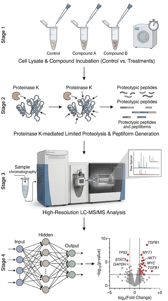
To overcome this, researchers applied the advanced LiP-Quant workflow. They incubated complex human cell lysates with several well-characterized kinase inhibitors across a full concentration gradient. They then performed strictly controlled limited proteolysis with Proteinase K. The resulting peptide mixtures were quantified using high-resolution, data-independent acquisition (DIA) mass spectrometry. Crucially, a novel machine learning (ML) algorithm (trained on hundreds of known protein-ligand interactions) was applied to the massive data array to extract true dose-response signatures, eliminate random structural fluctuations, and calculate specific, high-confidence "LiP scores."

Results

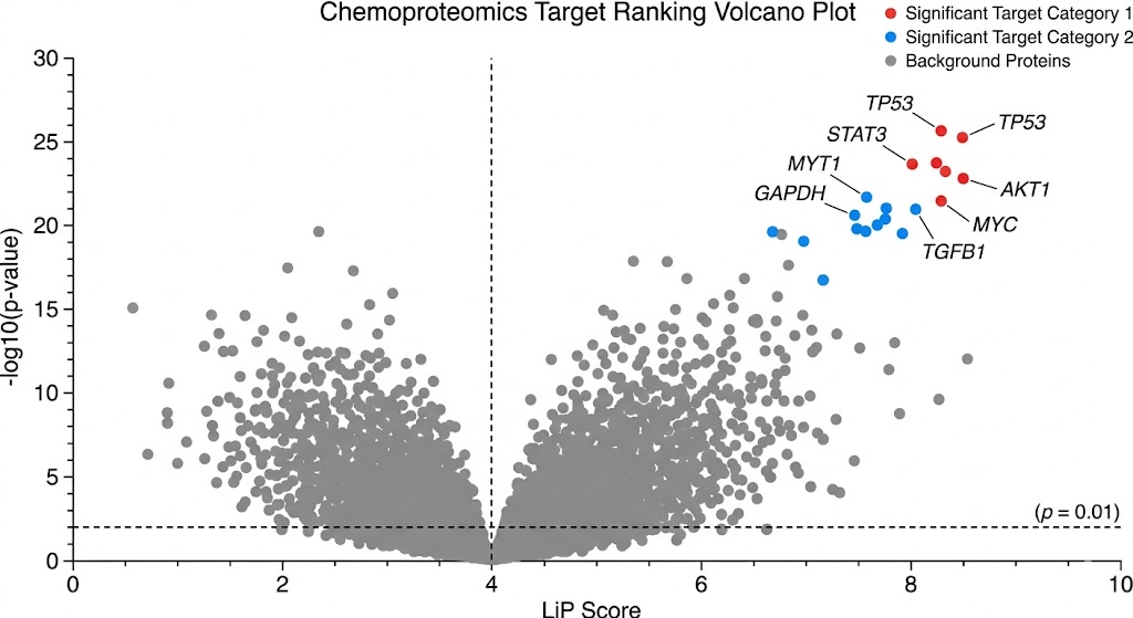
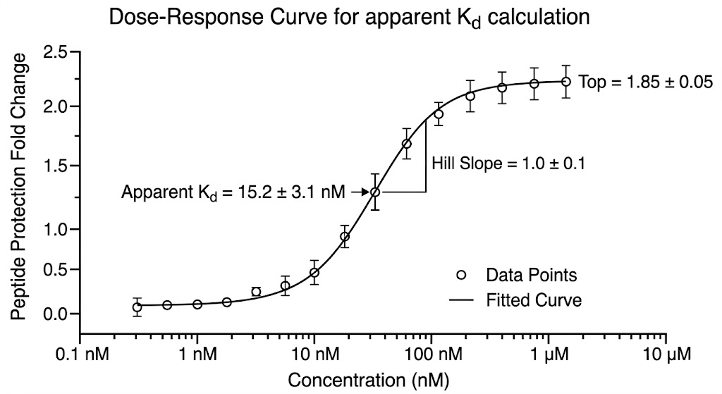
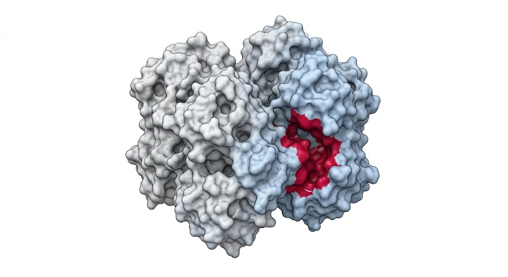
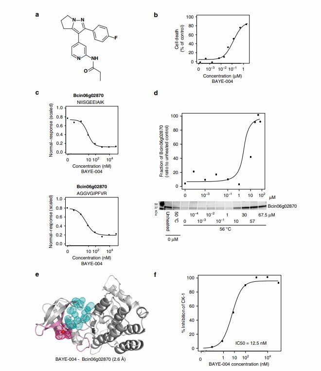
As shown in Figure 4 of the published study, the LiP-Quant platform successfully identified the specific binding targets of the kinase inhibitors. More importantly, the dose-response data accurately determined the apparent Kd values directly within the crude lysate, matching the known biochemical affinities of the drugs. The machine learning model perfectly distinguished the true targets from the high-abundance background proteome. Furthermore, the protected peptides identified by the mass spectrometer successfully mapped the exact ATP-binding pockets on the kinase targets when projected onto 3D protein models.

Conclusion

LiP-Quant, powered by machine learning, is an exceptionally robust label-free platform. It allows drug discovery teams to confidently deconvolve targets from phenotypic screens, comprehensively profile off-target effects across the proteome, and understand the molecular mechanism of action for early-stage drug candidates without the risk of destroying compound activity through chemical tagging.

Machine learning-based target deconvolution using LiP-Quant

LiP-Quant identifies binding targets and maps binding pockets at peptide-level resolution.

Bioinformatics & Data Deliverables (LiP Scores & 3D Mapping)

Our dedicated data analysis pipeline turns millions of raw mass spectrometry data points into a clear, prioritized list of targets ready for immediate biological validation.

Minimum Deliverables:

  • A comprehensive, proteome-wide target ranking list sorted by our high-confidence LiP Scores. We provide the False Discovery Rate (FDR) metrics to ensure statistical rigor.
  • Detailed dose-response curves with calculated apparent Kd values for your top-ranked targets, proving that the interaction is specific and concentration-dependent.
  • Peptide-level resolution tables showing exactly which protein regions were protected by your drug.
  • A full methodology and quality control report detailing the digestion efficiency and instrument performance.

Optional Add-ons:

  • 3D Structural Mapping: If a PDB crystal structure or a high-quality AlphaFold model is available for your identified targets, we will provide 3D visual mappings highlighting the exact binding pockets. We deliver high-resolution rendering files ready to be handed off to your molecular modeling and computational chemistry teams for immediate structure-activity relationship (SAR) optimization.
FAQ

Frequently Asked Questions

Q: Can LiP-Quant identify targets for complex natural products?

Yes. Because LiP-Quant is completely label-free, it is one of the best methods available for finding the targets of natural products, macrocycles, toxins, and complex metabolites that cannot be chemically modified with a tag without destroying their intricate stereochemistry and biological activity.

Q: Why is a compound concentration gradient (dose-response) required?

Using a single high concentration of a drug often leads to false positives (abundant proteins that stick to the drug non-specifically). By using a multi-point dose-response gradient, our machine learning software specifically looks for proteins that show a smooth, predictable increase in protection as the drug dose increases. This saturable binding behavior is the absolute hallmark of a true, specific biological interaction.

Q: How does your machine learning algorithm prevent false positives from highly abundant proteins?

Highly abundant proteins (like actin, tubulin, or heat shock proteins) often create massive background noise in structural mass spectrometry, masquerading as targets. Our machine learning algorithm is specifically trained to ignore random noise and strictly look for specific dose-dependent cleavage protection patterns that correlate with known thermodynamic binding principles. It calculates a LiP Score that effectively penalizes random fluctuations and filters out the background, leaving only high-confidence targets.

References

  1. A machine learning-based chemoproteomic approach to identify drug targets and binding sites in complex proteomes
  2. Target Deconvolution by Limited Proteolysis Coupled to Mass Spectrometry
  3. LiP-Quant, an automated chemoproteomic approach to identify drug targets in complex proteomes

Disclaimer: All services and products provided by Creative Proteomics are for Research Use Only (RUO) and are not intended for use in diagnostic procedures or clinical treatments.

Discover your true targets with the MassTarget™ team

Submit your project details and let our experts design a customized label-free target deconvolution strategy for your phenotypic hits.

Online Inquiry

Please submit a detailed description of your project. We will provide you with a customized project plan to meet your research requests. You can also send emails directly to for inquiries.

* Email
Phone
* Service & Products of Interest
Services Required and Project Description