Carboxyl Footprinting MS Services: Precision Mapping of Acidic Residues

Resolve the structural dynamics of acidic interfaces with our Carboxyl Footprinting MS services. By targeting aspartic acid, glutamic acid, and C-termini with residue-level precision, we provide critical insights into metal-binding sites and electrostatic interactions that traditional HDX-MS or hydroxyl radical methods simply cannot capture.

  • Residue-level mapping of Aspartic and Glutamic acid
  • Native-state chemical labeling with zero structural distortion
  • Expert 3D structural visualization and protection factor calculation
Carboxyl Footprinting MS Services: Precision Mapping of Acidic Residues
What is Carboxyl Footprinting?Service CapabilitiesTechnology ComparisonWorkflow & QC CheckpointsDemo ResultsSample RequirementsCase StudyBioinformaticsFAQ

What is Carboxyl Footprinting MS? (Precision Acidic Labeling)

Carboxyl Footprinting MS is an advanced structural mass spectrometry technique that utilizes carbodiimide chemistry to covalently label solvent-exposed aspartic and glutamic acid residues. By permanently modifying these acidic side chains, we achieve residue-level mapping of electrostatically driven protein interactions, metal-binding sites, and complex allosteric conformational changes.

Understanding protein conformation is rarely a one-size-fits-all endeavor. Approximately 20% of a typical protein's surface is composed of acidic amino acids. These negatively charged residues are the primary drivers of highly specific protein-protein interactions, the coordination of essential metal ions (like calcium, zinc, and magnesium), and the binding of positively charged small-molecule drugs. However, standard structural mass spectrometry tools have a massive biophysical blind spot when it comes to analyzing these critical anionic regions.

Standard HDX-MS measures the exchange of the amide backbone, completely missing the highly reactive side chains that directly participate in binding. Conversely, Hydroxyl Radical Footprinting (HRF-MS) excels at labeling aromatic and sulfur-containing residues but exhibits extremely poor reactivity toward carboxylic acids. Carboxyl Footprinting MS bridges this critical analytical gap. By utilizing carefully optimized EDC coupling chemistry, we covalently attach a stable mass tag—such as Glycinamide Ethyl Ester (GEE)—exclusively to the solvent-exposed carboxyl groups. Because this modification is entirely covalent and irreversible, it survives the harshest downstream enzymatic digestions and sample purifications, providing an unparalleled, high-resolution structural map of the acidic pockets that define your target's biological function.

Our Service Capabilities & Boundaries (Expert Insight)

Our structural mass spectrometry team specializes in utilizing Carboxyl Footprinting to solve complex biophysical puzzles that leave other analytical platforms struggling. We are highly consultative, ensuring this specialized technique is only deployed when it perfectly aligns with your specific molecular mechanism of action.

Projects We Excel At:

Positively Charged Ligand Engagement

Many potent antibiotics, polyamines, and novel small-molecule inhibitors carry a net positive charge. Their binding is heavily driven by electrostatic attraction to acidic pockets. We map these specific electrostatic interfaces, providing precise spatial coordinates needed to optimize drug affinity and residence time.

Ion Channels & Metalloproteins

Calcium, magnesium, and zinc ions almost exclusively coordinate with Asp and Glu residues within binding pockets. We utilize Carboxyl Footprinting to pinpoint exactly which residues act as the primary ion sensors, capturing subtle, long-range allosteric shifts that occur upon metal binding or ion channel activation.

Electrostatic Protein-Protein Interactions (PPIs)

Massive protein complexes, including viral capsids and transcription factor assemblies, often assemble through complementary charged surfaces. By comparing the footprinting profiles of the isolated monomeric proteins versus the assembled multimeric complex, we map the exact salt bridges and acidic contact points driving the macro-assembly.

PROTACs & HOS Orthogonal Proof

We deploy Carboxyl Footprinting to map the induced proximity interfaces of PROTAC ternary complexes, particularly when acidic patches mediate the neo-interaction. For biosimilars, we provide Carboxyl Footprinting as a complementary dataset alongside HDX-MS to prove that highly acidic domains fold consistently batch-to-batch.

Our Service Boundaries: To ensure the labeling chemistry proceeds flawlessly without denaturing your protein, the sample buffer environment must be strictly controlled. We cannot perform this assay if your sample is heavily buffered with primary amines (which compete with the nucleophile label) or carboxylates (which prematurely consume the EDC reagent). Furthermore, we require highly pure protein samples (>90%) to ensure quantitative accuracy during bioinformatics analysis.

Carboxyl Footprinting vs. HDX-MS vs. HRF-MS

Selecting the right structural mapping tool requires aligning the specific chemical labeling strategy with your exact biological question. We offer a comprehensive suite of structural MS services to ensure you never use the wrong tool for your pipeline.

FeatureCarboxyl Footprinting MSHDX-MSHRF-MS (Hydroxyl Radicals)
Primary TargetAcidic side chains (Asp, Glu, C-terminus)Amide backboneAromatic & sulfur side chains (Trp, Tyr, Met)
Labeling NatureIrreversible (Covalent +GEE mass tag)Reversible (Isotopic Deuterium exchange)Irreversible (Covalent +16 Da Oxygen)
Structural ResolutionResidue-level (Specific to acidic side chains)Peptide-level (Regional backbone dynamics)Residue-level (Broad side-chain coverage)
Downstream ProcessingHighly robust (Survives harsh digestion)Vulnerable to back-exchange (Requires fast, cold LC)Highly robust (Survives harsh digestion)
Ideal ApplicationMetal-binding, cationic drug interactions, acidic pocketsGeneral dynamic conformational mappingFast kinetic mapping, general solvent accessibility

Our Solution Selection Strategy:

  • Choose HDX-MS for the broad, routine mapping of overall protein dynamics, stability shifts, and general epitope mapping where peptide-level resolution is sufficient for your regulatory or design goals.
  • Choose HRF-MS when you need residue-level resolution across the broader protein surface, particularly in hydrophobic or aromatic-rich binding pockets where structural rearrangements occur rapidly.
  • Choose Carboxyl Footprinting MS when your primary binding mechanism involves metal ions, salt bridges, or positively charged ligands, and you need to definitively know which specific Asp or Glu residue is shielding the interaction interface.

Optimized End-to-End Workflow & QC Checkpoints

The biggest historical challenge with EDC-mediated carboxyl labeling is the risk of accidental protein cross-linking or chemical denaturation. We have completely mitigated this risk through a highly optimized, strictly timed workflow that utilizes Native MS validation principles to ensure your protein remains in its pristine, biologically active conformation during labeling.

1

Native Buffer Exchange & Incubation

We carefully exchange your protein into an optimized, amine-free and carboxylate-free physiological buffer (typically MOPS or MES). We then gently incubate the target with its small-molecule ligand or metal cofactor to achieve full binding equilibrium prior to labeling.

2

Controlled EDC/GEE Labeling

We introduce 1-ethyl-3-(3-dimethylaminopropyl)carbodiimide (EDC) alongside Glycinamide Ethyl Ester (GEE). EDC activates the exposed carboxyl groups, which are immediately trapped by the abundant GEE nucleophile, adding a stable +86 Da mass tag to the side chain. We strictly control the reaction time and reagent concentration to ensure only the most solvent-exposed residues are labeled, deliberately preventing any structural distortion or non-specific cross-linking.

3

Quenching & Denaturation

The chemical reaction is rapidly quenched to stop further labeling and preserve the native structural snapshot. The heavily stabilized, covalently modified protein is then fully denatured, reduced, and alkylated to prepare for enzymatic breakdown.

4

Complete Proteolysis

We utilize specific, high-efficiency proteases (such as Trypsin, Chymotrypsin, or Glu-C) to digest the protein into manageable peptides. Because the +86 Da label is permanent and covalent, we can use long digestion times to guarantee incredibly deep sequence coverage.

5

High-Resolution LC-MS/MS

The resulting peptide mixture is analyzed using our high-resolution Orbitrap mass spectrometers. We frequently employ specialized tandem mass spectrometry fragmentation techniques (like ETD or HCD) to pinpoint exactly which Asp or Glu residue on a given peptide holds the GEE tag.

Optimized EDC-mediated carboxyl footprinting workflow highlighting strict reaction control.

Demo Results: Visualizing Surface Accessibility

We translate highly complex mass spectrometry mass shifts into intuitive, actionable biophysical data. Our comprehensive reports provide clear evidence of target engagement, structural rearrangements, and precise binding site locations.

Residue-Level Protection Factor Bar Charts

Residue-Level Protection Factor Bar Charts

We plot the absolute modification extent of every detected Asp and Glu residue along the protein sequence. By quantitatively comparing the Apo (free) state to the Holo (ligand-bound) state, we calculate a distinct "Protection Factor." Residues that show a massive, statistically significant drop in labeling upon drug addition are immediately flagged as the primary binding coordinates.

3D Electrostatic Potential (ESP) Mapping

3D Electrostatic Potential (ESP) Mapping

We take the calculated protection factors and map them directly onto a 3D PDB crystal structure or AlphaFold model of your target. Because carboxyl groups dictate the protein's surface charge, we specifically project these results onto an Electrostatic Potential (ESP) surface map, visually highlighting exactly how your drug neutralizes or physically shields the negative acidic pockets in three-dimensional space.

Modification Kinetics Plots

Modification Kinetics Plots

To visually prove the rigorous quality and safety of our assay, we provide time-course or dose-response kinetic plots showing the rate of GEE incorporation. This confirms that our labeling followed true pseudo-first-order kinetics and did not artificially unfold or aggregate the protein during the experiment.

Sample Requirements & Technical Guidelines

Because Carboxyl Footprinting relies on highly specific carbodiimide coupling chemistry, the sample buffer environment must be absolutely free of competing chemicals. Even trace amounts of the wrong buffer components will rapidly consume the EDC reagent and result in a completely failed experiment. Please adhere strictly to the following sample preparation guidelines:

Sample TypeMinimum AmountStrict Buffer RestrictionsPurity Requirement
Purified Proteins / Biologics> 2 mg (Concentration > 1 mg/mL)NO Primary Amines: Tris, Glycine, or Ammonium salts are strictly forbidden. NO Carboxylates: Acetate or Citrate buffers are strictly forbidden. MOPS, MES, or simple Phosphate buffers are required.> 90% via SEC or SDS-PAGE
Ligands / Metal Ions / Drugs> 1 mgKeep DMSO concentrations as low as possible (< 2% final assay concentration). Must be fully soluble in aqueous solutions.> 95% purity

Note: Please ship all purified protein samples overnight on ample dry ice to preserve their structural integrity. If you are unable to exchange your protein into a completely compatible buffer, please notify our scientific team, and we can perform a rapid, native-friendly desalting step immediately prior to analysis.

Case Study: Mapping the HER2-HER3 Receptor Interface

Carboxyl Group Footprinting Mass Spectrometry and Molecular Dynamics Identify Key Interactions in the HER2-HER3 Receptor Tyrosine Kinase Interface. https://www.jbc.org/article/S0021-9258(20)49130-6/fulltext

Background

The HER2-HER3 receptor tyrosine kinase heterodimer is a critical, potent oncogenic driver in numerous breast and gastric cancers. Developing therapeutics that successfully block this specific dimerization is a major biopharmaceutical industry goal. However, capturing the highly dynamic, transient nature of the HER2-HER3 interface in solution using static X-ray crystallography is incredibly challenging. Researchers required an advanced, high-resolution footprinting method to map the precise electrostatic interactions driving the complex formation in a native liquid environment.

Methods

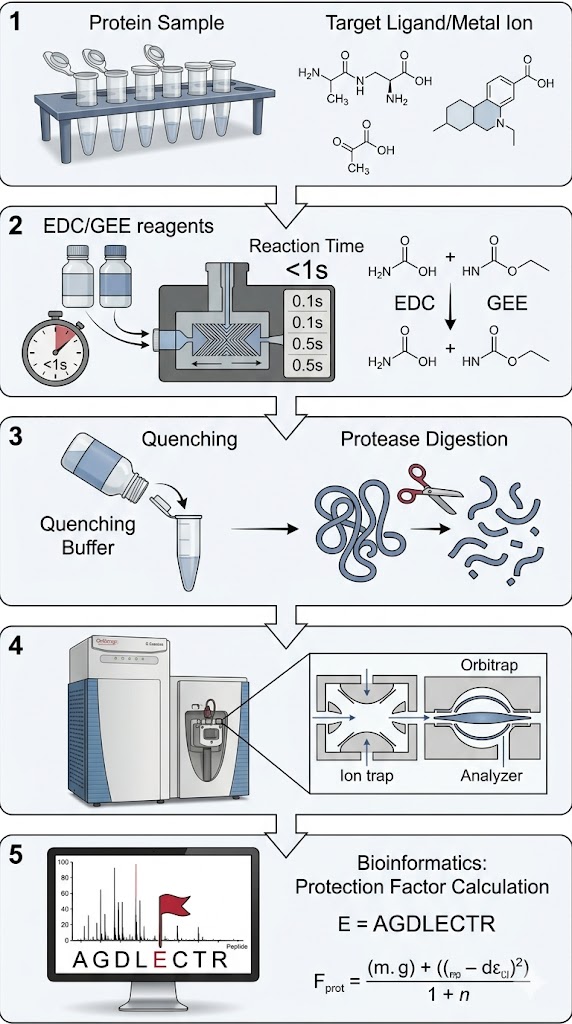
Researchers deployed Carboxyl Group Footprinting Mass Spectrometry, combined with computational Molecular Dynamics (MD) simulations, to deeply interrogate the acidic residues at the heterodimer interface. The isolated HER2 and HER3 kinase domains, as well as the fully assembled complex, were gently labeled in parallel using EDC and a stable nucleophile. The covalently labeled proteins were subsequently digested and analyzed via high-resolution LC-MS/MS to pinpoint and quantify the exact modification extent of the solvent-exposed Asp and Glu residues across the entire receptor.

Results

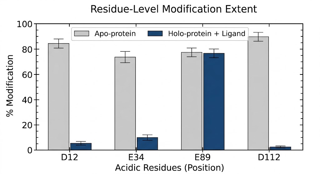
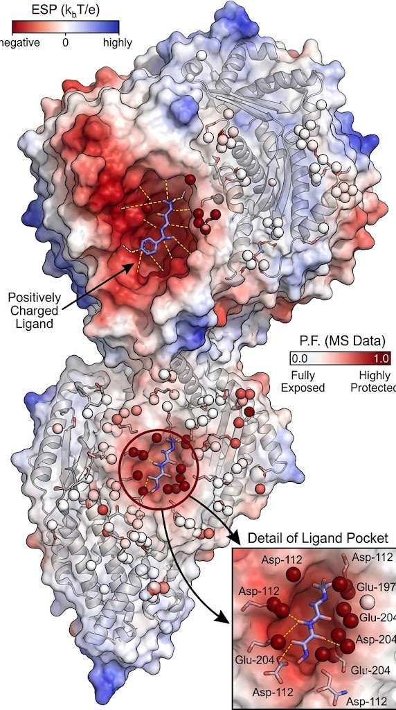
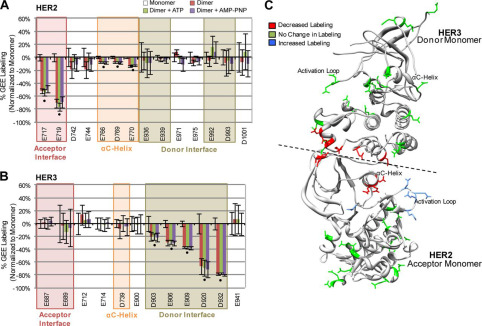
The advanced mass spectrometry data successfully pinpointed the key electrostatic "hotspots" driving the oncogenic dimerization. As specifically illustrated in Figure 2 of the referenced structural study, the footprinting analysis revealed significant, highly localized structural protection directly at the dimer interface. Crucially, the data achieved true single-residue resolution, demonstrating a massive, statistically significant decrease in labeling specifically at residues Glu-922 and Asp-948 when the distinct proteins formed the intact complex. These experimental mass spectrometry results perfectly correlated with the computational MD simulations, proving definitively that these two acidic residues act as essential, solvent-shielded structural anchors for the heterodimer.

Conclusion

Carboxyl Footprinting MS provided unprecedented, residue-level structural evidence of the HER2-HER3 interaction interface. By precisely locating the protected acidic residues in native solution, the biophysical study provided medicinal chemists with a highly accurate 3D roadmap for developing novel, allosteric small-molecule inhibitors designed to physically disrupt this critical oncogenic dimerization event.

Carboxyl Footprinting MS mapping of the HER2-HER3 receptor interface

Analysis of protection factors identified critical acidic residue hotspots at the heterodimer interface.

Bioinformatics: From Mass Shifts to Protection Factors

Our structural bioinformatics pipeline is expressly designed to eliminate the complexity of covalent label analysis and deliver clear, actionable mechanistic insights. The primary computational challenge in Carboxyl Footprinting is distinguishing the engineered +86 Da (GEE) or +57 Da (Glycinamide) mass shifts from naturally occurring endogenous post-translational modifications (PTMs), such as natural deamidation or oxidation artifacts.

Our proprietary structural algorithms solve this by utilizing advanced False Discovery Rate (FDR) control and strict PTM filtering against highly curated mass spectra libraries. We accurately quantify the area under the curve (AUC) for both the unmodified and modified versions of every specific acidic peptide identified by the mass spectrometer.

From this highly curated raw data, we calculate the absolute Fraction Unmodified (FU). By comparing the FU of the ligand-bound (Holo) state against the free protein (Apo) state, we generate highly accurate Protection Factors (PF). A high Protection Factor definitively proves that a specific Asp or Glu residue has been shielded from the surrounding solvent by the physical binding of your drug, mapping the active binding site with mathematical certainty and guiding your structure-activity relationship (SAR) campaigns.

FAQ

Frequently Asked Questions

Q: Does EDC labeling artificially change the protein's isoelectric point (pI) and cause it to unfold?

While neutralizing carboxyl groups does technically alter the local surface charge of the protein, we tightly control the reaction kinetics to prevent unfolding. We optimize the EDC concentration and reaction time so that only a small fraction (typically under 10-15%) of the total available carboxyl groups are modified on any single protein molecule. This extremely light chemical "grazing" ensures the overall macroscopic fold, stability, and native binding activity of the protein are perfectly preserved during the entire footprinting assay.

Q: Can you handle membrane proteins or targets solubilized in detergents?

Yes, but it requires careful upfront optimization. Many common detergents can heavily suppress mass spectrometry ionization. We have extensive experience utilizing MS-compatible, cleavable detergents and performing advanced sample cleanup post-labeling. Because the carboxyl footprint is completely permanent and covalent, we can aggressively wash the sample to remove interfering lipids and detergents prior to enzymatic digestion and LC-MS analysis.

Q: Why shouldn't I just use HDX-MS for my target?

If your primary goal is to map broad conformational changes, large allosteric shifts, or you are studying a highly hydrophobic pocket, HDX-MS is an excellent tool. However, HDX-MS only measures the isotopic exchange of the backbone amide hydrogens. If your drug specifically binds via an electrostatic salt bridge to a Glutamic Acid side chain extending out into the solvent, the backbone HDX signature might barely change at all, resulting in a false negative. Carboxyl Footprinting directly and exclusively probes that specific, functional acidic side chain.

Resolve acidic interfaces with the MassTarget™ team

Submit your target sequence and ligand properties, and let our experts design a customized Carboxyl Footprinting strategy to map your electrostatic interactions.


Online Inquiry

Please submit a detailed description of your project. We will provide you with a customized project plan to meet your research requests. You can also send emails directly to for inquiries.

* Email
Phone
* Service & Products of Interest
Services Required and Project Description