Photo-crosslinking Structural MS Service for Binding Site Mapping and Mechanism Studies

Capture ligand engagement, interpret binding regions, and support structure-aware project decisions.

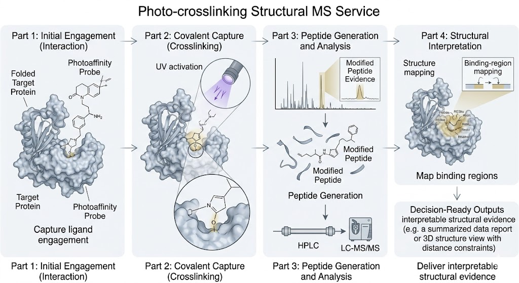
Photo-crosslinking Structural MS adds a direct evidence layer when a compound shows activity but the binding region is still unclear. By combining photoactivated capture with LC-MS/MS-based interpretation, we help you study ligand engagement, weak or transient interactions, and structure-aware mechanism questions in a format your team can review and act on.

This workflow is especially useful when standard function-only assays cannot provide enough spatial context, when interactions are difficult to stabilize, or when you need a method that bridges biochemical observations and deeper structural follow-up.

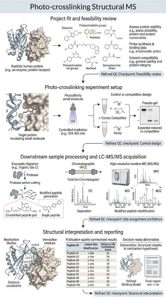
At Creative Proteomics, we do not treat photo-crosslinking as a standalone checkbox assay. We place it inside a broader MS evidence chain that can extend from hit support to target deconvolution, mechanism evidence, and orthogonal follow-up when your study needs more than one readout.

Key Advantages:

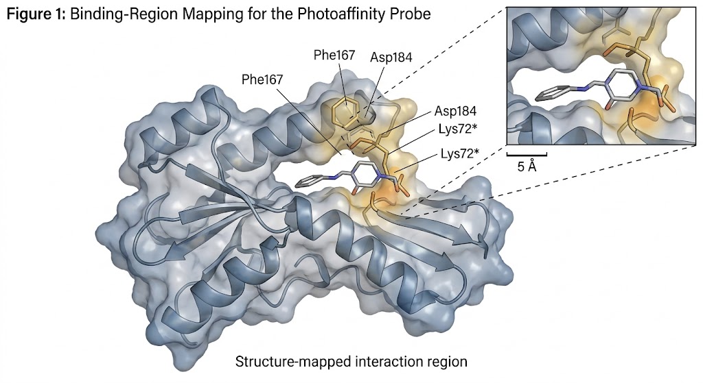
  • Binding-region-focused structural evidence for small-molecule engagement.
  • Strong utility for weak or transient interactions that are difficult to interpret with standard assays.
  • Explicit workflow and QC checkpoints from feasibility review to final reporting.
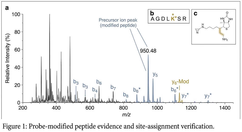
  • Decision-ready deliverables designed for technical review, not just raw spectra.
Photo-crosslinking Structural MS overview showing ligand engagement, photoactivation, LC-MS/MS interpretation, and binding-region mapping workflow.
What It Is Service Overview Comparison Sample Demo Case Study FAQ

What Is Photo-crosslinking Structural MS?

Photo-crosslinking Structural MS is a mass spectrometry-based workflow used to capture and interpret ligand–target interaction evidence after photoactivation. In practical drug discovery settings, it is most useful when your question is not only whether binding happens, but where the interaction is happening and how that interaction should be interpreted.

This service is commonly used for binding-region mapping, residue-level clues, competition-informed interpretation, and structure-aware support for mechanism studies. It can be especially valuable when interactions are weak, transient, or difficult to stabilize long enough for standard assay readouts.

It is not always the first method to choose. If your main question is only binding kinetics, a kinetic assay may be more direct. If your main question is broad target or off-target scope across the proteome, a broader chemoproteomic design may be more informative first. If you need near-atomic architecture, deeper structural follow-up may still be needed after this workflow.

Why This Workflow Matters for Discovery Teams

Direct binding-region evidence

When activity data is already available but the interaction region is still unclear, this workflow adds a more interpretable structural layer.

Useful for weak or transient interactions

Photoactivated capture can help preserve evidence from interactions that are difficult to stabilize or confirm using standard readouts alone.

Supports mechanism-focused projects

Outputs can help your team discuss hit triage, SAR, competition design, and structure-aware mechanism interpretation.

Built for technical review

We design the project around interpretable evidence views, structured result tables, and reporting language that supports internal decision-making.

When It Is Often a Strong Fit

We often recommend this service when you already have active compounds but need stronger structural evidence, when the interaction is difficult to capture in standard assays, or when you need a workflow that sits between biochemical evidence and deeper structural follow-up.

Service Overview – What We Can Execute for Your Project

We support drug discovery and early screening teams that need mass spectrometry-based evidence across hit, target, and mechanism questions. That broader capability matters because photo-crosslinking projects rarely stop at one modified peptide. They usually lead to decisions about selectivity, competition design, neighboring methods, or the next validation step.

We scope Photo-crosslinking Structural MS as part of a wider evidence strategy, not as an isolated readout. That means the project starts with fit assessment and ends with reporting that can feed directly into follow-up technical work.

MODE 1

Purified Protein Binding-Region Mapping

For projects that need direct interaction evidence on purified targets.

  • Suitable for site-aware interpretation of ligand engagement.
  • Useful when standard activity assays do not explain where binding occurs.
  • Can support SAR and hit triage discussions.
MODE 2

Protein Complex and Difficult Target Studies

Designed for systems where context matters for interaction interpretation.

  • Supports complexes and challenging target formats.
  • Useful when interaction stability is limited.
  • Can provide evidence that complements broader structural follow-up.
MODE 3

Mechanism-Focused Project Support

For studies where binding evidence needs to be discussed alongside project biology.

  • Supports competition-informed interpretation.
  • Useful for mechanism-of-action discussion.
  • Can help prioritize next-step orthogonal validation.
MODE 4

Integrated MS Evidence Strategy

For teams that need this workflow positioned within a broader MS evidence chain.

  • Connects photo-crosslinking outputs with adjacent MS methods.
  • Supports project decisions beyond a single modified feature.
  • Built for technical review and follow-up planning.

Photo-crosslinking Structural MS Workflow

The workflow combines technical execution and service-stage review from sample assessment to final reporting.

1

Feasibility review and project-fit assessment

We review the biological question, target context, ligand or probe status, expected comparison design, and any orthogonal evidence already available.

2

Photo-crosslinking design and control setup

We define the photoreactive strategy, control logic, competition options, and sample-context factors that may affect interpretation.

3

Capture, digestion, LC-MS/MS acquisition, and site review

After irradiation and downstream processing, the project proceeds through peptide-level acquisition and modified-feature review.

4

Structural interpretation and deliverable reporting

We assemble processed evidence views, structured result tables, interpretation notes, and reusable raw or semi-processed outputs where appropriate.

Vertical workflow diagram for Photo-crosslinking Structural MS showing project-fit review, photo-crosslinking experiment, LC-MS/MS acquisition, and structural interpretation.

Typical QC checkpoints include:

Target and sample identity context

Ligand or probe readiness

Control strategy for background interpretation

Modified-feature and site-assignment confidence

Structural interpretation scope

What the Results Can Look Like

The most useful demo results are not generic dashboards. They show the kinds of technical outputs your team can actually review, discuss, and use to plan the next experiment.

Modified-Peptide Evidence

This peptide-level view shows where a probe-modified signal or site-assignment clue is observed.

What it helps answer:

  • Did the experiment generate interpretable modification evidence?
  • Is there peptide-level support for site-aware interpretation?

Why it matters: it provides the first layer of confidence before structure mapping and condition comparison.

Binding-Region Mapping on a Structure

This view maps observed residues or clustered evidence back onto a protein surface or structural model.

What it helps answer:

  • Can the LC-MS/MS output be linked to an interpretable interaction region?
  • Does the result support structure-aware follow-up?

Why it matters: it moves the discussion beyond peptide lists and into project-level interpretation.

Condition or Competition Comparison

This view compares labeling behavior across control and experimental contexts.

What it helps answer:

  • Is the project generating mechanism-relevant evidence?
  • Do the observed signals behave in a way that supports the study question?

Why it matters: it helps distinguish isolated modified signals from interpretable project evidence.

Project Readiness Notes

The sample-submission guides you attached emphasize the same core ideas that matter for this service: consistency, rapid handling, prompt freezing, clear labeling, full matrix disclosure, and complete submission records. The metabolomics guide highlights representativeness, fast processing, flash-freezing, dry-ice shipment, and clear replicate labeling, while the proteomics guide reinforces sample representativeness, reproducibility, cryopreservation, and detailed notes for specially treated samples. :contentReference[oaicite:1]{index=1} :contentReference[oaicite:2]{index=2}

Project Readiness ItemWhy It MattersWhat to PrepareRecommended Handling
Project questionDefines whether photo-crosslinking is the right evidence layerBinding question, target context, comparison designSubmit with sample notes and prior assay context
Sample labelingPrevents confusion during acquisition and interpretationClear replicate names and group namingLabel before freezing and keep naming consistent
Special treatment historyHelps us interpret extraction and signal quality correctlyDrug treatment, stress condition, enrichment, or pre-processing notesDeclare on the submission form before project start
Matrix or buffer contextAffects compatibility and downstream interpretationBuffer composition, additives, solvent detailsProvide complete composition for liquid or pretreated samples

Technology Comparison: Photo-crosslinking Structural MS vs. Alternative Techniques

TechniqueCore Question AnsweredTypical ApplicationsKey StrengthsKey Limitations
Photo-crosslinking Structural MSWhere does a small molecule engage, and can direct interaction evidence be captured?Binding-region mapping, site-aware mechanism support, weak or transient interaction studies
  • Strong fit for binding-region-focused evidence
  • Useful for weak or transient interactions
  • Can support structure-aware interpretation
  • Does not by itself provide atomic-resolution structure
  • Interpretation depends on control design and site-assignment confidence
HDX-MSDoes ligand binding change solvent accessibility or conformational dynamics?Conformational analysis, allosteric interpretation, ligand-induced dynamics
  • Strong for conformational change questions
  • Useful for region-level dynamics
  • Binding-site localization is indirect
  • Less direct when the main question is site-specific engagement
XL-MSWhat spatial restraints or interaction architecture exist across proteins or assemblies?Protein complexes, assembly mapping, interface restraint analysis
  • Useful for assembly and architecture questions
  • Supports restraint-based interpretation
  • Less direct for small-molecule binding-region questions
PAL-ABPP / ChemoproteomicsWhat targets or off-targets are engaged across complex biological backgrounds?Target scope, off-target profiling, target deconvolution
  • Strong for proteome-wide target scope
  • Useful in complex biological backgrounds
  • Fine structural interpretation is more variable by design
cryo-EM or deeper structural follow-upWhat is the higher-resolution architecture of the bound state?Later-stage structural refinement and model building
  • Can provide high-resolution structural context
  • Strong for advanced structural follow-up
  • Not the most direct starting point for initial binding-region evidence
  • Sample suitability can be a major constraint

Sample Requirements

Sample TypeRecommended Starting PointContainerShippingNotes
Purified Protein150 µgLow-bind tubeDry iceProvide protein identity, buffer composition, and ligand or probe status; the proteomics guide lists 8 M urea as the preferred buffer for pure protein submission. :contentReference[oaicite:3]{index=3}
Cultured Cells5 × 106 cells1.5 mL low-bind tubeDry iceWash with pre-chilled PBS, collect pellets, and freeze quickly; the proteomics guide also recommends at least 2–3 tubes per sample with more than 50 µL pellet per tube. :contentReference[oaicite:4]{index=4}
Conservative Cell Reserve>1 × 10⁷ cells1.5 mL tubeDry iceThe metabolomics guide gives this as a higher-input benchmark that can be helpful when extra material is available for optimization. :contentReference[oaicite:5]{index=5}
Tissue100–200 mg for many routine tissue types; 200 mg for hard animal tissues in label-free proteomics guidanceCryovial or centrifuge tubeDry iceRemove non-target material, handle quickly, and freeze immediately after collection. :contentReference[oaicite:6]{index=6} :contentReference[oaicite:7]{index=7}
Plasma / Serum>100 µL in general MS submission guidance; 20 µL for standard label-free proteomics without depletion; 50–100 µL with depletion1.5 mL tubeDry iceAvoid repeated freeze-thaw cycles and note anticoagulant or depletion requirements clearly. :contentReference[oaicite:8]{index=8} :contentReference[oaicite:9]{index=9}
Culture Supernatant>2 mL in general MS submission guidance; 10 mL for label-free proteomicsScrew-cap tubeDry iceClarify medium composition and collection conditions before submission. :contentReference[oaicite:10]{index=10} :contentReference[oaicite:11]{index=11}
Saliva / Tears and Related Fluids>200 µL in general MS submission guidance; 1 mL for label-free proteomics saliva, tears, or milk1.5 mL tubeDry iceAliquot when possible and keep collection and handling consistent across groups. :contentReference[oaicite:12]{index=12} :contentReference[oaicite:13]{index=13}

Deliverables

  • Processed evidence views for key findings
  • Structured result tables for modified features or interpreted sites
  • Raw data or reusable source files where appropriate
  • Experiment and analysis notes tied to the project question
  • Optional structure-mapping views and follow-up interpretation support

Representative Photo-crosslinking Structural MS Demo Data

Annotated modified-peptide spectrum demonstrating probe-modified site evidence in Photo-crosslinking Structural MS.

Modified-peptide evidence view

Protein structure with highlighted ligand-binding region mapped from Photo-crosslinking Structural MS results.

Binding-region mapping view

Condition comparison plot showing interpretable labeling differences in Photo-crosslinking Structural MS.

Condition or competition comparison view

Case: Residue-Specific Distance Mapping with Diazirine Photo-crosslinking

Jiang Y., Zhang X., Nie H., et al. Dissecting diazirine photo-reaction mechanism for protein residue-specific cross-linking and distance mapping. Nature Communications (2024). Source article

Background

Diazirine-based photo-crosslinking can provide useful structural restraints, but ambiguous residue assignment has often limited confidence in downstream interpretation. That makes it directly relevant to teams who want site-aware evidence rather than only target capture.

Methods

In this study, the authors developed an in-line irradiation system and systematically modulated light intensity and irradiation time to evaluate diazirine photolysis and residue-specific photo-reaction behavior. They then mapped photo-crosslinking outputs onto protein structures to improve distance-based interpretation.

Results

The paper reported a two-step mechanism involving diazo and carbene intermediates and showed that the diazo intermediate preferentially targeted buried polar residues. Fig. 5f is the most useful figure for service-page communication because it maps PXL-derived information onto the BSA structure and shows how residue-specific photo-crosslinking can be translated into a structure-aware distance-mapping view rather than remaining a peptide-only observation. The companion panels in Fig. 5a–d strengthen that interpretation by showing residue preference and distance-distribution behavior.

Conclusion

This case supports using Photo-crosslinking Structural MS as a practical route to residue-aware structural evidence. It also reinforces why irradiation control, assignment confidence, and structure-mapping deliverables belong in the core workflow instead of being treated as optional extras.

Figure 5f from a diazirine photo-crosslinking study showing structure-mapped distance information on BSA.

Figure 5f illustrates structure-mapped distance information derived from residue-specific diazirine photo-crosslinking.

FAQ

Frequently Asked Questions

Q: When is Photo-crosslinking Structural MS more useful than a standard binding assay?

When you need direct evidence about where a molecule engages or whether condition-dependent labeling supports a mechanism hypothesis, this workflow can go beyond a yes-or-no binding answer.

Q: Can this help with weak or transient interactions?

Yes. That is one of the reasons photoaffinity workflows remain valuable, especially when standard assays struggle to stabilize or interpret the interaction.

Q: Do I need a finished photoaffinity probe before scoping a project?

Not always. Early scoping can still define whether the target question, target context, and intended evidence type are a good fit.

Q: What kinds of sample systems are suitable?

Purified proteins, cultured cells, tissue-derived material, and selected complex backgrounds can all be suitable, but the best choice depends on the biological question and control design.

Q: How do you control non-specific background labeling?

We control this through project-fit review, control strategy, sample-context review, and conservative interpretation of site evidence rather than overcalling isolated signals.

Q: What evidence do I receive beyond raw MS data?

You can receive processed evidence views, structured result tables, interpretive notes, and optional structure-linked reporting in addition to raw files.

Q: Can the outputs support medicinal chemistry or structural follow-up?

Yes. The most useful outputs are often the ones that narrow a binding region, prioritize follow-up conditions, or support a next-step structural decision.

Q: When should this be paired with HDX-MS, XL-MS, or chemoproteomics?

Pairing makes sense when the project needs a wider evidence chain, such as conformational change, complex architecture, or proteome-wide target scope.

Plan the Right Structural Evidence Path for Your Molecule and Target

If your current challenge is moving from activity data to interpretable binding-region evidence, we can help you evaluate whether Photo-crosslinking Structural MS is the right starting point, how the workflow should be scoped, and what evidence package is most likely to support your next project decision.

This service and all related deliverables are provided for research use only and are not intended for clinical or diagnostic use.

Online Inquiry

Please submit a detailed description of your project. We will provide you with a customized project plan to meet your research requests. You can also send emails directly to for inquiries.

* Email
Phone
* Service & Products of Interest
Services Required and Project Description