Molecular Glue Target Identification Service — MS-Based Neosubstrate Discovery from Phenotypic Hits to Preclinical Leads

Your molecular glue was designed to bring an E3 ligase and an unknown target together. The critical question — what is the target?

Molecular glues are monovalent small molecules that induce or stabilise protein-protein interactions between an E3 ubiquitin ligase and a previously non-interacting neosubstrate protein, leading to ubiquitination and proteasomal degradation of the neosubstrate. Unlike PROTACs — where both the target and E3 ligand are explicitly designed into a bifunctional molecule — molecular glues work by binding to the E3 ligase and modifying its surface to create a neo-interface that recruits a previously unrelated protein. This means that for most molecular glue programmes, the identity of the degraded target is unknown at the outset and must be discovered empirically.

Mass spectrometry-based proteomics is the only methodology capable of identifying the neosubstrate of a molecular glue in an unbiased, proteome-wide manner — without requiring prior knowledge of the target, specific antibodies, or genetic hypotheses. At Creative Proteomics MassTarget, we deploy an integrated MS-based target identification platform combining chemoproteomics (AP-MS interactomics), ubiquitinomics, quantitative proteomics, and limited proteolysis-mass spectrometry (LiP-MS) to identify the neosubstrate of any molecular glue, confirm its degradation mechanism, and profile selectivity across the proteome. For dedicated interactome mapping of E3 ligase-neosubstrate interactions, our interactomics (AP-MS / proximity labeling) platform provides the affinity purification-MS workflows for unbiased capture of molecular glue-induced protein-protein interactions in cellular lysates and live cells.

Key Advantages:

  • Unbiased neosubstrate discovery — no antibody, no genetic hypothesis, no prior target knowledge required.
  • Multi-platform orthogonal validation — AP-MS interactomics + ubiquitinomics + quantitative proteomics + LiP-MS under a single project.
  • Compatible with all major E3 ligases — established protocols for CRBN, VHL, MDM2, and novel E3 systems.
  • Works from limited compound — as little as 1–5 mg of molecular glue sufficient for a full target ID campaign.
  • Turnaround: 3–5 weeks depending on platform scope and number of compounds.
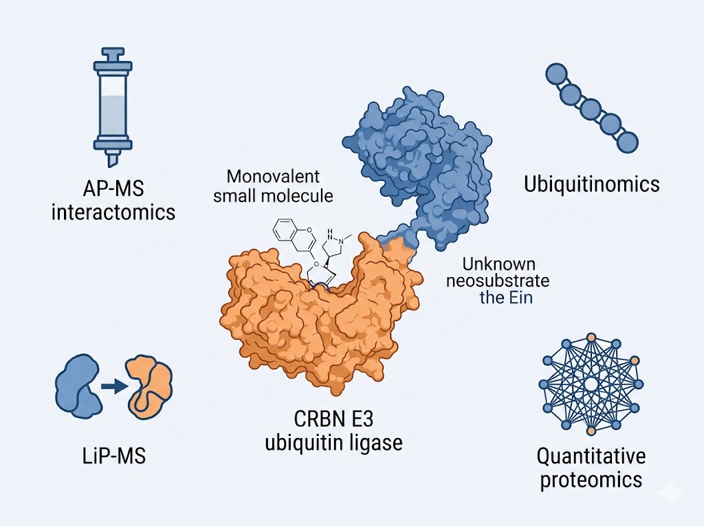
Molecular glue target identification service overview: molecular glue compound (monovalent small molecule) binding to CRBN E3 ubiquitin ligase (orange) and inducing a neo-interface that recruits an unknown neosubstrate protein (blue), analysed by AP-MS chemoproteomics, ubiquitinomics, quantitative proteomics, and LiP-MS for comprehensive neosubstrate discovery and degradation mechanism confirmation.
What Is MG Target ID Platform Suite Tech Comparison Sample Demo Case Study FAQ

What Is Molecular Glue Target Identification?

Molecular glue target identification is the process of determining which protein(s) a molecular glue recruits to an E3 ubiquitin ligase for ubiquitination and degradation. The challenge is fundamentally different from target identification for conventional inhibitors or PROTACs: because the molecular glue does not contain a dedicated target-binding moiety, the neosubstrate cannot be predicted from the compound structure and may belong to entirely different protein families than any previously known target of the same E3 ligase.

The core analytical problem is that the E3 ligase-molecular glue complex must first form, then create a new binding surface that is complementary to some surface on the neosubstrate. This induced-fit interaction is often of moderate affinity (high nM to low µM) and may depend on specific cellular conditions, post-translational modifications, or the presence of additional adaptor proteins. MS-based proteomics addresses this by providing an unbiased readout: AP-MS identifies proteins that co-purify with the E3 ligase only in the presence of the molecular glue; ubiquitinomics identifies proteins that receive ubiquitin upon glue treatment; quantitative proteomics identifies proteins whose abundance decreases; and LiP-MS identifies proteins whose conformation changes upon glue binding — each providing orthogonal evidence that converges on the neosubstrate identity.

What distinguishes MS-based target identification from alternative methods — such as CRISPR resistance screens or overexpression rescue — is its ability to provide direct molecular evidence of the glue-induced interaction. A CRISPR screen may identify genes whose knockout confers resistance, but it cannot distinguish direct neosubstrates from downstream signalling components or compensatory mechanisms. MS fills this gap by capturing the physical interaction and its immediate biochemical consequences at proteome scale.

Why MS-Based Target ID Is the Analytical Foundation of Molecular Glue Discovery

Identifies neosubstrates that other methods miss

CRISPR resistance screens are powerful but require that loss of the target gene produces measurable resistance, and they cannot distinguish between the drug's direct target and downstream resistance pathways. AP-MS and ubiquitinomics capture the molecular glue-induced E3-neosubstrate interaction directly and can identify targets that would not appear in a genetic screen — including essential genes, proteins with functional paralogues, and targets requiring multiple simultaneous mutations for resistance.

Maps the complete glue-induced interactome, not just one target

A molecular glue may recruit multiple neosubstrates to the same E3 ligase — some intended and some off-target. Unbiased AP-MS profiling with quantitative comparison across a concentration series reveals every protein whose interaction with the E3 ligase is enhanced by the compound, providing a complete recruitment landscape. This is essential for distinguishing the intended pharmacology from off-target degradation liabilities before committing to lead optimisation.

Confirms degradation mechanism through orthogonal evidence

A decrease in target protein abundance observed by quantitative proteomics is consistent with molecular glue-mediated degradation, but alternative explanations — transcriptional suppression, translational inhibition, secretion — must be ruled out. Ubiquitinomics provides the mechanistic confirmation: if the neosubstrate is ubiquitinated upon glue treatment, degradation proceeds through the intended ubiquitin-proteasome pathway. LiP-MS adds an additional orthogonal layer by detecting the conformational change in the neosubstrate induced by the glue-E3 complex.

Provides selectivity data as a natural byproduct

Proteome-wide quantitative proteomics, performed as part of the target ID workflow, automatically generates selectivity data for every protein in the cell. While the primary aim is to identify which protein is the most depleted (the intended neosubstrate), the same dataset reveals every other protein whose abundance changes upon glue treatment — providing off-target degradation liabilities, compensatory pathways, and downstream biological response data in a single experiment.

Works with endogenous protein expression levels

Unlike overexpression-based target ID approaches that can produce artefactual interactions, MS-based methods — particularly AP-MS from endogenous lysates and quantitative proteomics — operate at endogenous protein expression levels. This reduces false positives from overexpression artefacts and increases confidence that the identified target is relevant at physiological concentrations. Our LiP-MS and thermal proteome profiling platform adds the ability to detect target engagement and binding site information at endogenous protein levels without any labelling or genetic modification.

Delivers integrated data packages for candidate advancement

An integrated molecular glue characterisation report — combining AP-MS interactome evidence, ubiquitination confirmation, proteome-wide selectivity data, and LiP-MS binding site information — provides the comprehensive evidence package required for lead candidate selection, mechanism of action claims, and preclinical documentation. Each data layer can be presented independently or within an integrated narrative.

Our Molecular Glue Target ID Platform Suite

We deploy complementary MS platforms for molecular glue target identification, each producing a distinct layer of evidence that converges on the neosubstrate identity. Platform selection is matched to the programme stage — from early-stage unbiased discovery through lead candidate selectivity profiling. Our scientists recommend the optimal combination during project design.

PLATFORM 1

AP-MS Interactomics — Glue-Induced E3 Interactome Mapping

Affinity purification mass spectrometry captures the E3 ligase and its molecular glue-dependent interaction partners from cell lysates or live cells. A tagged E3 ligase complex (CRBN-DDB1, VHL-ElonginB/C, or customer-specified E3) is expressed in cells or added to lysates, treated with the molecular glue or vehicle, affinity-purified, and co-purifying proteins are identified and quantified by LC-MS/MS. Proteins that appear specifically in the glue-treated condition represent candidate neosubstrates.

  • Readout: protein interaction list with quantitative ratios (glue-treated vs vehicle); neosubstrate enrichment rank; dose-dependent recruitment curve
  • Detects: glue-dependent E3-interacting proteins; competing endogenous interactors displaced by glue treatment; concentration-dependent recruitment
  • Sample: E3-expressing cell lysates or live cells; molecular glue at 0.1–100 µM; 2 × 10⁷ cells per condition
  • Best for: unbiased neosubstrate discovery; multi-compound comparison for SAR; identifying off-target recruited proteins
  • Our interactomics (AP-MS) service provides the full workflow for bait optimisation, affinity purification, and MS acquisition
PLATFORM 2

Ubiquitinomics — Neosubstrate Ubiquitination Confirmation

To confirm that a candidate neosubstrate identified by AP-MS undergoes glue-induced ubiquitination, ubiquitin-modified peptides are enriched from glue-treated versus control cells using an anti-K-ε-GG antibody (diglycine remnant) following tryptic digestion. LC-MS/MS quantifies the ubiquitination status of every lysine residue on the candidate target, providing direct evidence that the molecular glue induced site-specific ubiquitination — the essential mechanistic step preceding proteasomal degradation.

  • Readout: ubiquitinated peptide list with site localisation probability scores; site-level fold-change across treatment conditions; comparison of ubiquitination efficiency across molecular glue concentrations and time points
  • Detects: ubiquitin-modified lysine residues on candidate neosubstrates with site localisation; global ubiquitinome changes reflecting broader pathway engagement
  • Sample: glue-treated cells (2 × 10⁷ per condition); 3–5 mg total protein input per condition for K-ε-GG enrichment
  • Best for: confirming glue-induced ubiquitination; distinguishing degradative from non-degradative neosubstrate engagement; identifying ubiquitination sites for mechanistic understanding
  • Our ubiquitinomics platform supports PROTAC and molecular glue evaluation with site-level ubiquitination quantification
PLATFORM 3

Quantitative Proteomics — Neosubstrate Degradation and Selectivity Profiling

Unbiased quantitative proteomics (DIA or TMT) compares the global proteome of glue-treated versus vehicle-treated cells across biological triplicates, quantifying 5,000–8,000 proteins. The most significantly depleted protein(s) represent the primary neosubstrate(s), with the depletion magnitude and significance providing a quantitative readout of degradation efficiency. The same dataset reveals all other abundance changes, delivering selectivity profiling as an integral component of target ID.

  • Readout: proteome-wide abundance table with fold-changes and FDR-adjusted p-values; ranked depletion list showing the most degraded protein vs all others; multi-dose comparison where applicable
  • Detects: degraded neosubstrate (intended target); off-target degraded proteins (unintended targets); compensatory protein abundance changes; pathway-level downstream effects
  • Coverage: 5,000–8,000 quantified proteins per experiment
  • Best for: neosubstrate identification by abundance depletion; proteome-wide selectivity assessment; multi-glue candidate ranking by degradation specificity
  • Our shotgun proteomics for drug effect profiling provides deep proteome coverage for unbiased degradation readouts in glue-treated cellular systems
PLATFORM 4

LiP-MS — Target Engagement and Binding Site Mapping

Limited proteolysis-mass spectrometry detects changes in protein conformation and solvent accessibility induced by molecular glue binding or glue-mediated E3 ligase recruitment. Proteins are subjected to limited proteolysis under native conditions; the resulting peptide fragments are quantified by MS. A protein that changes conformation upon glue treatment — either through direct binding to the E3 glue complex or through being recruited as a neosubstrate — will produce a different proteolytic pattern detectable at peptide-level resolution.

  • Readout: peptide-level LiP difference scores across treatment vs control; conformational change map per protein; binding site-inferred peptide regions
  • Detects: neosubstrate conformational changes induced by glue-E3 complex binding; off-target binding events; compound concentration-proteolysis relationships
  • Sample: cell lysates or purified systems; molecular glue at 0.1–100 µM; 1–2 mg total protein per condition
  • Best for: confirming target engagement at endogenous expression levels; mapping binding regions for structure-based design; discriminating direct neosubstrates from downstream abundance changes

For MG programmes entering hit-to-lead or lead optimisation phases where selectivity profiling is required across broader panels, our pharmaco-proteomics service provides integrated quantitative proteomic analysis for compound mechanism characterisation and off-target landscape mapping. For cellular model system validation across multiple treatment conditions, our cell-based MS drug screening platform supports multi-condition cellular degradation studies for selectivity interpretation.

Integrated Molecular Glue Target ID Workflow

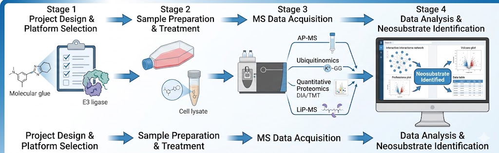
Four stages from compound receipt to comprehensive target identification report:

1

Project design and platform selection

Molecular glue properties (molecular weight, E3 ligase targeted, known SAR), programme stage, and available compound quantity are assessed. The optimal platform combination is selected — early-stage programmes with unknown targets start with AP-MS interactomics for broad neosubstrate discovery; confirmed hits advance to ubiquitinomics and quantitative proteomics for mechanistic validation; lead candidates receive LiP-MS for binding site mapping and full selectivity profiling. Known positive controls (e.g., pomalidomide for CRBN-recruiting glues) are selected based on the specific E3 ligase system. The project plan, concentration ranges, controls, and acceptance criteria are defined in approximately one week.

2

Sample preparation and treatment

For AP-MS: E3 ligase-expressing cell lines or lysates are treated with the molecular glue across a 6–8 point concentration series (0.01–100 µM) plus vehicle control and a reference glue positive control. For cellular platforms: cells are treated at specified concentrations and time points (4–24 h depending on expected degradation kinetics). DMSO vehicle control and a reference degrader positive control are included in every experiment. Triplicate biological replicates are prepared for all quantitative comparisons.

3

MS data acquisition

AP-MS: affinity purification (FLAG, GFP-Trap, or streptavidin pull-down) from cellular lysates followed by LC-MS/MS with TMT labelling for multiplexed quantitative accuracy across conditions. Ubiquitinomics: tryptic digestion, K-ε-GG antibody enrichment, single-shot or fractionated LC-MS/MS on Orbitrap Exploris 480. Quantitative proteomics: DIA acquisition with 4–6 m/z windows on Orbitrap platforms for deep proteome coverage. LiP-MS: limited proteolysis under native conditions followed by standard bottom-up proteomics for peptide-level quantification.

4

Data analysis and integrated reporting

AP-MS data are processed for protein identification and TMT quantification; glue-dependent E3 interactors are ranked by enrichment ratio. Ubiquitinomics data are searched for K-ε-GG modifications with site localisation scoring (class I sites, probability > 0.75). Proteomics data are processed for protein abundance quantification with statistical testing (Limma or t-test with FDR correction). LiP-MS data are analysed for peptide-level conformational differences. The integrated target identification report synthesises findings across all deployed platforms, presenting the converging lines of evidence that identify the neosubstrate and characterise the degradation mechanism, with supporting data tables and visualisations for each platform.

Molecular glue target identification workflow: project design and platform selection, sample preparation with molecular glue treatment across a concentration series, MS data acquisition across AP-MS/ubiquitinomics/quantitative proteomics/LiP-MS platforms, and integrated data analysis with neosubstrate identification and degradation mechanism characterisation.

Applications Across the Molecular Glue Discovery Pipeline

MS-based molecular glue target identification is most impactful where understanding the identity and behaviour of the neosubstrate determines programme direction and investment decisions.

De Novo Neosubstrate Discovery from Phenotypic Hits

The most common entry point: a phenotypic screen identifies a compound that produces a desired cellular phenotype (e.g., antiproliferative activity in a specific cancer cell line), but the molecular target is unknown. AP-MS interactomics across the glue-treated versus vehicle-treated E3 ligase purification identifies every protein whose interaction with the E3 is glue-dependent, providing the candidate neosubstrate list for downstream validation.

Output: ranked candidate neosubstrate list with quantitative enrichment ratios; prioritised targets for validation.

Multi-Compound SAR by Target Engagement Profiling

For molecular glue medicinal chemistry programmes, the ability to rank compounds by their ability to recruit the intended neosubstrate — and to identify which compounds recruit off-target proteins — is essential for synthetic prioritisation. AP-MS across a 10–50 compound panel identifies the recruitment profile of each analogue, directly guiding SAR without requiring a functional assay for each variant.

Output: compound-neosubstrate recruitment matrix; off-target recruitment fingerprint per compound; prioritised analogues for advancement.

Degradation Mechanism Confirmation

A candidate neosubstrate identified by AP-MS may be recruited to the E3 ligase but not necessarily ubiquitinated or degraded. Ubiquitinomics confirms glue-induced ubiquitination of the candidate target at specific lysine residues, providing the mechanistic evidence that recruitment leads to functional degradation. This is the critical go/no-go gate before committing resources to a target validation programme.

Output: ubiquitination site map on candidate neosubstrate; time-course and dose-response ubiquitination curves; mechanistic confirmation report.

Selectivity Profiling and Off-Target Identification

Before advancing a molecular glue lead candidate, unbiased proteome-wide selectivity data is essential. Quantitative proteomics across glue-treated versus control cells identifies all depleted proteins, distinguishing intended neosubstrate degradation from off-target liabilities — particularly important for CRBN-recruiting glues, which can recruit structurally related zinc finger proteins and cause unexpected degradation of transcription factors.

Output: proteome-wide degradation selectivity profile; ranked off-target list with quantitative fold-changes across the dose range.

Binding Site Mapping for Structure-Based Design

Once a neosubstrate is identified, understanding where the glue-E3 complex binds on the target surface enables structure-based optimisation of the glue and provides confidence in the proposed mechanism. LiP-MS identifies the protein regions that undergo conformational change upon glue-E3 complex binding, providing peptide-level binding site information without requiring a co-crystal structure.

Output: peptide-level conformational change map; inferred binding region on the neosubstrate; prioritised constructs for structural biology.

Differentiating Degradative from Non-Degradative Glue Engagement

A molecular glue can bind an E3 ligase and recruit a neosubstrate without triggering efficient ubiquitination — a non-degradative engagement that is often misinterpreted as target identification failure. The combination of AP-MS (detects recruitment) with ubiquitinomics (confirms ubiquitination) and quantitative proteomics (measures degradation) distinguishes true degraders from non-productive binders, enabling informed go/no-go decisions.

Output: integrated recruitment-ubiquitination-degradation classification for each candidate.

Technology Comparison: MS-Based MG Target ID vs Alternative Approaches

ApproachDetection PrincipleUnbiased DiscoveryNeosubstrate StoichiometryProteome CoverageWorks at Endogenous LevelsCellular Context
MS-Based MG Target ID (this service)AP-MS interactome + ubiquitinomics + quantitative proteomics + LiP-MSYes — no hypothesis requiredYes — from quantitative MS data5,000–8,000 proteinsYes (proteomics, LiP-MS)Yes — lysate and live cell
AP-MS (standalone)Affinity purification + LC-MS/MSYesLimited — from spectral countsE3 interactor-specificPartially — requires tagged baitLysate only
CRISPR Resistance ScreensgRNA depletion upon compound treatmentYes — geneticNoGene-levelYesYes
Ubiquitinomics (standalone)K-ε-GG enrichment + LC-MS/MSYesNo8,000–12,000 ubiquitination sitesYesYes
Western Blot (hypothesis-driven)Antibody signal intensityNo — requires antibody against suspected targetNo1–3 proteins per blotRequires specific antibodyYes
NanoBRET / AlphaLISAProximity luminescenceNo — requires known target pairNoSingle interactionRequires tagged proteinsLysate or purified
SPR (purified ternary complex)Refractive index changeNo — requires purified componentsNo — single interactionRequires purified E3 and candidate targetNoPurified only

Sample Requirements

ComponentFormat OptionsRecommended InputMinimum InputKey Notes
Molecular glue compoundPowder or DMSO stock5–10 mg (or 10 mM stock, 50 µL)1 mg (or 5 mM stock, 20 µL)Provide MW, known/predicted E3 ligase target; note light/oxygen sensitivity; DMSO stocks preferred for cellular assays
E3 ligase cell line — AP-MSStable or transient expression; frozen pellet2 × 107 cells per condition1 × 107 cells per conditionProvide E3 ligase identity (CRBN, VHL, MDM2, etc.); tag preferences (FLAG, GFP, Strep); confirm E3 expression by Western blot
Control cell lineParental or vehicle-treated; frozen pellet2 × 107 cells per condition1 × 107 cells per conditionIsogenic control line without E3 tag recommended for background subtraction; critical for distinguishing true interactors from bead non-specific binders
Cell lysate — LiP-MSFresh or flash-frozen lysate2 mg total protein per condition1 mg total protein per conditionAvoid freeze-thaw cycles; provide in native lysis buffer without denaturants; note protease inhibitor cocktail used
Reference glue (positive control)10 mM DMSO stock≥ 30 µL10 µLKnown degrader for the same E3 ligase recommended (e.g., pomalidomide for CRBN); provide known DC50 if available

All samples should be shipped on dry ice with completed sample submission forms. Biological triplicates are recommended for all quantitative comparisons; minimum two independent biological replicates for publication-grade target identification data. For AP-MS workflows, we recommend providing both E3-tagged and isogenic control cell lines to enable rigorous background subtraction.

Deliverables

  • AP-MS interactomics data: full protein identification list with TMT ratios; glue-dependent E3 interactor enrichment rank; dose-dependent recruitment curves for confirmed neosubstrates; interaction network comparison vs vehicle control
  • Ubiquitinomics data: K-ε-GG modified peptide list with site localisation scores (class I sites, probability > 0.75); site-level fold-change quantification across treatment conditions; comparison of ubiquitination efficiency across molecular glue concentrations and time points
  • Quantitative proteomics data: proteome-wide abundance table with protein-level fold-changes and FDR-adjusted p-values; ranked neosubstrate depletion list showing the primary target relative to all other quantified proteins; multi-glue comparison table where applicable
  • LiP-MS data: peptide-level LiP difference scores; conformational change map per candidate neosubstrate; inferred binding region coordinates
  • Raw MS data: full .raw or .mzML files for independent re-analysis or regulatory submission
  • QC report: protein coverage depth, CV distribution across replicates, replicate correlation heatmap, platform-specific quality metrics
  • Written interpretation report: integrated target identification narrative synthesising findings across all platforms with converging lines of evidence, candidate target prioritisation, and recommended validation experiments

Representative Results

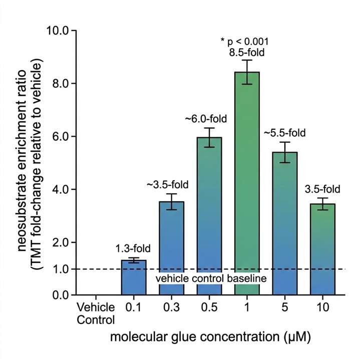
AP-MS interactome bar chart showing CRBN-DDB1 affinity purification from HEK293T lysates with a molecular glue across six concentrations from 0.1 to 10 µM plus vehicle control, demonstrating concentration-dependent neosubstrate enrichment with maximal 8.5-fold enrichment at 1 µM and hook effect at 10 µM.

AP-MS interactome: concentration-dependent neosubstrate recruitment

AP-MS analysis of CRBN-DDB1 affinity purification from HEK293T lysates treated with a CRBN-recruiting molecular glue (0.1–10 µM) versus vehicle control. TMT-based quantification reveals concentration-dependent enrichment of the candidate neosubstrate, with maximal enrichment at 1 µM (8.5-fold over vehicle, p < 0.001) and characteristic hook effect at 10 µM. The full interactome dataset identifies 15 proteins with significant glue-dependent enrichment (FDR < 0.05), from which the top 3 candidates are prioritised for ubiquitinomics validation.

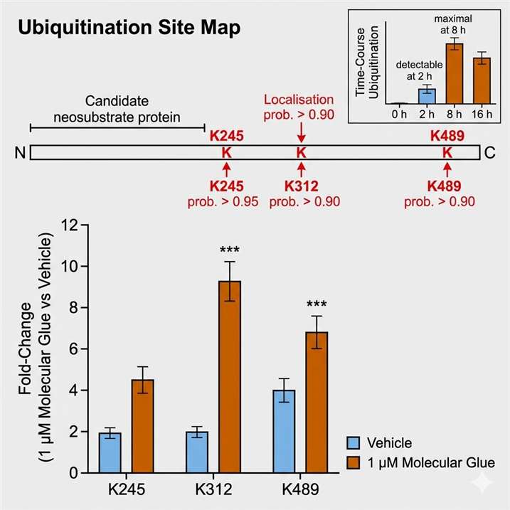
Ubiquitinomics bar chart showing K-epsilon-GG peptide enrichment from molecular glue-treated versus vehicle-treated cells, confirming site-specific ubiquitination of the candidate neosubstrate at three lysine residues (K245, K312, K489) with localisation probabilities above 0.90, time course from 2 to 24 hours.

Ubiquitinomics: site-specific neosubstrate ubiquitination confirmation

K-ε-GG peptide enrichment from glue-treated (1 µM, 8 h) versus vehicle-treated cells confirms site-specific ubiquitination of the candidate neosubstrate at three lysine residues (K245, K312, K489), all with localisation probability > 0.90. Ubiquitination is detectable at 2 h and maximal at 8 h of treatment. The global ubiquitinome analysis reveals that the glue does not induce widespread ubiquitination changes beyond the identified neosubstrate and a small number of closely related protein family members.

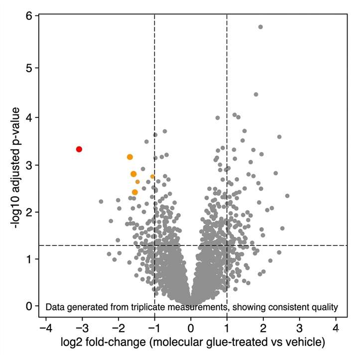
Volcano plot from quantitative DIA proteomics of molecular glue-treated versus vehicle-treated HCT116 cells showing log2 fold-change on x-axis versus -log10 p-value on y-axis, with the candidate neosubstrate highlighted in red at log2 fold-change minus 3.1 and two off-target depleted proteins in orange, across approximately 6,500 quantified proteins.

Quantitative proteomics: neosubstrate degradation selectivity fingerprint

DIA proteomics across glue-treated versus vehicle-treated HCT116 cells (1 µM, 24 h, biological triplicates) quantifies approximately 6,500 proteins. The candidate neosubstrate shows the largest abundance decrease at log2 fold-change −3.1 (p < 0.001). Two additional proteins show significant depletion (log2 FC < −1.5, FDR < 0.05) — representing off-target degradation liabilities — while all other quantified proteins remain unchanged. This unbiased selectivity fingerprint enables data-driven assessment of the glue's therapeutic window before in vivo studies.

Case Study: High-Throughput Chemoproteomics Maps the Hidden Interactome of CRBN Molecular Glues

Nat Commun. 2025;16:6831. https://doi.org/10.1038/s41467-025-62099-w (Open Access, CC BY 4.0).

Background

A fundamental challenge in molecular glue discovery is that the neosubstrate recruited to the E3 ligase cannot be predicted from the compound structure. CRBN-binding molecular glues — including lenalidomide, pomalidomide, and their derivatives — have been known to recruit neosubstrates from the C2H2 zinc finger family, but whether the full target landscape extends beyond this family had not been systematically investigated. Previous approaches relied on overexpression-based methods that could miss low-abundance targets and could not distinguish direct interactors from downstream effects.

Methods

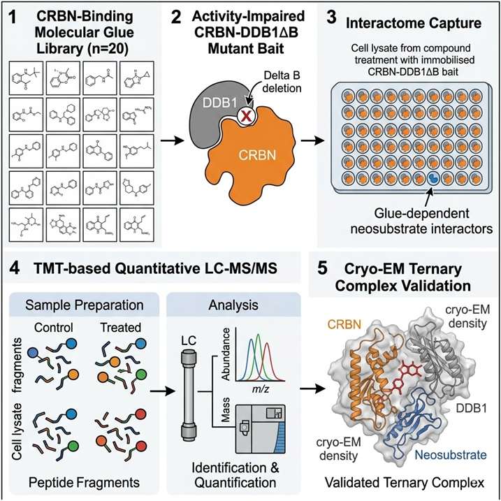
Baek et al. developed a high-throughput affinity proteomics workflow using an activity-impaired CRBN-DDB1ΔB mutant in cell lysates, enabling unbiased capture of molecular glue-induced neosubstrate recruitment without the confounding effects of degradation-dependent feedback. The workflow was applied to 20 CRBN-binding molecular glues spanning IMiDs (lenalidomide, pomalidomide), phenyl glutarimides (FPFT-2216, CC-885), and clinical-stage degraders. Each compound was assayed across a concentration gradient, and neo-interacting proteins were identified by TMT-based quantitative AP-MS. Hits were validated by cryo-EM, thermal stabilisation assays, and cellular degradation profiling.

Results

The study identified 298 protein targets across the 20-compound panel, of which 270 were previously unknown CRBN neosubstrates. Beyond the classic C2H2 zinc finger targets, the expanded landscape includes proteins from the cyclin, RRM, and PDZ domain families — substantially broadening the druggable scope of CRBN molecular glues. Cryo-EM structures of ternary complexes with PPIL4 (an RRM domain protein) and PDE6D revealed the structural basis of non-zinc finger recognition. A focused biochemical screen of approximately 6,000 IMiD analogues identified a selective lead compound targeting PPIL4, demonstrating that the expanded neosubstrate inventory can be translated into compound discovery campaigns targeting proteins previously considered undruggable by molecular glues.

Significance for Molecular Glue Target Identification

This study establishes several principles directly relevant to our target identification service. First, a well-designed AP-MS chemoproteomics workflow operating at scale can identify hundreds of neosubstrates across a compound panel, including targets from protein families not previously known to be CRBN-recruitable. Second, the use of an activity-impaired E3 ligase mutant eliminates degradation-dependent feedback that can obscure the direct interaction — a design principle we incorporate into our AP-MS platform for all E3 systems. Third, the integration of orthogonal validation (cryo-EM, thermal stabilisation, cellular degradation) confirms that AP-MS-identified interactors are genuine neosubstrates, not artefactual binders — the same multi-platform validation strategy we deploy across our AP-MS, ubiquitinomics, and quantitative proteomics pipeline.

Case study workflow diagram from the Baek et al. 2025 study (Nat Commun 2025, DOI: 10.1038/s41467-025-62099-w), showing the high-throughput AP-MS chemoproteomics workflow using activity-impaired CRBN-DDB1 Delta B mutant for unbiased identification of molecular glue-induced neosubstrates, with TMT-based quantification, cryo-EM validation, and cellular degradation profiling.

Figure from Baek et al. 2025 (Nat Commun, 2025, DOI: 10.1038/s41467-025-62099-w). High-throughput AP-MS chemoproteomics workflow using activity-impaired CRBN-DDB1ΔB mutant for unbiased molecular glue neosubstrate discovery. CC BY 4.0.

FAQ

Frequently Asked Questions

Q: What is the difference between molecular glue target identification and PROTAC ternary complex confirmation?

PROTAC ternary complex confirmation asks whether a known POI, known E3 ligase, and known PROTAC molecule form a pre-designed ternary complex — the target is known before the experiment. Molecular glue target identification asks which unknown neosubstrate protein a molecular glue recruits to the E3 ligase — the target must be discovered, not confirmed. The two services use related MS platforms but address fundamentally different questions: PROTAC profiling is confirmatory; MG target ID is discovery-oriented.

Q: How much compound is required for a full target identification campaign?

An AP-MS-only discovery campaign (the most common starting point) requires 1–3 mg of molecular glue. Adding ubiquitinomics and quantitative proteomics for mechanistic validation increases the requirement to 3–5 mg. A full multi-platform campaign including LiP-MS binding site mapping requires 5–10 mg. We optimise experimental design to extract maximum target ID data from available material and can advise on the minimum viable platform combination for early-stage programmes.

Q: Can you work with molecular glues targeting E3 ligases beyond CRBN?

Yes. Our AP-MS platform is E3-agnostic — we have established protocols for CRBN, VHL, MDM2, cIAP1, and RNF4 systems. For novel E3 ligases, we recommend confirming that the E3 complex can be expressed and purified in active form before initiating the target ID campaign. The MS detection is E3-agnostic — any E3 that can be expressed in the cellular system is detectable.

Q: How do you distinguish true neosubstrates from non-specifically binding proteins in AP-MS?

We apply a three-tier filter: (1) dose-dependent enrichment across multiple compound concentrations, eliminating proteins that appear only at a single high concentration; (2) parallel vehicle control and isogenic cell line comparison to identify proteins that co-purify with the E3 regardless of compound; (3) orthogonal validation by ubiquitinomics — a true neosubstrate should show glue-induced site-specific ubiquitination. Only proteins passing all three tiers advance as confirmed neosubstrates.

Q: What is the turnaround time for a molecular glue target ID project?

An AP-MS-only discovery campaign (neosubstrate identification without validation) takes 2–3 weeks. Adding ubiquitinomics and quantitative proteomics for mechanistic validation extends the timeline to 3–4 weeks. A full campaign including LiP-MS binding site mapping takes 4–6 weeks. Timelines are confirmed during project design and depend on cell culture requirements, number of compounds, and platform scope.

Q: Can you use endogenous E3 ligase without overexpression?

For AP-MS, some level of E3 tagging or overexpression is typically required for efficient affinity purification. However, our quantitative proteomics and LiP-MS platforms work entirely at endogenous protein levels without any genetic modification. The combination provides both the discovery power of AP-MS and the physiological relevance of endogenous detection. For E3 ligases where antibodies suitable for immunoprecipitation exist, endogenous AP-MS may be feasible.

Q: Can your service distinguish degradative from non-degradative neosubstrate engagement?

Yes — this is one of the key advantages of our multi-platform approach. AP-MS detects recruitment (binding), ubiquitinomics detects ubiquitination (the enzymatic consequence of recruitment), and quantitative proteomics detects degradation (the final functional outcome). A neosubstrate that is recruited but not ubiquitinated or degraded is classified as a non-degradative engagement — an important distinction that a single-platform approach would miss. This classification directly informs go/no-go decisions in the MG discovery pipeline.

Q: How is the hook effect handled in molecular glue target ID?

The hook effect — decreased neosubstrate recruitment at supra-optimal compound concentrations — is assayed across the full concentration series in both AP-MS and ubiquitinomics workflows. We report the optimal recruitment concentration alongside the hook concentration, ensuring that subsequent mechanistic studies are performed at the concentration producing maximal target engagement. The hook effect concentration is a standard reporting parameter in our AP-MS data package.

References

  1. Baek K., Metivier R.J., Roy Burman S.S., et al. Unveiling the hidden interactome of CRBN molecular glues with chemoproteomics. Nat Commun. 2025;16:6831.
  2. Merino-Cacho L., Barroso-Gomila O., Pozo-Rodríguez M., et al. Cullin-RING ligase BioE3 reveals molecular-glue-induced neosubstrates and rewiring of the endogenous Cereblon ubiquitome. Cell Commun Signal. 2025;23(1):101.
  3. Hu H., Ochoada J., Actis M., et al. Direct-to-biology enabled molecular glue discovery from large-scale combinatorial libraries. J Am Chem Soc. 2026;148(1):20–27.

Advance Your Molecular Glue Programme with MS-Based Target Identification

Submit your molecular glue compound and project background — our scientists will recommend the optimal platform combination and design a target identification strategy matched to your degrader's development stage, target class, and decision timeline.

For Research Use Only (RUO). Not intended for diagnostic, therapeutic, or clinical decision-making purposes. Creative Proteomics services are designed to support preclinical research, drug discovery, and targeted protein degradation studies only.

Online Inquiry

Please submit a detailed description of your project. We will provide you with a customized project plan to meet your research requests. You can also send emails directly to for inquiries.

* Email
Phone
* Service & Products of Interest
Services Required and Project Description