Photoaffinity Labeling (PAL-MS) Service

Decipher the exact mechanism of action for your phenotypic hits and molecular glues with our specialized Photoaffinity Labeling (PAL-MS) service. Identifying the true cellular target of a promising small molecule is often the most challenging hurdle in modern drug discovery.

We overcome the common challenges of low crosslinking yields and complex analytical fragmentation by pairing advanced Click chemistry enrichment protocols with custom bioinformatics deconvolution algorithms. From global target deconvolution to precise binding site mapping, we deliver unambiguous evidence straight from live cellular environments.

Photoaffinity Labeling (PAL-MS) Service platform highlighting live-cell target deconvolution and precise site mapping.
Overcoming Challenges Capabilities Enrichment Workflow Competitive Profiling Bioinformatics Probe Design & Samples Case Study FAQ

Overcoming the Challenges of Target Deconvolution

In modern drug discovery, researchers frequently utilize phenotypic screening to determine which compounds effectively alter a specific cellular behavior. This approach is incredibly powerful because it guarantees the compound is cell-permeable and functional within a real biological system. However, it leaves researchers with a major developmental bottleneck: elucidating the unknown target, referred to as target deconvolution.

Traditionally, scientists have relied on "pull-down" affinity chromatography. While historically useful, this method frequently fails because many small molecules exhibit transient or relatively weak interactions with their targets. When the solid support is subjected to stringent washing steps, the authentic target simply dissociates and is lost.

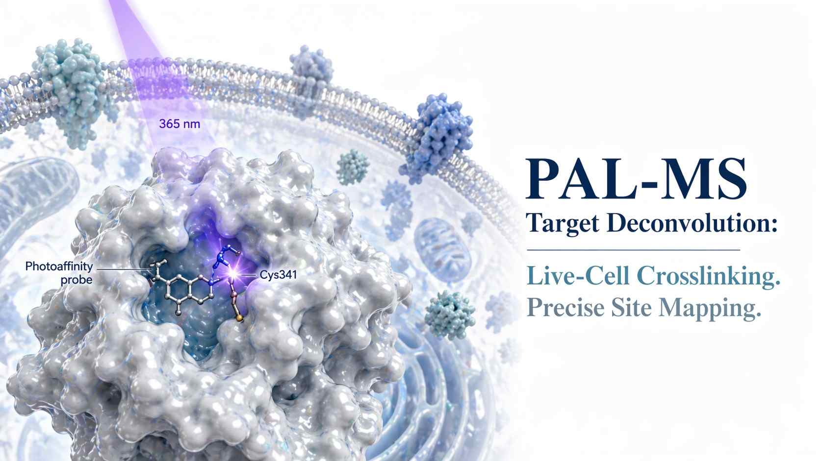
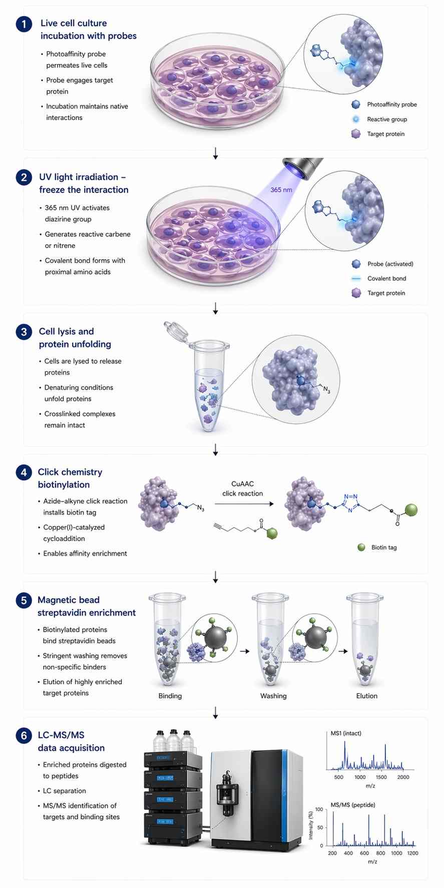
Photoaffinity Labeling (PAL) paired with Mass Spectrometry (MS) elegantly solves this dissociation problem. We synthesize a customized version of your active drug equipped with a photoreactive group. When we irradiate the live cells with ultraviolet (UV) light, this tag generates a highly reactive intermediate that forms a permanent, irreversible covalent bond with whatever amino acid residue it is physically touching at that exact microsecond. Because the interaction is now permanently locked, we can deploy extremely harsh denaturing washing conditions to quantitatively strip away all noisy background proteins, leaving only the true targets ready for identification.

Our PAL-MS Service Capabilities

We offer a highly specialized, dual-layered approach to cellular target discovery, providing both the definitive "who" (identifying the specific protein) and the exact "where" (mapping the specific amino acid residue) of your drug's complex interaction.

Global Target Identification

For phenotypic hits with unknown mechanisms, we incubate your photo-probe in live cell models, followed by UV crosslinking and robust enrichment. We scan the entire cellular proteome to provide a comprehensive list of all proteins that specifically interacted with your compound, uncovering novel targets and off-target liabilities.

High-Resolution Site Mapping

Knowing the identity of the target is only half the battle. By enzymatically digesting captured targets and utilizing customized LC-MS/MS, we can pinpoint the precise amino acid residue where the UV crosslinking event occurred. This structural "smoking gun" allows your team to computationally model the binding orientation.

Live-Cell Ternary Complex Validation

Molecular glues and PROTACs require the formation of a highly dynamic "ternary complex". Our live-cell PAL-MS workflows are heavily utilized to prove that these ternary complexes are actively forming inside live cells, validating your compound's true cellular target engagement and degradation potential.

Specialized Click Chemistry & Enrichment Workflow

The primary technical bottleneck hindering any PAL-MS project is the incredibly low chemical yield of the photoreaction itself. Typical photoreactive groups only successfully crosslink to their target protein approximately 1% to 5% of the time. To concentrate this rare fraction, we utilize a highly specialized Click chemistry enrichment workflow.

1

Live Cell Incubation

We introduce your synthesized photo-probe into a live cell culture system, allowing it to freely navigate the complex environment and locate its target naturally.

2

In-Situ UV Irradiation

We expose the cells to a precisely controlled wavelength of UV light to trigger the photoreactive group without inducing catastrophic cellular UV damage.

3

Harsh Cell Lysis & Denaturation

We lyse the cells using strong, denaturing detergents (e.g., SDS) to permanently disrupt all non-specific, sticky protein-protein interactions.

4

Click Chemistry Biotinylation

Using Copper-Catalyzed Azide-Alkyne Cycloaddition (CuAAC), we attach a Biotin-containing linker specifically to the bio-orthogonal handle on your photo-probe.

5

Stringent Streptavidin Enrichment

High-capacity streptavidin magnetic beads pull out only the covalently attached proteins, followed by aggressive washing to eliminate 99% of background proteins.

6

Enzymatic Digestion & LC-MS/MS

The highly purified, crosslinked proteins are enzymatically digested into peptides on-bead and analyzed via our ultra-sensitive mass spectrometers.

A longitudinal scientific flowchart in Nature journal style showing: Live cell incubation, UV irradiation, Cell lysis, Click chemistry biotinylation, Streptavidin enrichment, and LC-MS/MS.

Eliminating False Positives via Competitive Profiling

When the UV light flashes, the activated probe will blindly and rapidly attach to whatever molecule happens to be in its immediate vicinity. To guarantee that the targets we report are specifically driven by your compound's core scaffold, we employ a rigorous "competitive profiling" strategy using two strictly controlled groups:

  • The Probe Group: Cells are treated solely with your active photo-probe.
  • The Competitor Group (Control): Cells are pre-treated with a massive excess (usually 10x to 50x) of your original, unmodified parent drug, followed by the addition of the photo-probe.

Because the parent drug is present in overwhelming concentrations, it outcompetes the photo-probe and securely occupies all the true, specific binding pockets. The photo-probe is forced out of the active site and can only bind to random, non-specific bystander proteins. By analyzing both groups and mathematically comparing relative protein abundances, we generate a high-confidence Volcano Plot to definitively flag and eliminate non-specific background.

A competitive volcano plot highlighting true specific targets in red vs non-specific background alongside an annotated high-resolution MS2 fragmentation spectrum of a crosslinked peptide.
Differentiation of true targets via competitive profiling and confirmation via MS2 site mapping.

Bioinformatics: Decoding Complex Cross-Linked Peptides

Standard proteomics software pipelines are exclusively designed to read standard, unmodified peptides. When a peptide has a small-molecule drug permanently crosslinked to it, its mass is completely altered. Furthermore, during tandem mass spectrometry (MS/MS) high-energy collision, the attached drug often shatters unpredictably, creating a chaotic spectrum of fragments that standard software discards as noise.

Our bioinformatics team employs specialized algorithms designed exclusively for cross-linked peptides:

  • Precise Delta Mass Searching: Our software searches specifically for the unique mass addition (+ΔMass) generated by your exact probe structure, accounting for the loss of N2 or other leaving groups.
  • Diagnostic Ion Filtering: We explicitly program algorithms to identify specific "reporter fragments" that break off your probe's linker region during collision. Detecting these fragments acts as an absolute secondary confirmation.
  • Strict FDR Control: We utilize rigorous, target-decoy based False Discovery Rate (FDR) statistical models customized for crosslinked searches to ensure our peptide identifications hold up to the highest standards.

We can easily transition your project into our broader chemoproteomics services for further quantitative profiling once hits are confirmed.

Probe Design Guidelines & Sample Requirements

The ultimate success of a PAL-MS project begins with sound chemistry. If you are currently designing your probes, utilize the guide below to select the most appropriate photoreactive group for your target's microenvironment.

Comparison DimensionDiazirine (Aliphatic/Trifluoromethyl)BenzophenoneAryl Azide
Steric Bulk (Size)Smallest: Minimal disruption to binding affinity.Largest: May severely disrupt binding if pocket is tight.Medium: Moderate impact.
Excitation Wavelength~350-360 nm (Gentle on live cells)~350-360 nm (Gentle on live cells)~250-300 nm (Can cause severe UV damage to cells)
Reactivity SpeedFastest: Nanoseconds. Excellent for transient hits.Slower: Microseconds. Allows for reversible binding before trapping.Moderate: Microseconds to milliseconds.
Crosslinking RadiusShort reach. Reacts immediately within the binding pocket.Long reach. Highly flexible; reacts with C-H bonds outside pocket.Moderate reach. Prone to rearrangement.

The Strategy: For tight binding pockets, Diazirine is strongly preferred due to its tiny size and fast reaction time. If the binding site is highly flexible or solvent-exposed, Benzophenone may offer higher overall crosslinking yields.

Sample Requirements

All proprietary probe structures, ΔMass information, and target details are handled under strict Confidential Disclosure Agreements (CDA).

Sample TypeRecommended InputCondition / PrepNotes
Live Cells> 5 × 10⁷ cells per conditionIrradiated & Snap-frozenYou must provide the exact UV irradiation wavelength and exposure time used.
Photo-Probes> 2 mgHigh LC-MS puritySolubilized in dry DMSO. Specify the photoreactive group and click handle.
Parent Compound> 5 mgHigh purityRequired in large excess for the critical competitive control experiment.

Validated PAL-MS Target Identification Case Studies

Clickable Photoaffinity Labeling and Target Deconvolution

Reference link: https://pmc.ncbi.nlm.nih.gov/articles/PMC8209655/

Background

Identifying the unknown live-cell targets of a bioactive small molecule is notoriously difficult due to the high non-specific background typical of photoaffinity probes. Researchers needed a robust analytical way to filter out the massive proteomic noise and identify only the biologically relevant interactions driving the phenotype.

Methods

Researchers synthesized a photoreactive probe equipped with an alkyne click handle. This probe was incubated in live cells alongside a separate, rigorously matched control group that received both the probe and a massive excess of the parent compound (competitive control). Following precise UV irradiation and cell lysis, the proteins were tagged using Click chemistry, heavily enriched via streptavidin beads, and analyzed using quantitative high-resolution LC-MS/MS.

Results

The competitive chemical proteomics workflow successfully differentiated true specific targets from background noise. Proteins that were specifically competed away by the excess parent compound were identified with high statistical confidence, while bystander proteins were mathematically eliminated by the deconvolution algorithms.

Conclusion

This protocol definitively proves that combining carefully designed photo-probes with strict competitive controls and advanced MS proteomics provides a robust, reliable, and highly accurate pathway for target deconvolution in native cellular environments.

Figure 1 from PMC8209655 showing the protocol for clickable photoaffinity labeling and target deconvolution.

Figure 1: Schematic representation of the clickable photoaffinity labeling strategy.

FAQ

Frequently Asked Questions

Q: How do you distinguish true target binding from non-specific UV background?

We rely entirely on rigorous competitive profiling. By running a control sample where the cells are pre-treated with a large excess of the unmodified parent drug, we intentionally block the true binding sites. Any protein that is labeled by the UV probe in the normal sample, but not labeled in the competitive control sample, is confirmed as a true target.

Q: Our photo-probe yielded <5% crosslinking efficiency. Can your platform still detect it?

Yes. A 1% to 5% crosslinking yield is very standard for photoreactive groups like Diazirine. Our platform is specifically designed to overcome this limitation. By utilizing high-affinity Click chemistry and highly stringent streptavidin enrichment protocols, we concentrate that small fraction while washing away the 95% of unlinked background proteins, allowing our highly sensitive mass spectrometers to easily detect the isolated signal.

Q: What is the difference between your search algorithms and standard proteomics software?

Standard proteomics software only looks for known, natural peptide masses. When your probe crosslinks to a peptide, it adds a unique, unnatural weight (a ΔMass). Furthermore, the probe can shatter during MS/MS analysis. Our custom bioinformatics algorithms search specifically for these unnatural mass shifts and strictly filter the results using the unique "reporter fragments" generated by your specific chemical probe.

Q: Do we need to disclose the full chemical structure of our photo-probe?

No, your intellectual property remains fully protected. We do not need the full chemical structure of your proprietary molecule. To program our mass spectrometers and search algorithms, we only require the exact monoisotopic "ΔMass" (the weight added to the protein upon binding) and the identity of the Click handle (e.g., Alkyne or Azide).

References

  1. Protocol for clickable photoaffinity labeling and quantitative chemical proteomics.
  2. Development of Second-Generation Acyl Silane Photoaffinity Probes for Cellular Chemoproteomic Profiling.
  3. Photoaffinity Labelling Strategies for Mapping the Small Molecule-Protein Interactome.

Disclaimer: All services and data provided by our platform are for Research Use Only (RUO). Not for use in diagnostic procedures. The information provided is not intended to substitute for professional medical advice or clinical diagnosis.

Plan your PAL-MS campaign with the MassTarget™ team

Share your target background and photo-probe parameters, and our chemical biology experts will design a customized target deconvolution strategy for you.

Online Inquiry

Please submit a detailed description of your project. We will provide you with a customized project plan to meet your research requests. You can also send emails directly to for inquiries.

* Email
Phone
* Service & Products of Interest
Services Required and Project Description