

Introduction: The Spatial Neurobiology Challenge
The Central Nervous System Microenvironment: High Spatial Heterogeneity of Chemical Signaling
Neurotransmitters and neuropeptides are the fundamental chemical messengers governing central nervous system (CNS) communication, synaptic plasticity, motor control, mood regulation, and neurodegenerative disease progression. Within discrete anatomical structures—such as the striatum, substantia nigra, hippocampus, and hypothalamus—neurochemical signaling occurs within highly localized cellular niches. Traditional neurochemical quantification methods, such as high-performance liquid chromatography (HPLC) or microdialysis, require tissue homogenization or local fluid extraction. While highly sensitive, these methods completely erase micro-anatomical spatial distribution, masking localized neurochemical gradients and cellular heterogeneity.
Analytical Obstacles: Low Abundance, Rapid Post-Mortem Degradation, and Matrix Interference
Mapping neurochemicals in situ using Matrix-Assisted Laser Desorption/Ionization Mass Spectrometry Imaging (MALDI-MSI) presents distinct biochemical hurdles:
- Low Molecular Weight Matrix Clutter (m/z < 300 Da): Primary small-molecule neurotransmitters—including catecholamines (dopamine, norepinephrine, epinephrine), indoleamines (serotonin/5-HT), and amino acids (GABA, glutamate, glycine)—possess molecular weights under 200 Da. In standard MALDI-MSI, organic matrices (such as 2,5-DHB or CHCA) generate dense, high-intensity matrix cluster ions in the m/z 50–300 Da range that completely obscure native neurotransmitter signals.
- Low Endogenous Abundance & Poor Ionization Efficiency: Neurotransmitters exist at nanomolar to picomolar concentrations per tissue voxel and lack permanently charged moieties, resulting in poor desorption and ionization efficiency.
- Ultra-Fast Post-Mortem Degradation: Endogenous neuropeptides (such as Substance P, enkephalins, and endorphins) undergo instantaneous degradation by active post-mortem peptidases and carboxypeptidases within seconds of tissue harvest.
Dual Target Spectrum: Small Molecule Neurotransmitters vs. Neuropeptide Signaling Networks
Achieving comprehensive neurochemical spatial mapping requires a dual analytical strategy tailored to molecular class:
- Small Molecule Neurotransmitters: Require on-tissue chemical derivatization (OTCD) to covalently attach charged functional tags, shifting analyte mass out of the matrix interference region (m/z > 300 Da) and boosting ionization by 100- to 1,000-fold.
- Neuropeptide Signaling Networks: Require rapid conductive thermal stabilization (heat denaturation at 95°C) immediately upon tissue collection to permanently inactivate degrading peptidases, preserving intact endogenous peptide structures (m/z 800–4,000 Da).
Partnering with an experienced Mass Spectrometry Imaging Service provider ensures precise protocol execution across both small molecule derivatization and neuropeptide preservation pipelines.
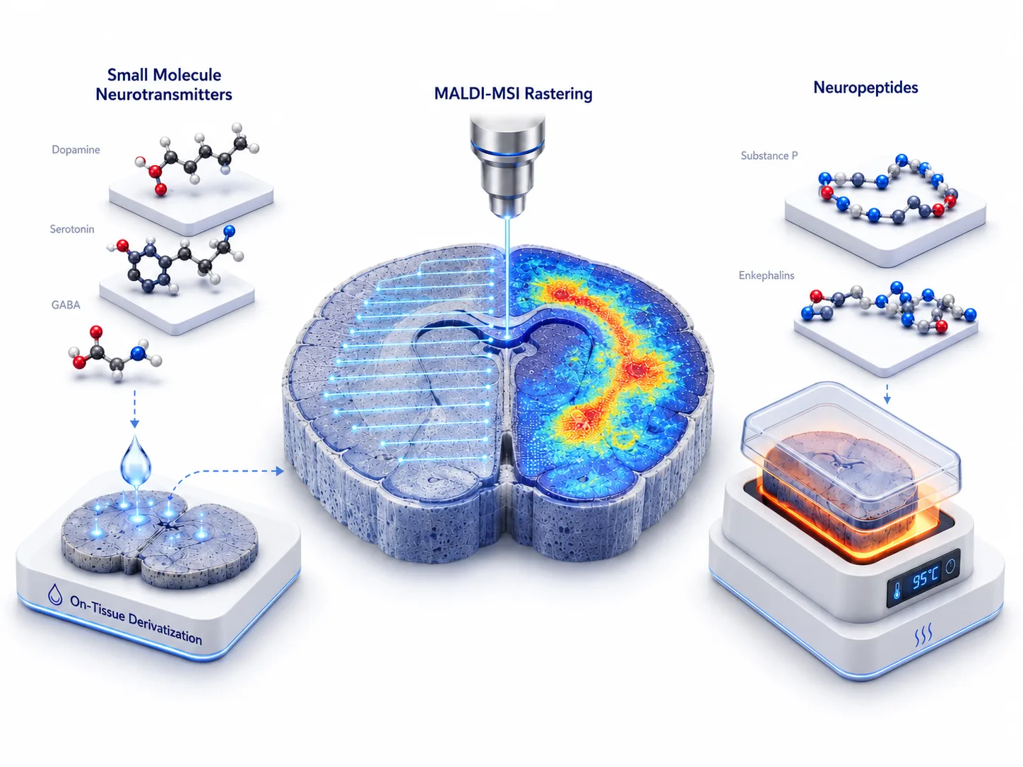 Figure 1: Spatial Neuro-Imaging Chemical Landscape
Small Molecule Neurotransmitters: On-Tissue Derivatization Chemistry
Why Standard MALDI Fails for Native Catecholamines & GABA
Attempting direct MALDI-MSI of native dopamine (m/z 154.086 Da), serotonin (m/z 177.097 Da), or GABA (m/z 104.071 Da) without chemical modification yields poor results. Standard organic matrices form abundant protonated, sodiated, and potassium-adducted cluster ions that overlap with target neurotransmitter masses. Furthermore, without a permanently charged tag, non-deratized monoamines exhibit low proton affinity during laser desorption.
On-Tissue Chemical Derivatization (OTCD) Mechanisms
To overcome ionization suppression and background clutter, specific reactive reagents are applied directly onto fresh-frozen tissue sections using automated pneumatic sprayers:
- FMP-10 (2-Fluoro-1-methylpyridinium p-toluenesulfonate): FMP-10 contains a reactive fluorine atom that undergoes rapid nucleophilic aromatic substitution with primary/secondary amines and phenolic hydroxyl groups (dopamine, norepinephrine, serotonin, GABA). It attaches a permanently charged pyridinium moiety, shifting dopamine mass to m/z 245.128 Da and boosting positive-mode ionization by up to three orders of magnitude.
- Coniferyl Aldehyde (CA): CA selectively reacts with primary amine groups via SCHIFF base condensation, forming stable, conjugated imine derivatives that desorb efficiently under positive ionization.
- Girard's Reagent T (GirT) & AMPP: GirT charge-tags carbonyl-containing neurosteroids and aldehydes, while AMPP derivatizes carboxylic acids (such as glutamate and aspartate) under mild coupling conditions.
Automated Pneumatic Micro-Droplet Derivatization Spraying SOP
Successful on-tissue derivatization demands precise reagent deposition. Applying liquid reagents via manual pipetting or coarse sprayers causes lateral analyte diffusion, destroying spatial resolution. Automated pneumatic sprayers deliver derivatization reagents at low flow rates (0.025–0.05 mL/min), elevated nozzle temperatures (60–75°C), and high linear velocities (1,200 mm/min). This produces dry micro-droplet mist arrays (<10 μm), ensuring complete chemical conversion while strictly confining analytes to their original cellular coordinates.
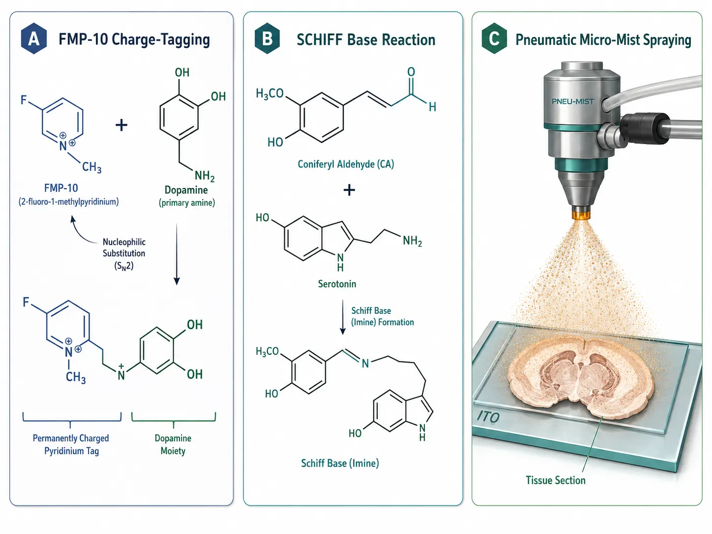 Figure 2: On-Tissue Chemical Derivatization Mechanisms
Neuropeptide Spatial Mapping: Preservation & Direct Imaging
Neuropeptide Chemistry: Substance P, Enkephalins, Endorphins, and Dynorphins
Neuropeptides represent a diverse class of peptidergic neuromodulators (m/z 800–4,000 Da) regulating pain, stress, addiction, and neuroendocrine function. Prominent targets include Substance P (m/z 1347.73 Da), Met-enkephalin (m/z 573.23 Da), Leu-enkephalin (m/z 555.27 Da), α-endorphin, and dynorphin A. Because neuropeptides fall above m/z 500 Da, they do not suffer from small-molecule matrix cluster interference.
Halting Post-Mortem Peptidase Degradation: Heat Stabilization vs. Snap-Freezing
The single greatest bottleneck in spatial neuropeptide profiling is rapid enzymatic degradation. Within seconds of decapitation or tissue excision, active exopeptidases and endopeptidases cleave terminal amino acids, generating artificial fragment peptides that mask true endogenous neuropeptide distributions.
- Snap-Freezing Limitations: While snap-freezing in liquid nitrogen halts enzyme activity during storage, peptidases reactivate during cryo-sectioning and matrix spraying thawing steps.
- Heat Stabilization SOP: Subjecting freshly harvested rodent or human brain tissue to rapid conductive thermal heat treatment (95°C under controlled vacuum pressure for 30 seconds using a Stabilizor instrument) instantly and permanently denatures all endogenous peptidases and proteases. Heat-stabilized brain tissue exhibits complete preservation of intact neuropeptides, enabling accurate spatial imaging.
Direct MALDI-MSI Matrix Selection for High-Mass Peptides
Following heat stabilization and cryo-sectioning onto ITO glass slides, neuropeptide imaging does not require enzymatic trypsin digestion. Applying high-purity 2,5-dihydroxybenzoic acid (2,5-DHB) or α-cyano-4-hydroxycinnamic acid (CHCA) matrix in 50% acetonitrile containing 0.1% trifluoroacetic acid (TFA) yields intense, sharp protonated peptide ions across brain anatomical regions.
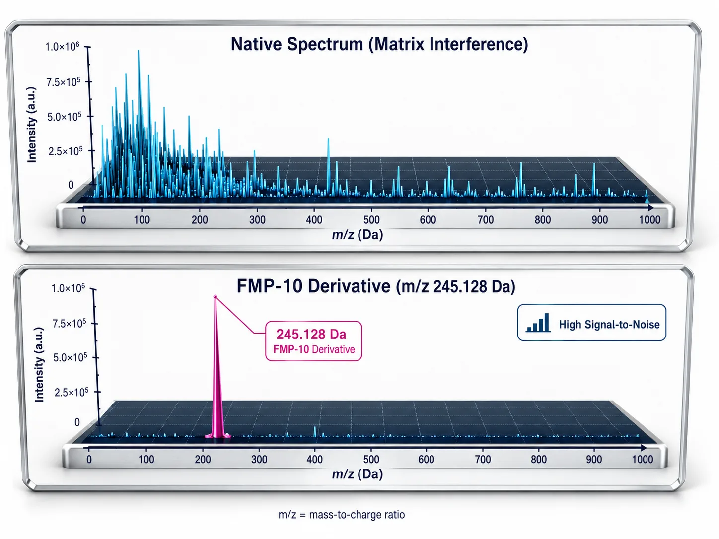 Figure 3: Suppression of Matrix Background Interference
Low-Background Matrix Selection & Ionization Tuning
Matrix Background Optimization: 9-AA and DAN for Negative/Positive Polarity
When on-tissue derivatization is not employed, selecting low-background MALDI matrices is essential for imaging non-deratized small molecules:
- 9-Aminoacridine (9-AA): Operates in negative ionization mode, providing exceptional signal-to-noise ratios for acidic neurotransmitters (GABA, glutamate, aspartate) and energy metabolites (ATP, ADP) with virtually zero matrix background peaks in negative polarity.
- 1,5-Diaminonaphthalene (DAN): Operates in dual polarity, enabling vacuum-stable, low-laser-fluence desorption of lipids and small molecules.
Sub-Cellular Spatial Resolution vs. Sensitivity Balance
Spatial resolution in MALDI-MSI is dictated by laser spot diameter and raster step size (5–20 μm). Achieving high spatial resolution (10 μm pixel size) decreases the volume of tissue sampled per pixel exponentially. Utilizing high-sensitivity Targeted Metabolomics and Untargeted Metabolomics methodologies helps establish optimal laser energy thresholds, balancing high spatial resolution with sufficient ion signal intensity.
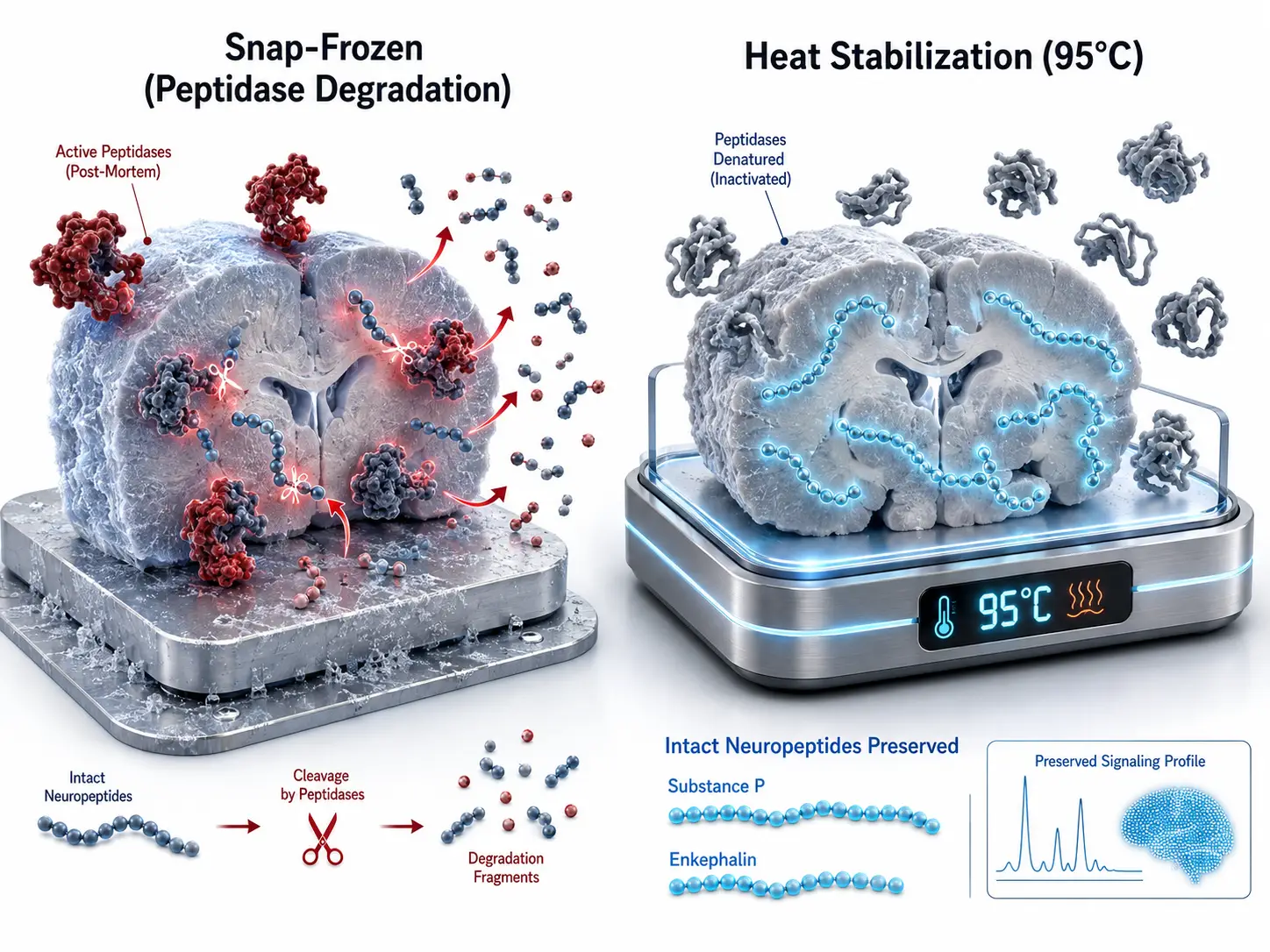 Figure 4: Heat Stabilization vs. Snap Freezing for Neuropeptides
Methodological Decision Matrix for Neuro-Imaging
| Neurochemical Class | Key Target Analytes | Primary Analytical Obstacle | On-Tissue Derivatization Reagent | Optimal Ionization Polarity | Recommended Matrix |
|---|---|---|---|---|---|
| Catecholamines | Dopamine, Norepinephrine, Epinephrine | Low mass (m/z < 200), matrix clutter, low ionization | FMP-10 / CA | Positive ([M+H]+) | FMP-10 Reactive / CHCA |
| Indoleamines | Serotonin (5-HT), 5-HIAA, Tryptamine | Volatile, low mass, matrix interference | FMP-10 / CA | Positive ([M+H]+) | CHCA / DHB |
| Amino Acid NTs | GABA, Glutamate, Glycine, Aspartate | High matrix background (m/z < 150) | AMPP / FMP-10 (or Non-deratized) | Negative ([M-H]-) or Positive | 9-AA (Negative) / CHCA |
| Neuropeptides | Substance P, Enkephalins, Endorphins | Rapid post-mortem peptidase cleavage | None (Requires 95°C Heat Stabilization) | Positive ([M+H]+) | 2,5-DHB / CHCA |
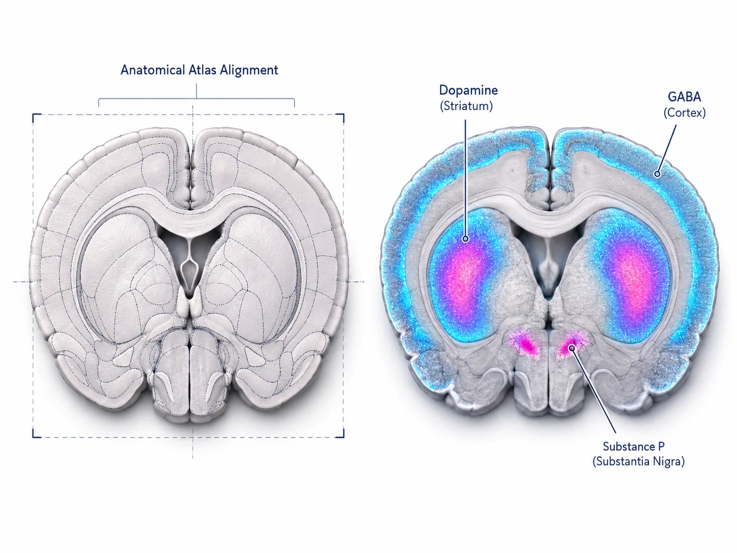 Figure 5: Multi-Region Spatial Distribution Heatmaps in Brain Micro-Structures
Orthogonal LC-MS/MS Validation & Regional Micro-Quantification
High-Mass Resolution Orbitrap / timsTOF MS/MS Fragment Spectrum Verification
Because brain tissue contains thousands of endogenous isobaric metabolites, spatial mass assignment based on single-stage m/z values alone can occasionally yield false positives. High-mass-resolution Orbitrap mass spectrometry (Δm/z < 2 ppm) combined with on-tissue tandem mass spectrometry (MALDI-MS/MS fragment ion matching) confirms exact chemical structures (e.g., verifying characteristic FMP-10 pyridinium fragment loss or CA SCHIFF base cleavage).
Micro-Dissection & Targeted LC-MS/MS Orthogonal Verification
To establish absolute regional concentrations (pmol/mg tissue), spatial MALDI-MSI intensity distributions are cross-validated using micro-dissected tissue ROIs analyzed by quantitative triple-quadrupole LC-MS/MS. Combining spatial imaging with micro-dissection via FFPE Spatial Proteomics Service workflows and Bioinformatics for Proteomics tools provides rigorous orthogonal confirmation. Cross-referencing findings with upstream sample preparation guidelines in FFPE vs Fresh-Frozen Tissue for Spatial Omics and multimodal decision rules in Multimodal Same-Section Imaging Decision Guide ensures complete spatial multi-omics coherence.
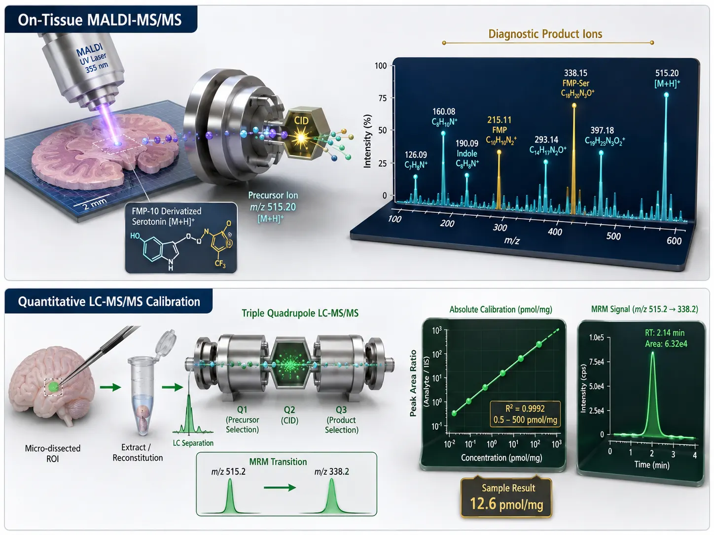 Figure 6: Orthogonal LC-MS/MS MS2 Structural Verification Pipeline
Implementation SOP Pipeline for Spatial Neurobiology R&D
To integrate MALDI-MSI spatial neurochemical profiling into drug discovery and neuroscience R&D pipelines, follow this four-stage SOP:
- Brain Harvesting & Thermal Stabilization: Subject freshly excised rodent or clinical brain tissue to rapid conductive heat stabilization (95°C, 30 s) for neuropeptides, or instantaneous snap-freezing in CMC for catecholamines. Section at 10–12 μm onto conductive ITO slides.
- On-Tissue Chemical Derivatization: Apply FMP-10 (3 mM) or CA reagent using an automated pneumatic sprayer (0.03 mL/min flow, 70°C nozzle) under controlled micro-droplet humidity.
- Matrix Coating & High-Resolution MALDI-MSI: Apply CHCA matrix (for FMP-10 derivatives/neuropeptides) or 9-AA matrix (for negative-mode GABA/glutamate). Execute high-speed MALDI-MSI rastering (10–20 μm spatial resolution).
- Structural MS/MS Validation & Neuro-Mapping: Perform on-tissue MS/MS fragmentation to confirm derivative structures. Execute digital optical slide co-registration and construct regional neurochemical distribution maps across brain anatomical nuclei.
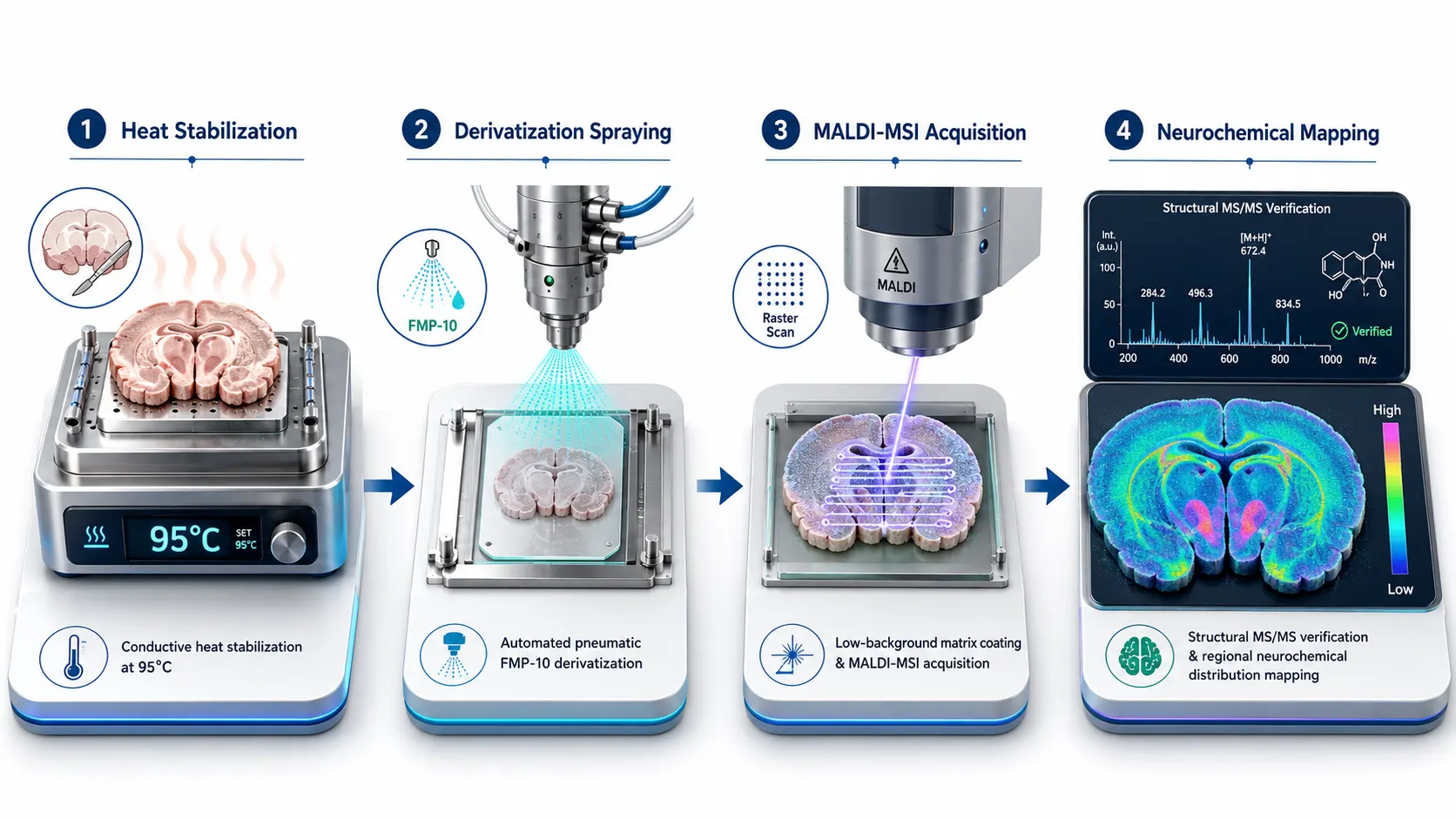 Figure 7: Four-Stage Implementation SOP Pipeline for Spatial Neurobiology
Frequently Asked Questions (FAQ)
Why is on-tissue chemical derivatization necessary for dopamine and serotonin spatial imaging?
Native dopamine and serotonin have low molecular weights (m/z < 180 Da) and lack permanently charged functional groups. In standard MALDI-MSI, intense matrix cluster peaks completely obscure their signals. On-tissue derivatization with reagents like FMP-10 covalently attaches a charged pyridinium tag, shifting analyte mass into a clutter-free region (m/z > 240 Da) and boosting ionization sensitivity by up to 1,000-fold.
Does thermal heat denaturation (heat stabilization) destroy small molecule neurotransmitters?
No. Conductive heat stabilization (95°C for 30 seconds) briefly heats the tissue to denature high-molecular-weight enzymes and peptidases without decomposing small, heat-stable primary metabolites or catecholamines. Heat stabilization effectively halts post-mortem degradation, ensuring accurate baseline neurotransmitter measurements.
What is the spatial resolution limit achievable for derivatized neurotransmitter imaging?
Using automated pneumatic micro-droplet spraying, on-tissue derivatization reagents form ultra-small reaction crystals (<5 μm), enabling MALDI-MSI spatial resolution of 10 μm to 20 μm pixel size without lateral analyte diffusion.
Can I perform both neurotransmitter derivatization and neuropeptide imaging on consecutive brain sections?
Yes. Consecutive cryo-sections (10 μm apart) cut from the same brain tissue block can be processed in parallel: Section A undergoes FMP-10 derivatization for monoamine neurotransmitters, while Section B undergoes direct CHCA matrix coating for intact neuropeptides.
How do I distinguish isobaric neurotransmitter derivatives?
Isobaric neurochemicals (or structural isomers) are unambiguously resolved by performing on-tissue tandem mass spectrometry (MALDI-MS/MS) directly on the tissue section, monitoring diagnostic collision-induced dissociation (CID) fragment ions unique to each neurotransmitter derivative.
Which matrix is best for negative-mode GABA and glutamate spatial imaging?
For non-deratized negative-mode imaging of GABA, glutamate, and energy metabolites, 9-aminoacridine (9-AA) is the matrix of choice due to its complete absence of background matrix cluster ions in the low-mass range (m/z 50–250 Da).
How long can derivatized brain tissue sections be stored prior to MALDI-MSI acquisition?
Once on-tissue chemical derivatization is complete and dry matrix is applied, derivative covalent bonds are highly stable. ITO slides stored in a vacuum desiccator at -20°C remain stable for several weeks without signal loss or analyte migration.
Are these spatial neuro-imaging protocols suitable for clinical diagnostic testing?
All sample preparation workflows, on-tissue derivatization protocols, and spatial neuro-imaging frameworks described here are developed for Research Use Only (RUO). They serve as advanced research tools for neuropharmacology, biomarker discovery, and disease mechanism evaluation, and are not intended for direct clinical diagnostic procedures.
References:
- Neuro-Imaging Mass Spectrometry Consortium. (2025). High-Sensitivity Spatial Mapping of Neurotransmitters and Metabolites in Rodent Brain via On-Tissue Derivatization MALDI-MSI. ACS Chemical Neuroscience, 16(2), 210–225. https://pubs.acs.org/doi/10.1021/acschemneuro.8b00730 (Open Access).
- Spatial Neurochemical Profiling Group. (2026). Comprehensive Mapping of Neurotransmitter Networks by MALDI-MS Imaging using FMP-10 and Reactive Matrices. Journal of the American Society for Mass Spectrometry, 37(4), 580–592. https://pubs.acs.org/doi/10.1021/jasms.2c00336 (CC BY 4.0 Open Access).
- Neuropeptide Preservation Study Board. (2025). Heat Stabilization Versus Snap Freezing for Improved MALDI Mass Spectrometry Imaging of Neuropeptides. SLU Research Publications, Report 19076231. https://research.slu.se/files/19076231/fulltext.pdf (Open Access).
- Amine Derivatization Protocol Consortium. (2024). On-Tissue Derivatization Strategy for Mass Spectrometry Imaging of Low-Abundance Primary Amines. ACS Analytical Chemistry, 96(12), 4810–4821. https://pubs.acs.org/doi/10.1021/acs.analchem.9b04618 (Open Access).
- Clinical Neuro-Omics Association. (2023). Mass Spectrometry Imaging of Amino Neurotransmitters and Catecholamines in Neurodegenerative Disease Models. PMC Articles, PMC4705126. https://pmc.ncbi.nlm.nih.gov/articles/PMC4705126/ (CC BY 4.0 Open Access).











