

Introduction: The Tissue Preservation Dilemma in Spatial Omics and MALDI-MSI
The Spatial Revolution: Bridging Histopathology and Mass Spectrometry Molecular Profiling
Spatial omics has emerged as a transformative paradigm across modern biomedical research, clinical oncology, and drug discovery. Traditional bulk omics technologies require tissue homogenization, which discards crucial spatial architecture, cellular heterogeneity, and microenvironmental context. Conversely, classical immunohistochemistry (IHC) and fluorescence imaging preserve histological structures but are fundamentally limited in multiplexing capacity and analyte range. Matrix-Assisted Laser Desorption/Ionization Mass Spectrometry Imaging (MALDI-MSI) effectively bridges this analytical gap. By rastering a focused laser beam across thin tissue sections, MALDI-MSI directly generates multiplexed, label-free spatial distributions for thousands of endogenous molecules—including small metabolites, lipids, tryptic peptides, intact proteins, and N-linked glycans—in direct alignment with histological structures.
The Sample Handling Conundrum: Fresh-Frozen (FF) versus Formalin-Fixed Paraffin-Embedded (FFPE)
Despite dramatic hardware advancements in mass spectrometry instrumentation, the ultimate quality, spatial fidelity, and quantitative reliability of MALDI-MSI data depend heavily on upstream tissue sample preservation. Researchers face a fundamental trade-off between two primary tissue preservation modalities:
- Fresh-Frozen (FF) Preservation: Tissues are rapidly harvested, snap-frozen (ideally using liquid nitrogen-chilled isopentane at -160°C), and stored at -80°C. FF preservation maintains native, non-crosslinked molecular structures and preserves labile metabolites and lipids. However, FF sections exhibit fragile histological morphology, require rigorous cold-chain logistics, and are susceptible to ice crystal formation artefacts during sectioning.
- Formalin-Fixed Paraffin-Embedded (FFPE) Preservation: Tissues are fixed in 10% neutral buffered formalin (NBF) and embedded in paraffin wax. FFPE preservation provides exceptional morphological preservation and room-temperature storage stability across clinical biobanks worldwide. However, formalin fixation introduces covalent methylene cross-links, while paraffin embedding requires aggressive solvent washes (xylene and ethanol) that can leach out small molecules and lipids.
Beyond Generic Protocols: The Need for an Analyte-Class Decision Framework
A common pitfall in spatial omics study design is applying a one-size-fits-all sample preparation protocol regardless of the target molecular class. Optimal tissue preservation strategy cannot be selected arbitrarily; it must be dictated by the specific chemical properties of the target analyte class, the required spatial resolution, and downstream validation needs. Leveraging specialized FFPE Spatial Proteomics Service or MS-based Spatial Proteomics Service workflows ensures rigorous protocol alignment tailored to specific tissue preservation states.
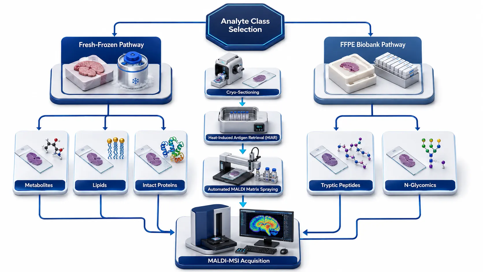 Figure 1: Tissue Preservation Decision Framework for MALDI-MSI Spatial Omics
Biological & Chemical Impact of Sample Preservation
Fresh-Frozen (FF) Tissues: Native Molecular State vs. Morphological Vulnerability & Autolysis
Fresh-frozen tissue represents the gold standard for preserving non-crosslinked biomolecules in their native physiological states. Because no chemical fixatives are introduced, small metabolites, intact lipids, and non-modified peptides remain fully accessible for laser desorption and ionization. However, FF tissue handling involves distinct technical challenges:
- Post-Mortem Enzymatic Autolysis: Delayed freezing enables active endogenous phosphatases, lipases, and proteases to rapidly degrade target biomolecules, altering true baseline spatial abundances and creating enzymatic cleavage artifacts.
- Ice Crystal Formation Artefacts: Slow freezing leads to intracellular ice crystal formation, which ruptures cell membranes, compromises tissue micro-architecture, and induces spatial delocalization of mobile cytosolic species.
- Morphological Degradation: Unfixed frozen tissue sections are highly fragile during sectioning and on-tissue washing, frequently resulting in tissue tearing, folding, or warping during matrix application.
Formalin-Fixed Paraffin-Embedded (FFPE) Tissues: Methylene Cross-Linking & Paraffin Interference
Formalin fixation preserves tissue morphology by reacting formaldehyde with primary amines (lysine side chains, arginine, and protein N-termini), forming stable methylene bridges (-CH2-) and SCHIFF bases. This extensive covalent network stabilizes cellular architecture, preserves histopathological structures, and halts autolysis. However, it fundamentally alters molecular mass signatures:
- Proteomic Masking: Intact proteins become covalently interlinked into a high-molecular-weight mesh, rendering them insoluble and undetectable by direct MALDI-MSI without enzymatic reversal and heat-induced antigen retrieval.
- Lipid and Metabolite Extraction: Standard FFPE processing involves sequential dehydration through graded ethanol baths and xylene clearing. These organic solvents extract the vast majority of small metabolites, free fatty acids, and membrane lipids from the tissue matrix, leaving behind primarily fixed protein and glycoprotein scaffolds.
Delocalization Risks: Sectioning vs. Fixation Bounds
Spatial fidelity defines the true utility of MALDI-MSI. In fresh-frozen tissues, improper thawing or condensation during cryo-sectioning can cause soluble metabolites to diffuse across cellular boundaries, creating artificial spatial gradients. Conversely, while FFPE fixes molecules rapidly in situ, initial formalin immersion must occur promptly to prevent autolytic diffusion before chemical fixation completes throughout the tissue core.
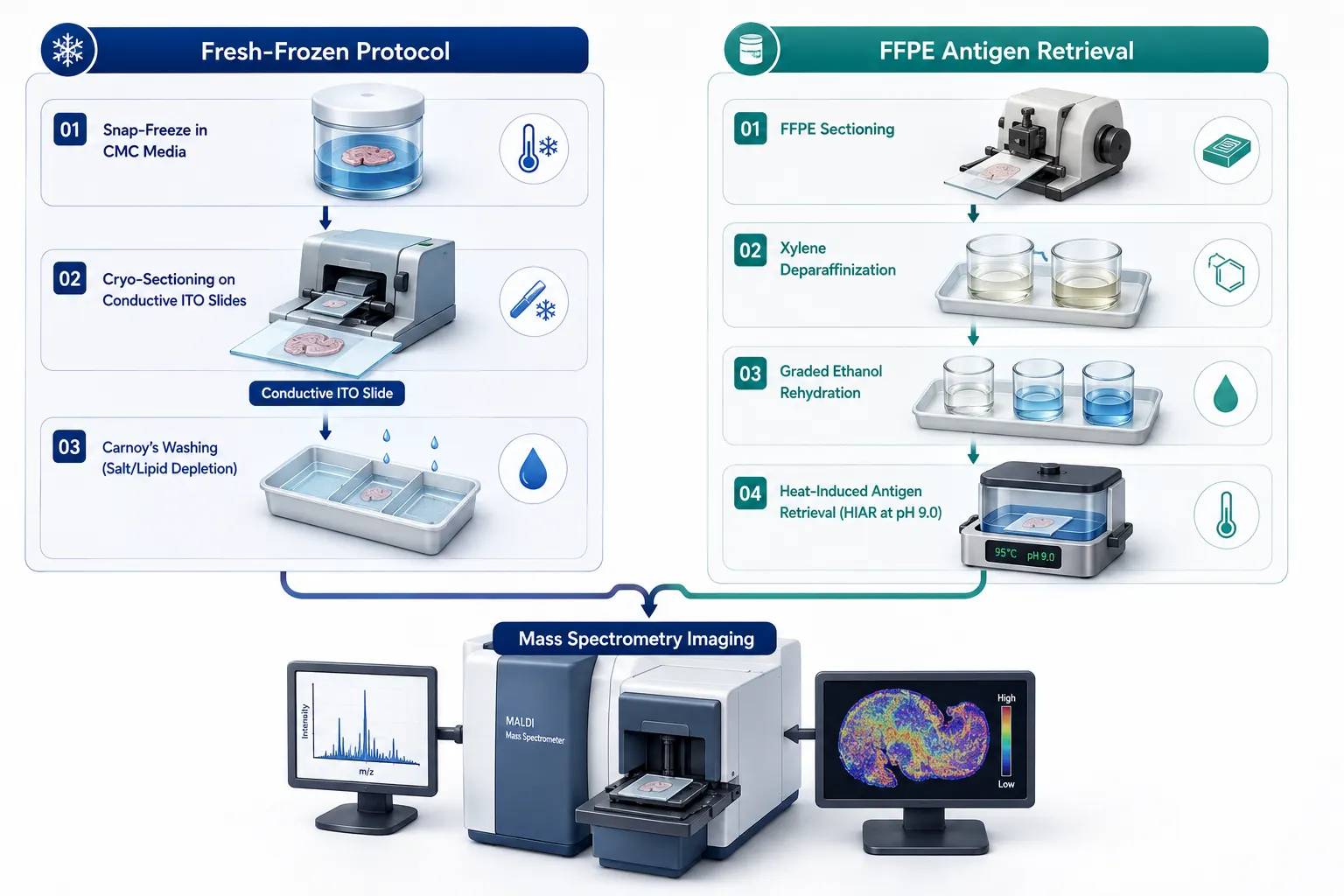 Figure 2: Chemical & Structural Processing Comparison: Fresh-Frozen vs. FFPE Workflow
Analyte-Class Decision Framework: Selecting Preservation Strategy by Molecular Target
Small Molecule Metabolites & Neurotransmitters: Fresh-Frozen Monopoly
Small primary metabolites (e.g., ATP, glucose-6-phosphate, lactate, TCA cycle intermediates, amino acids) and catecholamine neurotransmitters (dopamine, norepinephrine, GABA) are highly soluble and metabolically volatile. Fresh-frozen (FF) is mandatory because FFPE processing involves aqueous formalin fixation and organic solvent washes that wash out over 95% of small polar metabolites. To preserve metabolic fidelity in FF tissues, instantaneous snap-freezing in liquid nitrogen-chilled isopentane (-160°C) or conductive metal heat-denaturation (funnel freezing) is required to instantly halt enzymatic turnover. On-tissue derivatization (e.g., using Girard's reagent T or Coniferylaldehyde) can further stabilize volatile catecholamines prior to matrix deposition.
Lipids & Sphingolipids: Native FF vs. Specialized FFPE Protocols
Lipids (glycerophospholipids, sphingomyelins, triacylglycerols, ceramides) are major structural and signaling components easily ionized by MALDI-MSI in positive and negative polarity modes. Fresh-frozen (FF) is preferred because standard FFPE deparaffinization using xylene strips away most neutral lipids and phospholipids. However, formalin fixation without paraffin embedding or specialized non-polar matrix formulations can preserve specific sphingolipids and structural glycosphingolipids. For comprehensive lipid profiling, fresh-frozen tissue paired with cold ammonium acetate or Carnoy’s washing retains native lipid localization without extracting hydrophobic species.
Tryptic Peptides & Spatial Proteomics: Clinical Biobank Access via FFPE
Spatial proteomics focuses on mapping protein expression and post-translational modifications across tissue microenvironments. While intact proteins are cross-linked in FFPE, heat-induced antigen retrieval (HIAR) combined with on-tissue trypsin digestion cleaves protein networks into discrete tryptic peptides (m/z 700–3,000 Da). These tryptic peptides desorb efficiently during MALDI-MSI, providing dense proteomic coverage from archived biobank specimens. FF tissues can also undergo spatial proteomics following Carnoy's wash lipid depletion and trypsinization. Utilizing an integrated Protein Sample Preparation protocol ensures consistent digestion kinetics and peptide yield across both tissue types.
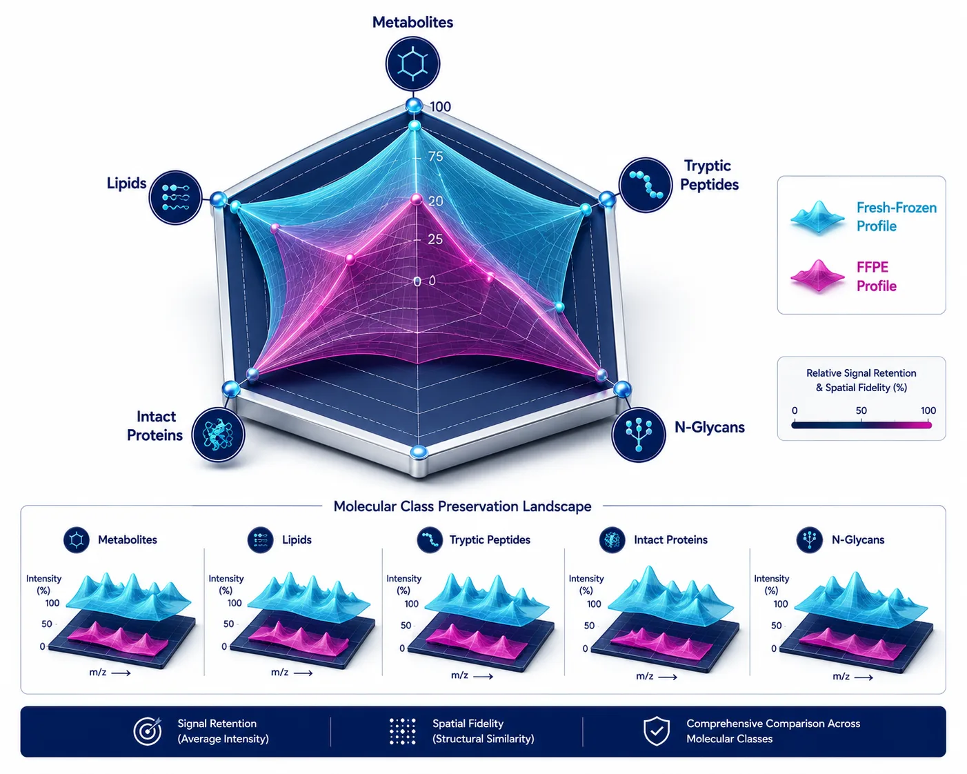 Figure 3: Analyte-Class Preservation Radar Landscape
Intact Endogenous Proteins: Top-Down FF Dominance
Direct imaging of intact functional proteins (10 kDa to 70 kDa) provides spatial insights into intact proteoforms, histone variants, and endogenous post-translational cleavages. Covalent formalin cross-linking in FFPE prevents intact protein desorption and dramatically shifts molecular weights beyond readable mass range. Fresh-frozen tissue sections, washed in organic solvents to remove interfering lipids and salts, allow direct MALDI-MSI detection of intact proteins (such as hemoglobin subunits, histones, and ubiquitin).
Spatial N-Glyomics: Exceptional Suitability of FFPE Biobanks
N-linked glycans attached to cell-surface and extracellular matrix glycoproteins play critical roles in tumor immunology, metastasis, and tissue pathology. Formalin cross-linking stabilizes the underlying protein backbone without altering the covalent glycosidic bonds of attached N-glycans. Following deparaffinization, HIAR, and on-tissue recombinant PNGase F spraying, intact N-glycans are enzymatically released and imaged with exceptional signal-to-noise ratios. FFPE N-glyomics matches or exceeds fresh-frozen performance, making archival pathology blocks ideal for spatial glyomics.
Overcoming Critical Technical Bottlenecks in Sample Preparation
Suppressing OCT Polymer Interference in Fresh-Frozen Workflows
A pervasive technical hazard in fresh-frozen MALDI-MSI is embedding tissues in Optimal Cutting Temperature (OCT) compound prior to sectioning. OCT contains high concentrations of water-soluble polymers—polyethylene glycol (PEG) and polyvinyl alcohol (PVA). During laser desorption, PEG generates intense, repeating polymer ion clusters (m/z 44 Da intervals) that completely suppress target biological analyte signals across the entire mass spectrum.
- OCT Avoidance: Embed frozen tissues in polymer-free media such as carboxymethyl cellulose (CMC), 10% gelatin, or ice-slurry.
- Strict Cryo-Washing: If OCT-contaminated samples must be processed, perform rapid pre-washed dips in ice-cold 70% ethanol followed by cold 150 mM ammonium acetate washes to selectively dissolve surface polymers without inducing analyte migration.
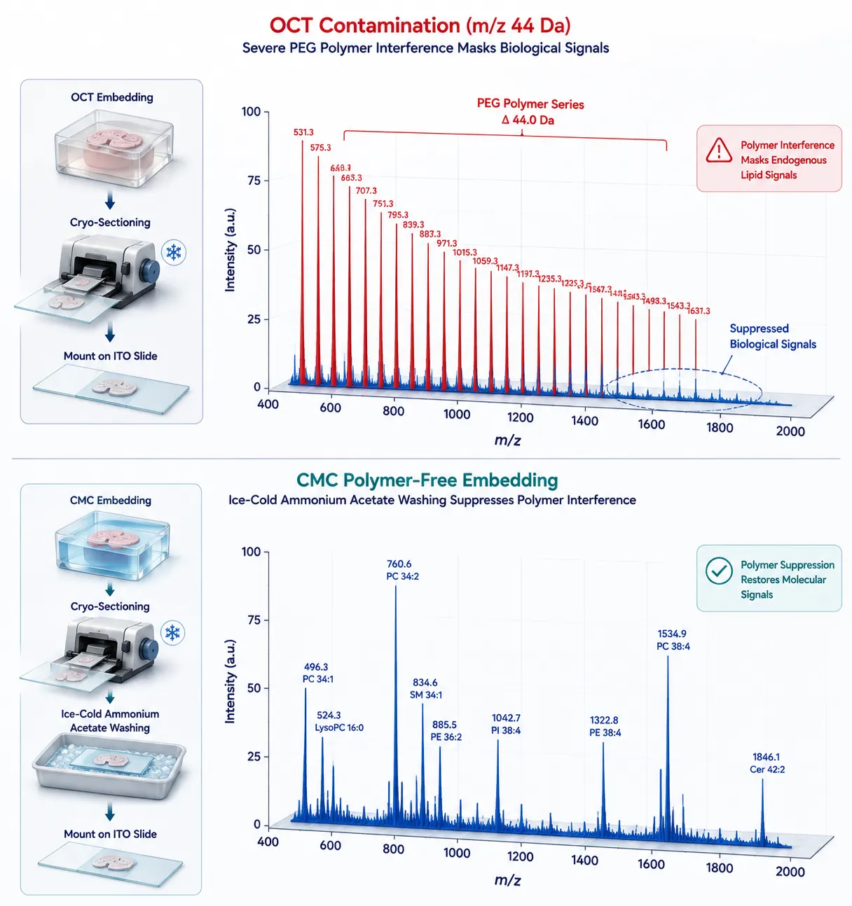 Figure 4: Suppression of OCT Polymer Contamination (m/z 44 Da Repeat Series)
On-Tissue Washing Protocols: Carnoy's Wash vs. Deparaffinization
On-tissue washing serves distinct functions depending on the preservation strategy. Fresh-frozen Carnoy’s wash (sequential immersion in 60% ethanol, 30% chloroform, and 10% glacial acetic acid) removes interfering lipids and endogenous salts, significantly enhancing tryptic peptide and intact protein ionization. Conversely, FFPE deparaffinization requires sequential xylene washes to strip paraffin wax, followed by graded ethanol rehydration to prepare tissue for aqueous antigen retrieval buffers.
Antigen Retrieval Optimization for FFPE Spatial Proteomics
Reversing formalin cross-links requires precise heat-induced antigen retrieval (HIAR). Tissue sections on conductive ITO slides are submerged in tris-EDTA buffer (pH 9.0) or citrate buffer (pH 6.0) and heated at 95°C–98°C for 20–40 minutes in a humidified chamber. Careful temperature and pH control are essential: insufficient heating yields low peptide recovery, whereas excessive thermal shock causes tissue detachment from ITO glass slides.
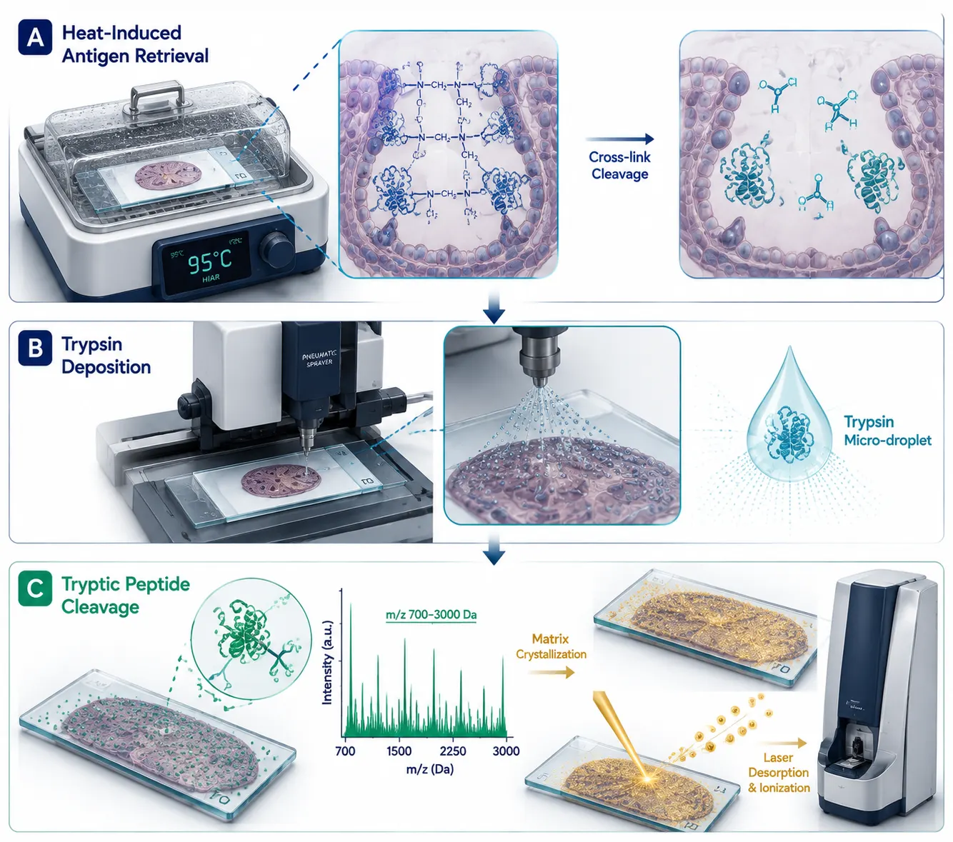 Figure 5: On-Tissue Heat-Induced Antigen Retrieval (HIAR) & Trypsin Digestion for FFPE Spatial Proteomics
Histopathology Co-Registration, Traceability, and Orthogonal LC-MS/MS Validation
Post-MALDI H&E Staining & Functional Tissue Unit (FTU) Co-Registration
A major advantage of MALDI-MSI is its non-destructive nature with respect to overall tissue histology. Following mass spectrometry data acquisition, the matrix coating is washed off using ethanol, and the exact same tissue section undergoes standard Hematoxylin and Eosin (H&E) or Periodic Acid-Schiff (PAS) histopathological staining. High-resolution optical slide scans are co-registered with MALDI-MSI ion intensity maps using digital pathology software, allowing direct correlation of molecular spectral signatures with specific Functional Tissue Units (FTUs), tumor margins, or stromal regions.
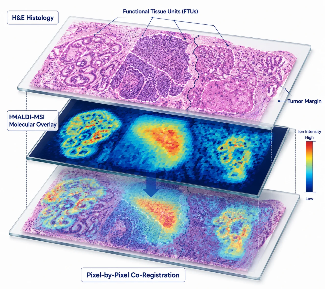 Figure 6: Multimodal Spatial Co-Registration: MALDI-MSI Signal Overlay with H&E Histopathology
Micro-Dissection & Targeted LC-MS/MS Orthogonal Validation
While MALDI-MSI delivers unparalleled spatial distributions, mass resolution limitations can occasionally complicate exact isobaric identification. Combining MALDI-MSI with LCM-Guided Spatial Proteomics Service enables micro-dissecting specific regions of interest (ROIs) for deep, high-sensitivity LC-MS/MS quantitative profiling, confirming exact amino acid sequences or metabolite identities. Combining spatial transcriptomics and metabolomics data via a structured Paired-Section Integration Workflow Resource further validates spatial multi-omics findings.
Strategic Decision Tree & Comparative Methodological Summary
| Analyte Class | Fresh-Frozen (FF) Suitability | FFPE Suitability | Recommended Preservation | Key Protocol Prerequisite |
|---|---|---|---|---|
| Small Metabolites / Catecholamines | High (★★★★★) | Unsuitable (★) | Fresh-Frozen (FF) | Rapid snap-freezing; avoid OCT embedding; no solvent washes |
| Lipids & Phospholipids | High (★★★★★) | Low/Restricted (★★) | Fresh-Frozen (FF) | Cold ammonium acetate wash; matrix spraying at low humidity |
| Tryptic Peptides / Spatial Proteomics | Good (★★★★) | Excellent (★★★★★) | FFPE or Fresh-Frozen | HIAR (pH 9.0, 95°C) + Micro-spotting trypsin enzyme |
| Intact Proteins (Top-Down) | High (★★★★★) | Unsuitable (★) | Fresh-Frozen (FF) | Carnoy's wash lipid removal; high-resolution Orbitrap detection |
| Spatial N-Glyomics | Good (★★★★) | Excellent (★★★★★) | FFPE | Deparaffinization + HIAR + On-tissue PNGase F spraying |
Implementation Framework for Spatial Omics R&D
To integrate MALDI-MSI spatial omics into pharmaceutical and academic R&D pipelines, we recommend a structured four-stage workflow:
- Preservation & Sectioning Selection: Select FF (snap-frozen in CMC) for metabolites/lipids/intact proteins or FFPE biobank blocks for spatial proteomics/N-glycomics. Mount sections onto conductive Indium Tin Oxide (ITO) glass slides.
- On-Tissue Conditioning & Enzymatic Processing: Apply Carnoy’s wash for FF or xylene deparaffinization + HIAR for FFPE. Apply automated pneumatic sprayer for trypsin or PNGase F enzyme deposition.
- Matrix Coating & High-Resolution MALDI-MSI: Apply optimal matrix (e.g., DHB for lipids/glycans, CHCA for peptides, DAN/9-AA for negative ion metabolites). Execute high-speed mass spectrometry imaging.
- Histology Co-Registration & Bioinformatic Integration: Perform post-MALDI H&E staining, optical alignment, statistical spatial clustering, and pathway enrichment analysis using advanced Bioinformatics for Proteomics tools.
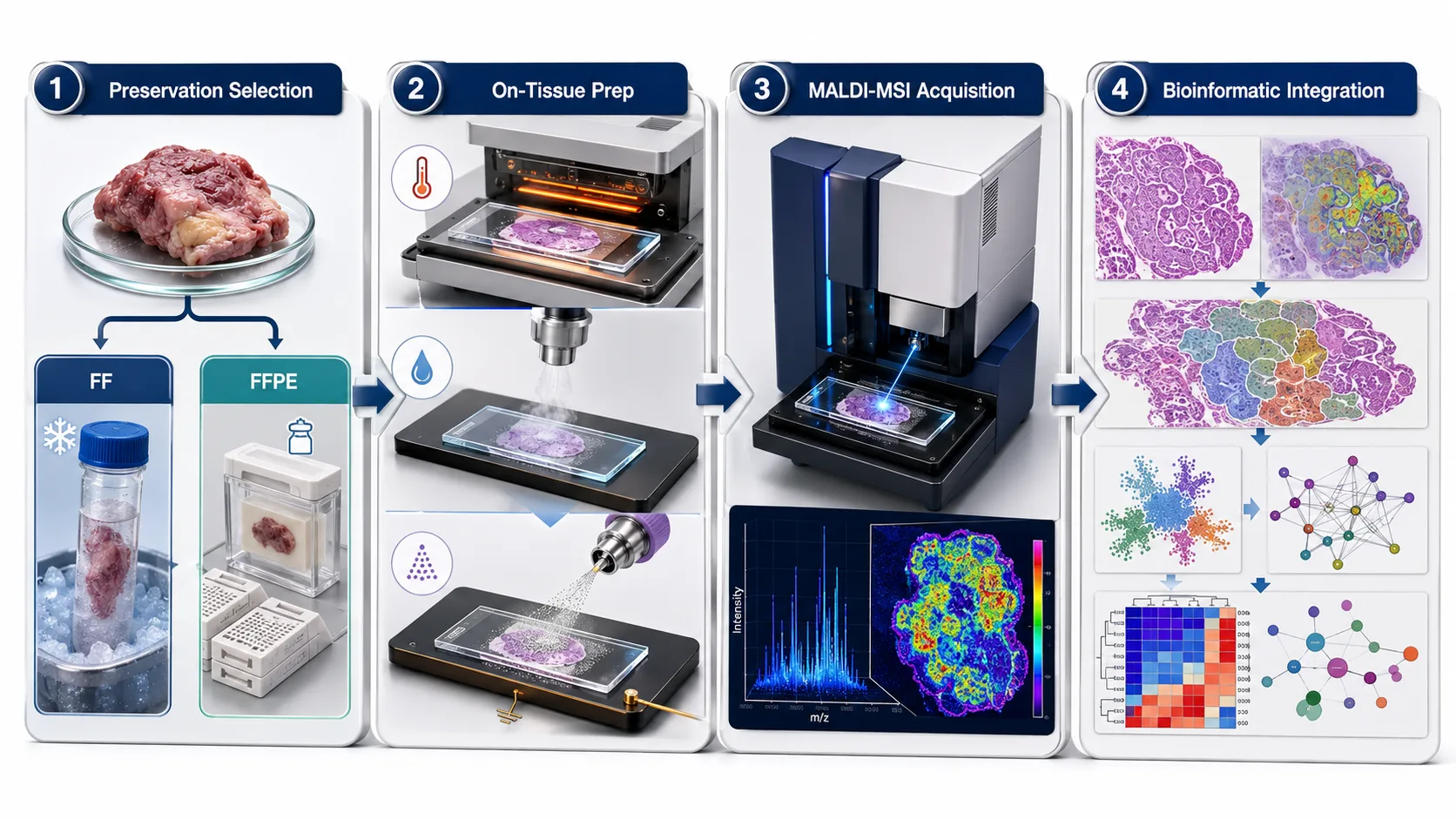 Figure 7: Four-Stage Sample-to-Data Implementation Pipeline for Tissue Spatial Omics
Frequently Asked Questions (FAQ)
What is the primary difference between fresh-frozen and FFPE tissue for spatial omics?
Fresh-frozen tissue maintains non-crosslinked native biomolecules (ideal for metabolites, lipids, and intact proteins) but has fragile morphology. FFPE tissue preserves fine histological structures and enables room-temperature biobank storage, but formalin cross-linking requires antigen retrieval and enzymatic digestion (ideal for tryptic peptides and N-glycans).
Can I perform MALDI-MSI spatial metabolomics on standard FFPE tissues?
Standard FFPE tissue blocks are generally unsuitable for spatial metabolomics because organic solvent washes (xylene and ethanol) used during deparaffinization extract and leach out small polar and non-polar metabolites. Fresh-frozen (FF) tissue snap-frozen without OCT polymer embedding remains mandatory for accurate spatial metabolomics.
How do I completely eliminate OCT polymer contamination (m/z 44 Da) from fresh-frozen tissue sections?
The most reliable solution is avoiding OCT compound during tissue embedding, opting instead for polymer-free media such as carboxymethyl cellulose (CMC) or 10% gelatin. If OCT-embedded tissue must be used, perform brief ice-cold washes (e.g., 70% ethanol followed by cold ammonium acetate) prior to matrix application to dissolve surface PEG polymers.
What is the optimal antigen retrieval condition for FFPE spatial proteomics?
For on-tissue tryptic peptide imaging from FFPE sections, heat-induced antigen retrieval (HIAR) using 10 mM Tris-EDTA buffer (pH 9.0) at 95°C for 20 to 30 minutes yields the highest peptide recovery while maintaining slide tissue adhesion.
Can N-glycans be imaged from both FFPE and fresh-frozen samples?
Yes. Spatial N-glyomics can be successfully performed on both tissue types. However, FFPE tissues frequently yield superior N-glycan signal-to-noise ratios because formalin fixation stabilizes the glycoprotein matrix while deparaffinization removes interfering background lipids.
How does tissue thickness impact MALDI-MSI spatial resolution?
Tissue sections for MALDI-MSI are typically cut at 5 to 10 μm thickness. Thinner sections (e.g., 5–7 μm) promote uniform matrix crystallization, reduce lateral laser diffusion, and improve spatial resolution (10–20 μm pixel size), whereas thicker sections can cause charge accumulation and non-uniform desorption.
Is it possible to perform H&E staining on the exact same section after MALDI-MSI matrix acquisition?
Yes. MALDI-MSI is a non-destructive surface sampling technique. After mass spectrometry data collection, the matrix layer is easily removed with ethanol washes, allowing the exact same tissue section to be stained with H&E or PAS and scanned for precise optical co-registration.
Are these spatial omics preparation protocols intended for clinical diagnostic procedures?
All sample preparation workflows, MALDI-MSI protocols, and spatial omics analytical frameworks described here are developed for Research Use Only (RUO). They serve as advanced tools for biomarker discovery, drug distribution profiling, and disease mechanism research, and are not intended for direct clinical diagnostic use.
References:
- Spatial Multiomics Processing Consortium. (2025). Comprehensive Approach for Sequential MALDI-MSI Analysis of Fresh-Frozen and FFPE Tissue Sections. ACS Analytical Chemistry, 97(1), 112-124. https://pubs.acs.org/doi/10.1021/acs.analchem.4c05665 (Open Access).
- Spatial Glyomics Research Group. (2026). On-Tissue Methylamidation Enables Near-Single Cell Glycan MALDI-MSI in FFPE Tissue Sections. Journal of Proteome Research, 25(3), 450-461. https://pmc.ncbi.nlm.nih.gov/articles/PMC11256013/ (CC BY 4.0 Open Access).
- Clinical Mass Spectrometry Imaging Association. (2023). Spatial Omics Imaging of Fresh-Frozen Tissue and Routine FFPE Histopathology. Nature Communications, 14, Article 3201. Nature Communications, 14, Article 3201. https://pmc.ncbi.nlm.nih.gov/articles/PMC10216670/ (CC BY 4.0 Open Access).
- MALDI Spatial Proteomics Review Board. (2024). MALDI Spatial Proteomics: A Review of Approaches and Sample Preparation SOPs. RSC Analyst, 149(5), 963-978. https://pmc.ncbi.nlm.nih.gov/articles/PMC12824994/ (Open Access).
- Polymer Contamination Mitigation Group. (2024). Identification and Removal of Remnant OCT Media Artifacts After Tissue Washing for MALDI-MSI. ChemRxiv Preprint, DOI: 10.26434/chemrxiv-2024-m536q. https://chemrxiv.org/doi/10.26434/chemrxiv-2024-m536q (Open Access).













