Mass Spectrometry Imaging Service
Creative Proteomics offers mass spectrometry imaging (MSI) services, specializing in molecular profiling, tissue imaging, and drug discovery. They analyze biomolecules such as lipids, proteins, peptides, and metabolites in biological samples, providing insights into diseases, drug distribution, and pharmacokinetics.
Submit Your Request Now
×
- Define
- Principle
- What We Provide
- Research Applications
- Ion Sources
- Advantages
- Sample Requirements
- Demo
- FAQs
- Case
What is Mass Spectrometry Imaging?
Mass Spectrometry Imaging (MSI) is a molecular imaging technique based on mass spectrometry analysis. Its main features include label-free, high-throughput, high sensitivity, and no need for complex sample preparation. MSI achieves imaging analysis of the spatial distribution of various molecules by ionizing molecules on the surface of the sample and collecting mass spectrometry data from each pixel.
MSI technology can simultaneously detect and record the spatial distribution information of multiple molecules on the sample surface, including metabolites, peptides, proteins, lipids, and glycosylated compounds. This technology utilizes different ionization methods, such as Matrix-Assisted Laser Desorption Ionization (MALDI), Secondary Ion Mass Spectrometry (SIMS), and Desorption Electrospray Ionization (DESI), to ionize and detect the molecules.
The application range of MSI is very broad. It is not only used in biomedical research to analyze the distribution of elements, metabolites, proteins, and drugs and their metabolites in tissues, but also shows great potential in fields such as chemistry, pharmacy, and environmental science. In addition, MSI technology has been widely applied in tumor research, providing information about the heterogeneity of the tumor microenvironment, and helping to study the spatial distribution and dynamic changes of functional molecules in tumors.
The Principle of Mass Spectrometry Imaging
The operation of Mass Spectrometry Imaging revolves around the coupling of ionization techniques with high-resolution mass spectrometry analysis. The basic principle involves the analysis of ions generated from a surface or section of a sample. The spatial location of these ions is recorded, which, when mapped, creates an image that reflects the molecular distribution within the sample.
- Microprobe Technique: A focused ion beam is used to analyze a specific region of the sample. This process generates a mass spectrum, which is stored along with the spatial coordinates of the analyzed region. By scanning multiple regions and combining all the spectra, a spatial distribution map is created, offering a detailed molecular image.
- Microscope Technique: A 2D position-sensitive detector is employed to determine the spatial origin of ions generated by the ion optics. The resolution depends on the magnification of the microscope, the quality of ion optics, and detector sensitivity. This method is efficient for analyzing smaller areas of the sample but is limited by the finite depth of vision.
At Creative Proteomics, we use the most advanced MSI techniques to ensure the highest quality and resolution of molecular imaging, providing accurate and insightful results.
 Principle of MALDI Imaging mass spectrometry ( Figure from Aichler, Laboratory investigation, 2015).
Mass Spectrometry Imaging Service We Can Provide
At Creative Proteomics, we provide comprehensive Mass Spectrometry Imaging (MSI) services tailored to a variety of research needs. Our expertise allows for precise molecular profiling, tissue imaging, and drug discovery support. We specialize in high-resolution analysis of biological samples, enabling detailed mapping of biomolecules such as lipids, metabolites, and proteins. Our MSI services are instrumental in studying complex diseases like cancer and Alzheimer's, as well as assessing drug distribution and pharmacokinetics. Additionally, we offer advanced quantitative analysis and integrate MSI with other techniques like genomics and transcriptomics, providing a multi-omics approach for deeper insights into biological processes and personalized medicine.
Molecules We Can Analyze
- Proteins and Their Modifications: MSI allows for the analysis of proteins and their modifications, such as post-translational modifications, across tissue samples.
- Peptides: It can provide detailed imaging of peptides within complex biological samples, useful in proteomics research.
- Lipids: MSI is effective for lipidomic analysis, mapping the distribution of various lipid species in biological tissues.
- Metabolites: Metabolic profiling, including the localization and distribution of metabolites, is another key application of MSI in understanding metabolic networks.
- Carbohydrates: MSI can also analyze carbohydrates and glycans, providing insights into their distribution in tissues and cells.
- Hormones and Drug Metabolites: MSI is used in drug development for studying pharmacokinetics and pharmacodynamics, helping to understand the distribution of drugs and their metabolites within tissues.
- Environmental Systems and Small Proteins: In environmental studies, MSI supports research into the distribution of small proteins and their proteoforms.
- Cell-Specific Metabolites: It can quantify metabolites at the single-cell level, providing insights into cell signaling and communication.
- Phytohormones and Microbial Compounds: MSI is used to study plant systems, including the distribution of phytohormones and microbial compounds in plant tissues and the surrounding environment.
Research Applications of Our Mass Spectrometry Imaging Service
Disease Characterization and Biomarker Investigations
MSI is crucial in understanding disease mechanisms and identifying biomarkers for conditions such as cancer, inflammatory diseases, bacterial infections, and neurological disorders. It allows for the examination of tissue samples to map the distribution of biomarkers and molecular changes associated with disease progression.
Tumor Heterogeneity Studies
MSI helps analyze inter- and intra-tumor heterogeneity, providing insights into molecular differences within tumor tissues. This is essential for personalized cancer treatment and understanding how tumors evolve.
Pharmacokinetics and Pharmacodynamics
MSI is used in drug development to track the spatial distribution of drugs and their metabolites within tissues. This helps in understanding the pharmacokinetics (movement of drugs within the body) and pharmacodynamics (effects of drugs on tissues and cells).
Cell Signaling and Metabolic Networks
MSI supports research in metabolic networks and cell signaling by providing detailed molecular maps of metabolites and signaling molecules in cells or tissue communities, facilitating the study of cellular communication and metabolism.
Rhizosphere Function Studies
In plant research, MSI is employed to examine the localization of molecules in plant tissues and their surrounding environments. This is useful for studying phytohormones, organic acids, and other molecules involved in plant growth and interactions with microorganisms.
Environmental Systems Research
MSI aids in understanding small proteins and their proteoforms in environmental systems, offering insights into molecular distributions in complex environments.
Ion Sources for Mass Spectrometry Imaging
At Creative Proteomics, we use a variety of ionization sources to ensure the best results for our clients' diverse needs. Our primary ionization techniques include:
Matrix-Assisted Laser Desorption/Ionization (MALDI)
MALDI is widely used for analyzing large biomolecules such as peptides, proteins, and lipids. It offers high sensitivity and spatial resolutions up to 20 µm.
Secondary Ion Mass Spectrometry (SIMS)
SIMS provides high-resolution imaging with subcellular resolution (down to 50 nm), allowing detection of elemental ions, small molecules, and lipids. It is perfect for detailed molecular distribution mapping on solid surfaces and thin films.
Desorption Electrospray Ionization (DESI)
DESI is a softer ionization technique suitable for analyzing a wide range of biological and organic compounds without extensive sample preparation. It is ideal for large-area scans but offers lower resolution compared to MALDI and SIMS.
Laser Ablation Electrospray Ionization (LAESI)
LAESI combines laser ablation with electrospray ionization, providing high-resolution, sensitive molecular analysis from tissue sections and environmental samples.
| Ionization Method | Spatial Resolution | Application Fields | Data Processing Methods |
|---|---|---|---|
| Matrix-Assisted Laser Desorption/Ionization (MALDI) | 5–10 μm | Proteomics, lipidomics, biomarker discovery, spatial metabolomics, plants, animals, microorganisms, clinical medicine | Mass spectral signal reconstruction, 2D or 3D distribution maps |
| Secondary Ion Mass Spectrometry (SIMS) | ~100 nm, optimized to ~50 nm | High-resolution imaging, biomolecule detection, solid surfaces, thin films, elemental ions, small molecules, and lipids | Mass spectral analysis and imaging |
| Desorption Electrospray Ionization (DESI) | 5 μm or higher | Single-cell level studies, life activity patterns, disease mechanisms, drug-targeted therapy, large-area scans | Mass spectral analysis and imaging |
| Laser Ablation Electrospray Ionization (LAESI) | High-resolution (similar to MALDI) | Tissue sections, environmental samples, sensitive molecular analysis | High-resolution image reconstruction and analysis |
Advantages of Mass Spectrometry Imaging
- Label-free Analysis: MSI enables the analysis of molecules without the need for labels or tags, preserving the integrity of the sample.
- High Sensitivity: The technique allows for the detection of a wide range of molecules, even those present in low abundance, with high sensitivity.
- Spatial Resolution: MSI provides excellent spatial resolution, with some techniques achieving up to 1 µm, enabling precise mapping of molecular distribution in tissues.
- Multiplexing: MSI can detect hundreds of molecules simultaneously in a single sample, allowing for comprehensive molecular analysis.
- Comprehensive Molecular Profiling: MSI can analyze a diverse array of biomolecules, including proteins, lipids, metabolites, and more, in a single experiment.
- Minimal Sample Preparation: MSI requires minimal sample preparation, reducing the risk of sample degradation and preserving the natural state of the molecules.
- Versatility: MSI can be applied to a wide range of biological and environmental samples, including tissues, cells, and plant materials.
- Real-time Data: MSI provides real-time molecular data, which is valuable for dynamic studies such as tracking drug distribution or monitoring metabolic pathways.
Sample Requirements for Mass Spectrometry Imaging Analysis
| Requirement | Details |
|---|---|
| Sample Type | Tissues (e.g., brain, liver, cancer tissues), cells, plant material, microbial samples, organs |
| Sample Size | Typically 1-5 mm² for small samples; larger samples may require sectioning for analysis |
| Sample Preparation | Minimal preparation (e.g., tissue freezing or fixation) depending on ionization method used |
| Thickness of Sections | 10-100 µm (for tissue or cell sections); thinner sections may be required for high-resolution imaging |
| Surface Cleanliness | The surface must be clean and free from contaminants; cleaning protocols depend on sample type |
| Storage and Handling | Samples should be stored frozen or in a stable condition to avoid degradation before analysis |
| Moisture Content | Low moisture content is preferred to prevent ion suppression in the ionization process |
| Homogeneity | Samples should be as homogeneous as possible to avoid inconsistent ionization and analysis |
Demo
Figure from Louie, Katherine B., et al., Scientific reports, 2013
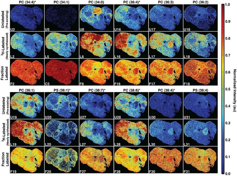
Intensity images of twelve phospholipids, showing unlabeled (top), 2H-labeled (middle), and fraction labeled (bottom) levels.
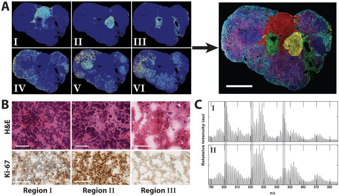
Spatial distribution of tumor phospholipids (A), H&E and Ki-67 stains (B), and average spectra for K-means regions I and II in the 2H-labeled tumor (C).
FAQ of Mass Spectrometry Imaging Analysis
What types of samples can be analyzed using MSI?
MSI is highly versatile and can analyze a wide range of biological and environmental samples. Common sample types include tissue sections (e.g., brain, liver, cancer tissue), cells, plant materials, microbial samples, and organs. The sample must be prepared to ensure proper ionization, such as tissue freezing or fixation, depending on the ionization method used.
How is sample preparation for MSI different from other techniques?
MSI requires minimal sample preparation compared to other techniques, preserving the integrity of the sample. Tissues are often frozen or fixed to maintain their molecular structure. For some ionization techniques, such as MALDI, the sample may require application of a matrix, while other techniques, like DESI, may require less pre-treatment. The primary goal is to maintain the natural state of the molecules for accurate molecular profiling.
Can MSI detect low-abundance molecules in complex samples?
Yes, MSI is highly sensitive and capable of detecting molecules present in low abundance. With high sensitivity and advanced ionization techniques, MSI can identify and map even trace amounts of biomolecules, such as metabolites or drug metabolites, in complex biological tissues.
What is the spatial resolution of MSI, and how does it impact results?
MSI provides excellent spatial resolution, with some techniques achieving up to 1 µm, depending on the ionization method and the instrument used. This level of resolution allows for precise localization of molecular distributions within tissues, providing valuable insights into the heterogeneity of tissues, such as tumor microenvironments, and enabling detailed mapping of biological processes at cellular and subcellular levels.
Can MSI be used for quantitative analysis of molecular distributions?
Yes, MSI supports quantitative analysis. By measuring the intensity of mass spectral peaks, MSI can quantify molecular abundance across different regions of a tissue. This is particularly useful for assessing molecular changes, such as those associated with disease progression or drug metabolism, providing deeper insights into biological processes.
Is MSI suitable for single-cell analysis?
Yes, MSI can be adapted for single-cell analysis, particularly with high-resolution ionization methods such as MALDI. This allows for the precise localization of metabolites and lipids at the single-cell level, facilitating the study of cellular heterogeneity and the molecular mechanisms behind cell signaling, metabolism, and disease processes.
Learn about other Q&A.


