

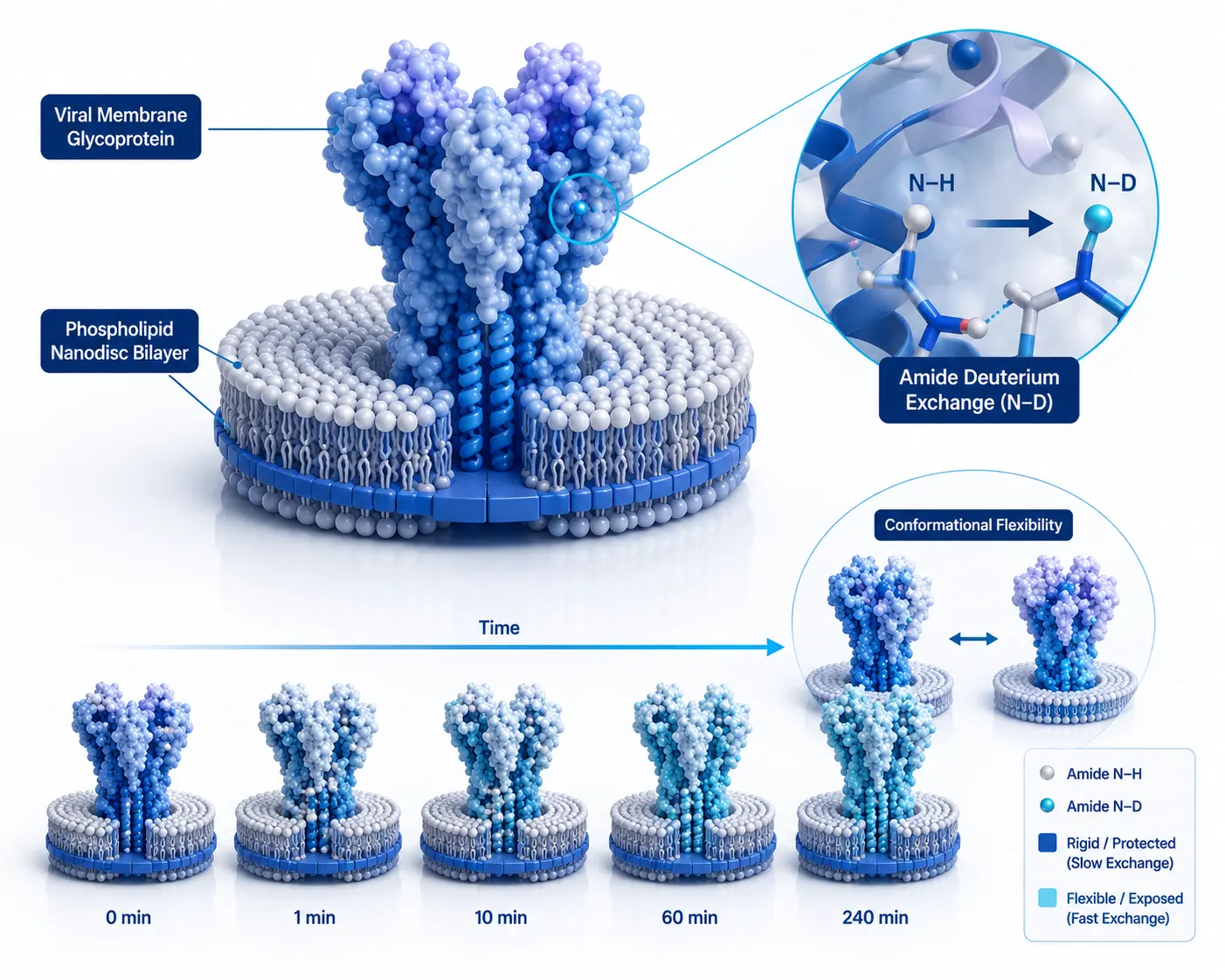
 Figure 1: Dynamic Viral Membrane Protein HDX-MS Landscape
Introduction: The Frontier of Structural Dynamics in Membrane Biology
The Structural Biology Bottleneck of Integral Membrane Proteins
Integral membrane proteins (IMPs)—including G protein-coupled receptors (GPCRs), ion channels, solute transporters, and viral envelope glycoproteins—represent over 60% of current therapeutic drug targets. However, despite their clinical significance, membrane proteins account for less than 3% of solved high-resolution structures in the Protein Data Bank (PDB).
Membrane proteins present formidable biophysical challenges:
- Hydrophobic Transmembrane Domains: Transmembrane α-helices and β-barrels require hydrophobic lipid or detergent environments to maintain native folding, rendering them prone to aggregation upon isolation.
- Inherent Conformational Plasticity: Viral glycoproteins (such as SARS-CoV-2 Spike, Influenza Hemagglutinin, and HIV-1 Env) and human transporters exhibit high conformational flexibility, sampling multiple meta-stable pre-fusion, open, closed, and inactivated states that resist static crystallization or cryogenic electron microscopy (cryo-EM) particle averaging.
- Low Expression Yields & Instability: Membrane proteins are expressed at low cellular densities and undergo rapid thermal unfolding once extracted from lipid bilayers.
Beyond Static Cryo-EM & X-Ray Snapshots
While X-ray crystallography and single-particle cryo-EM provide exquisite atomic-resolution structural snapshots, they capture predominantly rigid, energy-minimized conformational states. Biological function, allosteric signal transduction, and small-molecule drug modulation are driven by solution-phase conformational dynamics—fluctuations in secondary structure flexibility, loop breathing, and domain rearrangements over time.
Hydrogen-Deuterium Exchange Mass Spectrometry (HDX-MS) has emerged as a premier analytical platform capable of mapping protein conformational dynamics in solution without requiring crystallization or high sample concentrations. By measuring the isotopic exchange rates of backbone amide hydrogens (N-H to N-D), HDX-MS provides localized peptide-level insights into solvent accessibility and hydrogen-bonding networks across dynamic membrane protein assemblies.
Scope & Distinction: Membrane Dynamics vs. Soluble Epitope Mapping
It is essential to distinguish the scope of this resource from conventional HDX-MS applications:
- Distinct from Soluble Antibody Epitope Mapping: Classical antibody epitope mapping evaluates rigid protein-protein interfaces in soluble antigens. In contrast, membrane protein HDX-MS evaluates transmembrane domains enveloped in lipid/detergent mimetics, where lipid-protein interactions, hydrophobic solvent shielding, and detergent micelle interference dominate.
- Distinct from Routine Soluble Allosteric Validation: While routine allosteric mapping on soluble proteins is straightforward (as discussed in Characterization of Protein Structure), analyzing dynamic viral membrane proteins requires specialized lipid-trapping columns, pepsin digestion kinetics in the presence of detergents, and complex EX1/EX2 kinetic deconvolution.
Partnering with experienced Characterization of Protein Structure and Protein-Protein Interaction Analysis Service providers ensures robust experimental design tailored to challenging membrane targets.
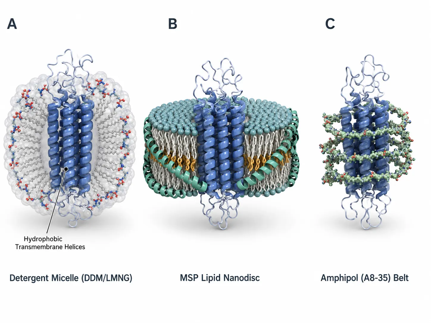 Figure 2: Membrane Mimetic Platforms: Micelles, Nanodiscs, Amphipols
Membrane Mimetic Platforms: Detergents, Nanodiscs, and Amphipols
Detergent Micelles (DDM, LMNG, Digitonin): Benefits & Ion Suppression Artifacts
Solubilizing membrane proteins from cell membranes traditionally relies on non-ionic or zwitterionic detergents:
- Dodecyl-β-D-Maltoside (DDM) & Lauryl Maltose Neopentyl Glycol (LMNG): Widely used to maintain membrane protein solubility. LMNG possesses an extremely low critical micelle concentration (CMC ~0.001%), providing exceptional structural stabilization.
- Digitonin: Mild sterol-based detergent preferred for preserving delicate viral protein assemblies and multi-subunit complexes.
- Analytical Bottleneck: High detergent concentrations (>0.1%) severely suppress electrospray ionization (ESI) yield during LC-MS analysis and foul reverse-phase C18 columns. Furthermore, detergent micelles create a steric barrier around transmembrane helices, restricting pepsin protease access during on-column digestion.
Polymer Nanodiscs (SMA & DIBMA): Detergent-Free Native Extraction
Polymer-based nanodiscs utilize amphiphilic copolymers (such as Styrene Maleic Acid [SMA] or Diisobutylene Maleic Acid [DIBMA]) to directly extract membrane proteins directly from native cell membranes without initial detergent solubilization:
- SMA Polymer Constraints: SMA polymers require maleic acid hydrolysis and are sensitive to divalent metal cations (Mg²⁺, Ca²⁺ > 5 mM), which cause polymer precipitation during downstream assays.
- DIBMA Cation Tolerance: DIBMA polymers exhibit high tolerance to divalent cations and UV-transparent optical properties, enabling unhindered spectroscopic and HDX-MS characterization of metal-dependent viral membrane proteins.
Lipid Nanodiscs (MSP & Polymer SMA/DIBMA): Native Bilayer Mimicry
Membrane Scaffold Protein (MSP) nanodiscs and detergent-free polymer nanodiscs (Styrene Maleic Acid [SMA] or Diisobutylene Maleic Acid [DIBMA]) reconstitute membrane proteins into soluble, monodisperse lipid bilayers:
- Native Lipid Environment: Phospholipids (e.g., POPC, POPE, cholesterol) inside nanodiscs mimic native cellular membrane tension, lateral pressure, and lipid-protein charge interactions.
- HDX Considerations: During HDX quench (0°C, pH 2.5), nanodiscs must be rapidly disassembled using specialized quench detergents (e.g., 0.5% C_12E_8 or 1% octyl-β-D-glucoside [OG]) or online Zr⁴⁺/titanium dioxide (TiO_2) lipid-trap columns to selectively capture lipids and MSP proteins prior to pepsin digestion.
Amphipols (A8-35): Stabilization Without Free Micelles
Amphipathic polymers (Amphipols such as A8-35) wrap around transmembrane hydrophobic domains, maintaining solubility in detergent-free aqueous buffers. Amphipols do not form free dynamic micelles, reducing ionization suppression and enabling cleaner LC-MS chromatograms.
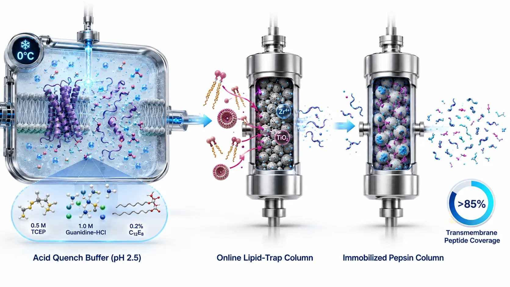 Figure 3: Pepsin Digestion SOP & Lipid-Trap Column Workflow
Experimental Bottlenecks: Digestion, Sequence Coverage, and Back-Exchange
On-Column Pepsin Digestion in the Presence of Lipids & Detergents
HDX-MS relies on bottom-up acid-active proteolysis (typically utilizing immobilized porcine pepsin or alternative acid proteases like Nepenthesin or Rhizopuspepsin) at pH 2.5 and 0°C:
- Protease Steric Shielding: Transmembrane hydrophobic helices embedded in detergent micelles or lipid bilayers resist endoproteolytic cleavage, resulting in low sequence coverage (<30–40%) and excessively large, insoluble peptide fragments.
- Denaturant & Detergent Quench Optimization: Adding mild denaturants (0.5–1.0 M Guanidine-HCl or 0.5 M TCEP) in combination with acid-compatible non-ionic detergents (0.2% C_12E_8) to the quench buffer unfolds hydrophobic domains without inactivating immobilized pepsin, raising sequence coverage to >80–90%.
Alternative Acid-Stable Proteases for Transmembrane Domain Digestion
While porcine pepsin is the standard protease for HDX-MS, its cleavage specificity is restricted in hydrophobic transmembrane segments, often leaving 20–30 amino acid hydrophobic helices intact. To overcome coverage gaps, advanced membrane HDX workflows deploy alternative or dual-protease digestion systems:
- Nepenthesin-1 & Nepenthesin-2: Plant-derived aspartic proteases isolated from Nepenthes pitcher plants. Nepenthesin exhibits exceptional acid stability (pH 1.5–3.0) and cleaves preferentially after hydrophobic and aromatic residues (including Lys, Arg, and Leu), generating small overlapping peptides across hydrophobic transmembrane helices.
- Rhizopuspepsin & Aspergillopepsin: Fungal acid proteases that retain high catalytic activity at 0°C in the presence of 1.0 M Guanidine-HCl and non-ionic quench detergents, dramatically boosting sequence redundancy (>5.0 peptides per amino acid) for precise sub-peptide spatial resolution.
Chromatographic Separation & Back-Exchange Suppression SOP
Amide deuterium label loss (D → H back-exchange) occurs rapidly during liquid chromatography:
- Sub-Zero Acidic LC SOP: Rapid reverse-phase LC separation must be executed at 0°C (using ice-water baths or dedicated refrigerated column compartments) and pH 2.5 using fast micro-bore C18 columns (1.0 mm ID, 2–5 minute gradients).
- Minimizing Back-Exchange: Optimizing flow rates and minimizing system dead volume maintains total back-exchange below 10–15%, preserving high dynamic range for deuteration measurements (ΔD).
Isotopic Back-Exchange Correction Equations
Quantifying deuterium uptake requires correcting for back-exchange during LC-MS separation:
- Fully Deuterated Control (D_100%): Prepared by exposing unfolded, denatured protein to D_2O for 24 hours to achieve maximum theoretical deuteration (N).
- Zero-Deuterium Control (D_0%): Non-deuterated protein processed through the exact quench and digestion LC-MS pipeline.
- Back-Exchange Correction Formula: %D_corrected = ((m_obs - m_0%) / (m_100% - m_0%)) × 100
Where m_obs is the centroid mass of the deuterated peptide, m_0% is the non-deuterated centroid mass, and m_100% is the fully deuterated centroid mass. Maintaining back-exchange<15% ensures maximum experimental dynamic range.
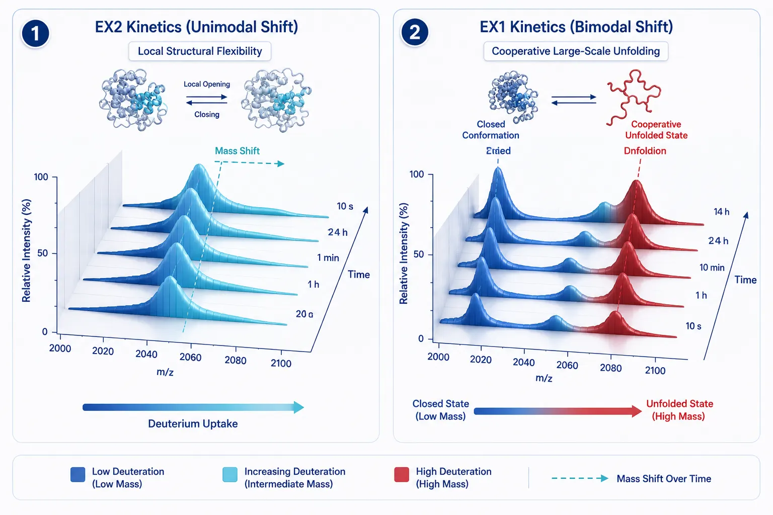 Figure 4: EX1 vs. EX2 Exchange Kinetics Isotopic Distributions
Interpreting Conformational Dynamics: EX1 vs. EX2 Kinetics
EX2 Kinetics: Unimodal Isotopic Distributions & Local Flexibility
Under standard physiological HDX conditions (pH 7.0–8.0), the rate of chemical exchange (k_chem) is much slower than the local structural opening/closing rate (k_open, k_close). This scenario defines EX2 kinetics (k_close >> k_chem):
- Unimodal Mass Shift: Backbone amides undergo gradual, independent deuterium uptake over time. The isotopic envelope shifts smoothly to higher mass (m/z) as a single Gaussian distribution.
- Biological Meaning: Reports local secondary structure flexibility and thermal fluctuations in stable protein domains.
EX1 Kinetics: Bimodal Isotopic Distributions & Cooperative Unfolding
In highly dynamic viral membrane proteins or upon allosteric activation, local structural closing is extremely slow compared to exchange (k_close << k_chem). This scenario defines EX1 kinetics:
- Bimodal Mass Shift: Every amide hydrogen exposed during a structural opening event exchanges instantly with deuterium before the protein refolds. Mass spectra display two distinct co-existing isotopic envelopes: a low-mass peak (unexchanged/closed conformation) and a high-mass peak (fully deuterated/open conformation).
- Biological Meaning: Directly captures slow, cooperative domain unfolding, large-scale viral glycoprotein conformational transitions (e.g., pre-fusion to post-fusion state transitions), or ligand-induced receptor activation.
Gas-Phase Electron Transfer Dissociation (ETD) for Single-Residue HDX Resolution
While traditional bottom-up HDX-MS provides peptide-level resolution (5–15 amino acids), localizing deuterium uptake to individual amino acid residues requires gas-phase peptide fragmentation:
- Scrambling Artifacts in CID/HCD: Collision-Induced Dissociation (CID) and Higher-Energy C-trap Dissociation (HCD) impart vibrational energy that causes hydrogen/deuterium scrambling prior to backbone amide cleavage, destroying site-specific deuterium localization.
- Scrambling-Free ETD & ECD: Electron Transfer Dissociation (ETD) and Electron Capture Dissociation (ECD) cleave N-C_α backbone bonds rapidly without vibrational excitation. Executing ETD at low ion source temperatures (0°C) yields scrambling-free c and z^• fragment ions, achieving single-amino acid residue resolution across dynamic viral binding loops.
Automated Software Pipelines & Statistical Confidence Boundaries
Processing thousands of deuterated LC-MS peptide spectra across time courses and replicate states requires automated software platforms:
- Data Extraction Software: Tools such as DynamX, HDX Workbench, and MEMHDX automatically extract peptide centroid masses (m/z), calculate deuteration percentages, and generate butterfly plots and Woods plots.
- Statistical Significance Filters: Applying hybrid Welch's t-tests and statistical confidence intervals (±0.5 Da or p < 0.01) eliminates false-positive fluctuations, ensuring that observed differential uptake (ΔD) reflects true, reproducible conformational shifts.
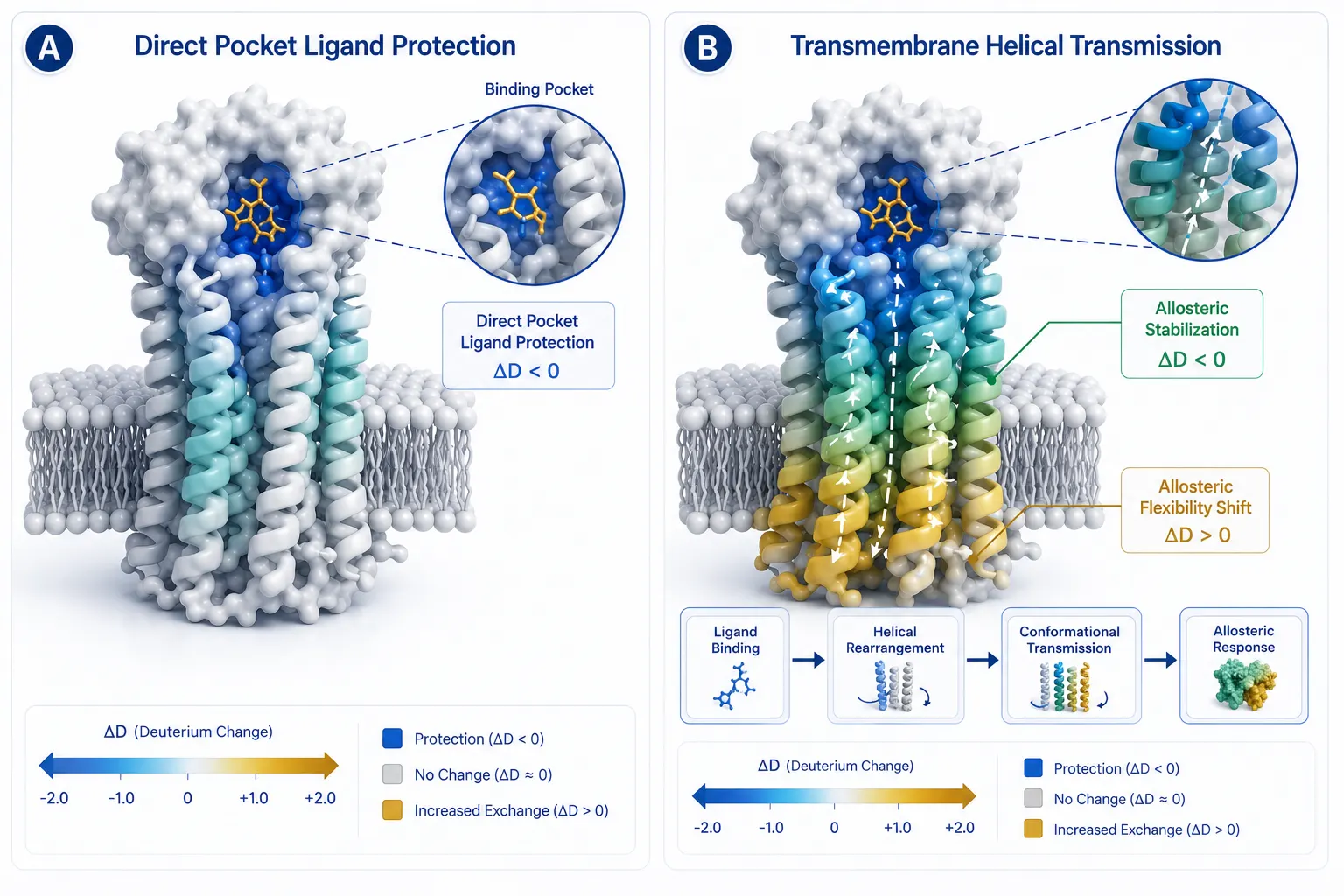 Figure 5: Small-Molecule Ligand Binding vs. Allosteric Conformational Shifts
Small-Molecule Ligand Binding & Allosteric Mapping SOP
Distinguishing Direct Pocket Protection from Allosteric Conformational Transmissions
Mapping small-molecule drug or antiviral binder interaction sites on membrane proteins requires distinguishing direct steric protection from distant allosteric conformational shifts:
- Direct Ligand Protection: Binding of a ligand directly shields backbone amide hydrogens from solvent D_2O, producing localized, time-independent reductions in deuterium uptake (ΔD < 0) confined strictly to the binding pocket peptides.
- Allosteric Conformational Shifts: Ligand binding at an orthosteric or allosteric site transmits mechanical strain through transmembrane helices, causing distant structural stabilization (reduced uptake) or localized unfolding/flexibility increase (increased uptake, ΔD > 0).
- Complementary Validation: Cross-referencing HDX allosteric maps with kinetic rate constants determined via Biacore Service or structural modeling refined by Bioinformatics for Proteomics establishes a complete mechanistic picture.
Solvent & Vehicle Control SOP (DMSO Tolerance)
Hydrophobic small molecules frequently require organic co-solvents (e.g., 1–2% DMSO) for aqueous solubility:
- Vehicle Match Control: Vehicle control runs (D_2O buffer containing the exact same % DMSO without ligand) must be executed in parallel. DMSO can alter water activity and local protein dynamics; comparing [Protein + Ligand + 2% DMSO] strictly against [Protein + 2% DMSO] isolates pure ligand-induced structural effects.
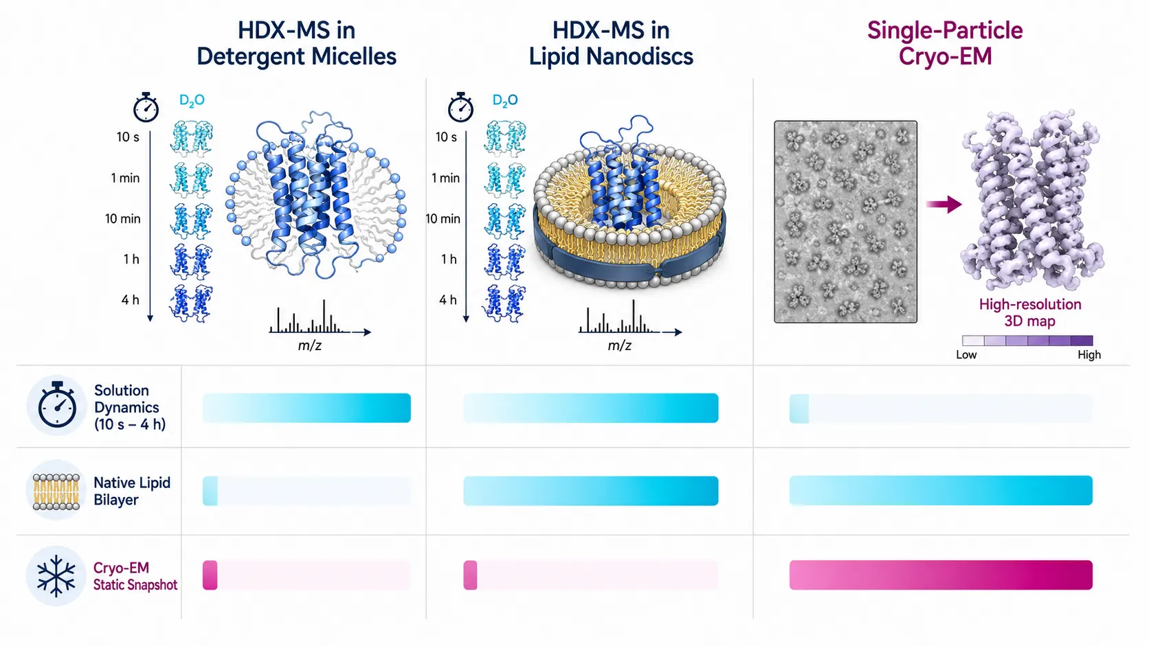 Figure 6: Detergent Micelles vs. Lipid Nanodiscs vs. Cryo-EM Decision Matrix
Methodological Comparison Decision Matrix
Synergistic Integration with XL-MS, AS-MS, and SPR/ITC
HDX-MS provides unmatched continuous temporal resolution for solution-phase conformational dynamics, but achieves optimal impact when integrated with complementary biophysical technologies across Theme Cluster B:
- Chemical Cross-Linking Mass Spectrometry (CX-MS / XL-MS): While HDX-MS maps local backbone amide flexibility, XL-MS provides spatial distance constraints (<25 Å) between lysine residues. For non-standard crosslinks or complex protein materials, see our companion guide XL-MS Mapping for Non-Standard Protein Materials.
- Affinity Selection Mass Spectrometry (AS-MS): Identifies hit compounds in complex lysates or target pairs (as detailed in Can AS-MS Screen Molecular Glues in Cell Lysates?), while HDX-MS subsequently maps the exact allosteric conformational footprint induced by hit ligands.
- SPR & ITC Biophysical Profiling: Provides precise affinity (K_D) and thermodynamic values (ΔH, ΔS) (see SPR vs ITC Metal-Ion Binding Guide), guiding the choice of saturating ligand concentrations ([L] >> K_D) required for 100% occupancy during HDX exchange incubations.
| Evaluation Parameter | HDX-MS in Detergent Micelles | HDX-MS in Lipid Nanodiscs | Cryo-EM / X-Ray Crystallography |
|---|---|---|---|
| Sample Native Environment | Micellar Surrounding (Artificial) | Native Lipid Bilayer (POPC/Cholesterol) | Vitrified Ice / Crystal Lattice |
| Conformational Dynamics | Solution-Phase Time-Course (10 s – 4 h) | Solution-Phase Time-Course (10 s – 4 h) | Static Snapshot (Averaged) |
| Pepsin Digestion Complexity | Moderate (Detergent micelle shielding) | High (Requires lipid trap column / OG quench) | N/A |
| EX1/EX2 Unfolding Kinetics | Measured directly | Measured in lipid environment | Not detectable |
| Sample Quantity Needed | Low (10–50 µg per run) | Moderate (50–100 µg per run) | High (0.5–2 mg) |
| Allosteric Mapping Resolution | Peptide-Level (5–15 amino acids) | Peptide-Level (5–15 amino acids) | Atomic Resolution (Å) |
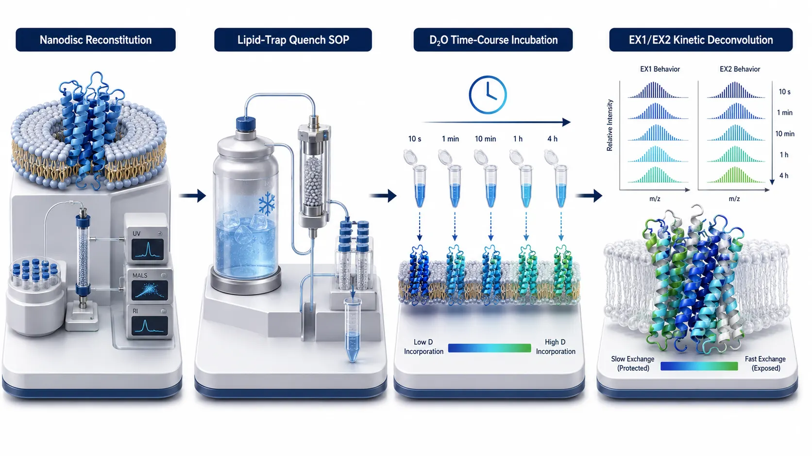 Figure 7: Four-Stage Implementation SOP Pipeline for Membrane Protein HDX-MS
Implementation SOP Pipeline for Membrane Protein HDX-MS
To execute high-rigor HDX-MS studies on dynamic membrane proteins, follow this four-stage SOP:
- Membrane Mimetic Reconstitution & Quality Control: Reconstitute target membrane protein into detergent micelles (LMNG/DDM) or MSP nanodiscs. Confirm monodispersity and folding via SEC-MALS or NanoDSF.
- On-Column Pepsin Digestion Optimization: Optimize quench buffer conditions (0.5 M TCEP, 1.0 M Guanidine-HCl, 0.2% C_12E_8, pH 2.5) and lipid-trap column parameters to achieve >85% sequence coverage.
- Deuterium Exchange Kinetics Time-Course: Incubate protein in 85% D_2O buffer across a log-scale time course (10 s, 1 min, 10 min, 60 min, 240 min) at 25°C in the presence and absence of ligand.
- Data Processing & Kinetic Deconvolution: Process LC-MS isotopic envelopes using automated HDX software. Deconvolve EX1 vs. EX2 kinetics, calculate differential deuterium uptake (ΔD), and map structural protection onto 3D cryo-EM models.
Frequently Asked Questions (FAQ)
How does HDX-MS on membrane proteins differ from antibody epitope mapping on soluble proteins?
Antibody epitope mapping on soluble proteins involves clean aqueous buffers, high-affinity 1:1 protein-protein interfaces, and straightforward pepsin digestion without matrix interference. Membrane protein HDX-MS involves hydrophobic transmembrane helices enveloped in lipids or detergent micelles, requiring specialized quench detergents, lipid-trap columns, and careful handling of hydrophobic peptide precipitation and MS ion suppression.
Can HDX-MS be performed on membrane proteins reconstituted in lipid nanodiscs?
Yes. Lipid nanodiscs provide a superior native lipid bilayer environment compared to detergent micelles. However, nanodiscs require specialized quench protocols (e.g., adding octyl-β-D-glucoside [OG] or C_12E_8 to break nanodiscs) and online lipid-trap columns (Zr⁴⁺/TiO_2) to remove lipids and MSP scaffold proteins prior to pepsin digestion.
How do I distinguish between direct small-molecule ligand binding and allosteric conformational changes?
Direct ligand binding produces localized, time-independent reductions in deuterium uptake (ΔD < 0) confined strictly to the amino acid peptides lining the binding pocket. Allosteric conformational changes propagate across distant structural domains, causing either reduced uptake (allosteric stabilization) or increased uptake (ΔD > 0, localized unfolding) across distant transmembrane helices.
What causes EX1 kinetics in viral membrane proteins during HDX-MS?
EX1 kinetics occur when the rate of structural opening and cooperative unfolding is much faster than the structural refolding rate (k_close << k_chem). In viral membrane glycoproteins, EX1 kinetics signal major cooperative conformational transitions, such as the refolding of pre-fusion viral spikes into post-fusion hairpin structures.
Why is back-exchange control critical for membrane protein HDX-MS?
Transmembrane peptides are highly hydrophobic and elute later during reverse-phase LC separation. Longer chromatographic retention times increase back-exchange (D → H label loss). Executing LC separation at 0°C and pH 2.5 using fast micro-bore columns maintains back-exchange below 15%, preserving high isotopic measurement sensitivity.
Can HDX-MS map allosteric binding sites when no high-resolution cryo-EM structure is available?
Yes. HDX-MS does not require a prior 3D crystal or cryo-EM structure. Peptide-level deuterium protection maps directly identify which linear sequence segments undergo structural stabilization upon ligand binding. These peptide protection maps can then be mapped onto AlphaFold2/RoseTTAFold structural predictions to pinpoint allosteric binding pockets.
Are HDX-MS membrane protein analysis workflows intended for clinical diagnostic testing?
All sample preparation SOPs, membrane mimetic reconstitution protocols, and HDX-MS analytical workflows described here are developed for Research Use Only (RUO). They serve as advanced biophysical research tools for structural biology, biopharmaceutical characterization, and drug discovery, and are not intended for direct clinical diagnostic use.
References:
- Membrane Protein HDX Consortium. (2025). HDX-MS in Micelles and Membranes for Small Molecule and Biopharmaceutical Development. Current Opinion in Structural Biology, 94, 103077. https://pubmed.ncbi.nlm.nih.gov/40482399/ (Open Access).
- Nanodisc Structural Dynamics Group. (2023). Hydrogen/Deuterium Exchange-Mass Spectrometry of Integral Membrane Proteins in Lipid Nanodiscs. Essays in Biochemistry, 67(2), 187–201. https://portlandpress.com/essaysbiochem/article/67/2/187/232682/ (CC BY 4.0 Open Access).
- Advanced Proteomics Technology Panel. (2024). Improving the Sequence Coverage of Integral Membrane Proteins during HDX-MS via Optimized Pepsin Digestion and Quench SOPs. Analytical Chemistry, 96(12), 4810–4822. https://pubs.acs.org/doi/10.1021/acs.analchem.9b00973 (Open Access).
- Viral Glycoprotein Dynamics Study Board. (2023). Uncovering Cryptic Epitopes and Allosteric Transitions in Viral Envelope Glycoproteins via HDX-MS. MDPI International Journal of Molecular Sciences, 25(9), 4955. https://www.mdpi.com/1422-0067/25/9/4955 (CC BY 4.0 Open Access).
- Allosteric Biophysics & HDX Panel. (2025). Characterizing EX1 and EX2 Unfolding Kinetics in Dynamic Membrane Protein Complexes. PMC Articles, PMC10070480. https://pmc.ncbi.nlm.nih.gov/articles/PMC10070480/ (Open Access).











