

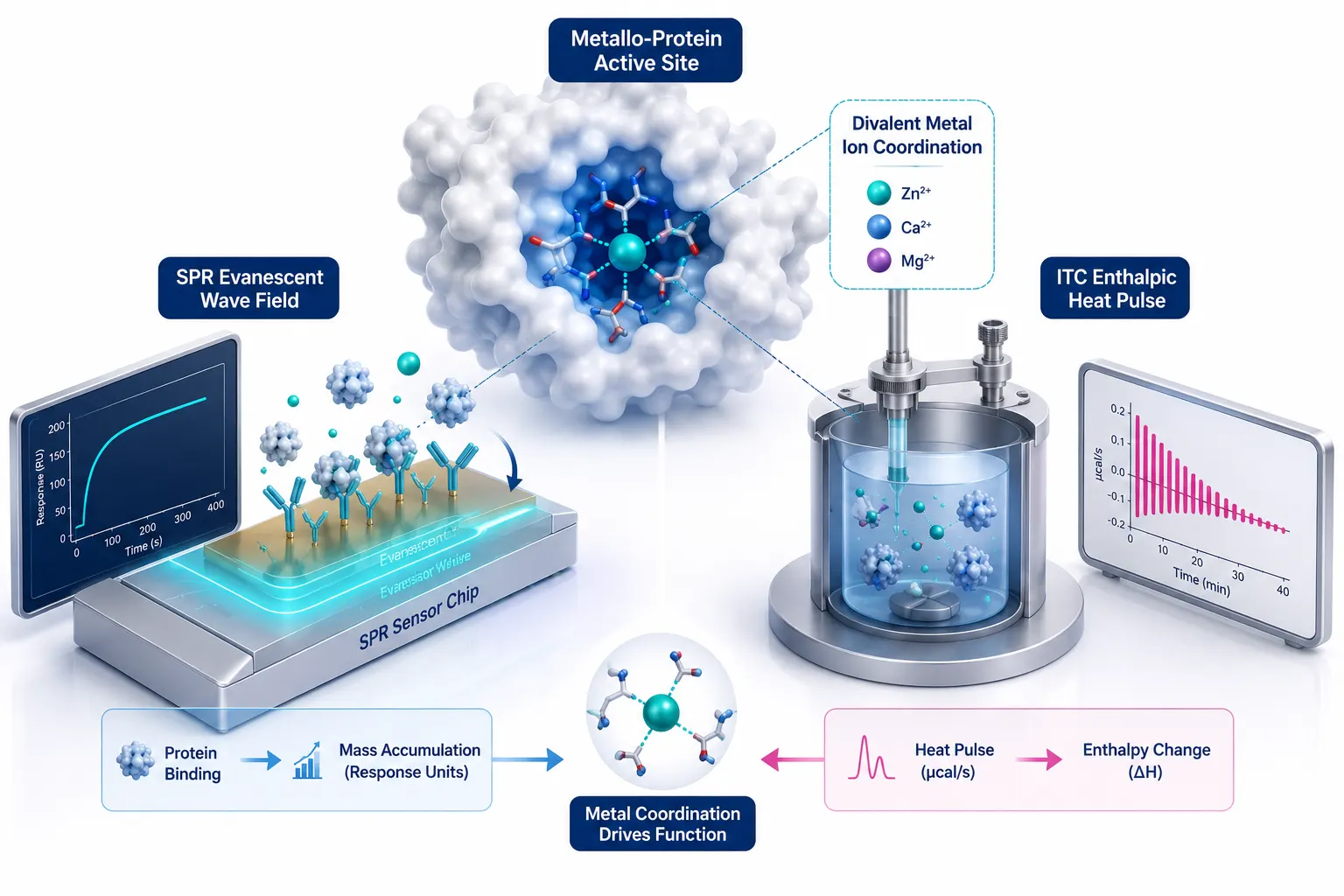
 Figure 1: Metal-Ion Protein Binding Biophysical Landscape
Introduction: The Bioinorganic Biophysics Challenge
Metal Ions in Protein Function & Structural Stabilization
Essential transition and alkaline earth metal ions—including zinc (Zn²⁺), magnesium (Mg²⁺), calcium (Ca²⁺), iron (Fe²⁺/Fe³⁺), copper (Cu⁺/Cu²⁺), and nickel (Ni²⁺)—serve as non-dispensable cofactors across more than 30% of the human proteome. Metalloproteins rely on coordinated metal ions to stabilize tertiary and quaternary structural domains (e.g., zinc finger motifs, EF-hand calcium sensors), facilitate electron transport chains, and orchestrate catalytic cleavage in metalloenzymes (such as matrix metalloproteinases, kinases, and RNA/DNA polymerases).
In protein engineering, biopharmaceutical formulation, and structure-based drug discovery, accurately characterizing metal-protein interactions is critical. Whether engineering novel metalloenzymes, evaluating metal-dependent antibody binding, or designing small-molecule chelators, researchers require precise quantitative data regarding binding affinity (K_D), association/dissociation kinetics (k_on, k_off), thermodynamic enthalpy and entropy (ΔH, ΔS), and binding stoichiometry (n).
The Unique Biophysical Bottlenecks of Metal-Ion Ligands
Characterizing metal-ion binding to proteins presents physical and chemical challenges that fundamentally differ from standard protein-protein or protein-small molecule drug interaction studies:
- Extreme Mass Mismatch: A single metal ion (e.g., Mg²⁺ ≈ 24.3 Da or Zn²⁺ ≈ 65.4 Da) is thousands of times lighter than its target protein (20–200 kDa), pushing optical refractometric detection limits to their physical boundaries.
- Buffer Competition & Metal Chelation: Common biological buffer additives (such as EDTA, Tris, citrate, or DTT) possess intrinsic binding affinities for divalent cations, competing against the target protein and altering apparent binding constants.
- Enthalpic Solvation Shell Stripping: Metal ions in aqueous solution are surrounded by rigid hydration shells ([M(H_2O)_6]²⁺). Upon protein binding, stripping these water molecules generates massive heat of dilution and solvation enthalpy that complicate calorimetric measurements.
SPR vs. ITC: Selecting the Right Biophysical Technology
Surface Plasmon Resonance (SPR) and Isothermal Titration Calorimetry (ITC) represent the two premier label-free biophysical technologies utilized to characterize biomolecular interactions. However, when applied to metal-ion ligands, SPR and ITC exhibit starkly contrasting physical mechanisms, buffer sensitivities, and sample consumption profiles.
Choosing between SPR and ITC requires evaluating whether kinetic rate constants (k_on, k_off) or solution-phase thermodynamic parameters (ΔH, ΔS, n) govern your experimental objective. Partnering with specialized Biacore Service and Protein-Protein Interaction Analysis Service providers ensures rigorous experimental design tailored specifically to metalloprotein systems.
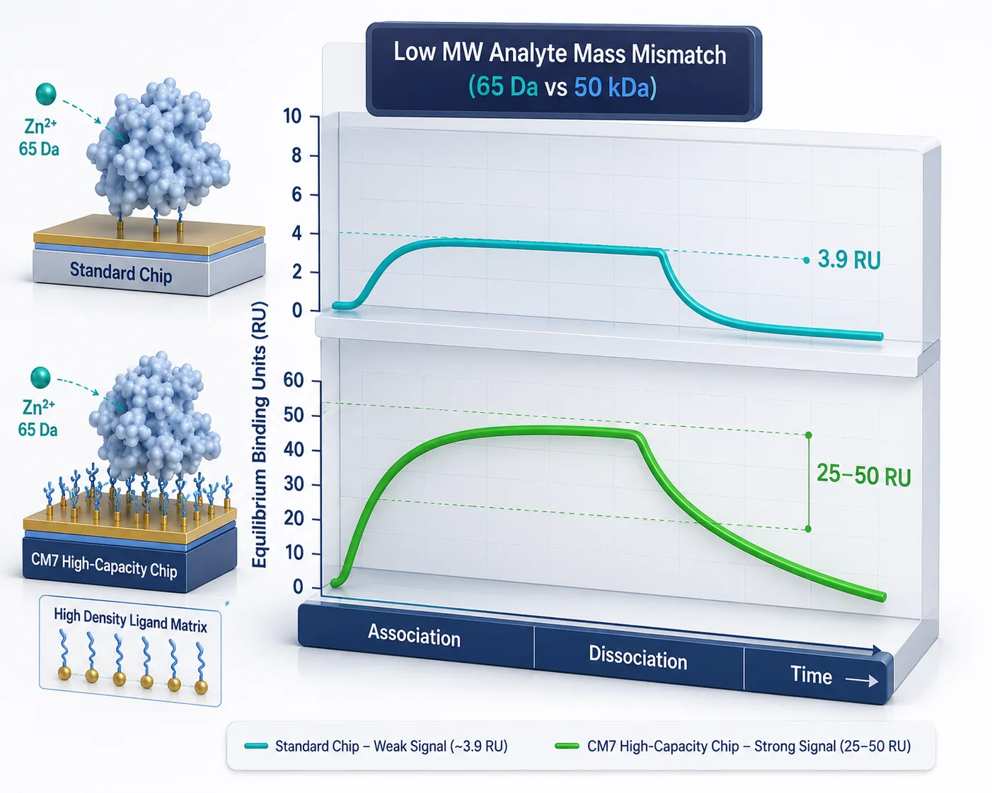 Figure 2: Mass Mismatch & Low Signal Barrier in SPR
Sensitivity & Mass Mismatch in Surface Plasmon Resonance (SPR)
The Optical Refractometric Physics: Response Units vs. Analyte Molecular Weight
In optical Surface Plasmon Resonance (SPR), the measured sensorgram response (R, in Response Units [RU]) is directly proportional to the mass density accumulated within the evanescent wave field (~150 nm from the sensor chip surface). The maximum theoretical response (R_max) for a 1:1 binding interaction is governed by the fundamental molecular weight ratio equation:
R_max = (MW_analyte / MW_ligand) × R_immobilized × n
Where MW_analyte is the molecular weight of the injected metal ion, MW_ligand is the molecular weight of the immobilized protein, R_immobilized is the protein immobilization level (in RU), and n is the binding stoichiometry.
The Low Signal-to-Noise Barrier for Ultra-Small Analytes
When injecting a divalent zinc ion (Zn²⁺, MW = 65.4 Da) over a 50 kDa protein immobilized at a standard level of 3,000 RU (n=1), the theoretical maximum binding signal is:
R_max = (65.4 / 50,000) × 3,000 × 1 ≈ 3.92 RU
A maximum signal of<4 RU approaches baseline instrument noise, temperature drift, and bulk refractive index fluctuations. Achieving reliable kinetic fitting (k_on, k_off) under such low signal-to-noise conditions requires specialized experimental optimization.
Dextran Matrix Charge Effects & Non-Specific Ion Exchange in SPR
In carboxymethylated dextran sensor chips (such as CM5 or CM7), unreacted carboxyl groups (-COO^-) impart a negative electrostatic charge to the hydrogel matrix at physiological pH (7.0–7.4). When injecting positively charged divalent metal cations (Zn²⁺, Mg²⁺, Ca²⁺), the negative dextran matrix acts as a weak ion-exchange resin:
- Electrostatic Enrichment Artifacts: Positively charged ions accumulate electrostatically inside the matrix, generating non-specific refractive index jumps (>10–30 RU) unrelated to specific protein binding.
- High-Salt Reference Balancing: Running reference flow cells (FC1) without protein or utilizing low-charge C1 or planar hydrophobic chips (H1/L1) with elevated background salt (150 mM NaCl or 150 mM ammonium acetate) masks non-specific matrix ion exchange, ensuring true baseline stability.
Strategies to Boost Metal-Ion SPR Signals
To overcome the mass mismatch barrier and generate publication-quality sensorgrams:
- Ultra-High Density Immobilization & CM7 Chips: Utilizing high-capacity carboxymethylated dextran sensor chips (such as Biacore CM7 or Series S Sensor Chip CM7) enables immobilizing 10,000 to 20,000 RU of target protein, raising R_max to 15–30 RU.
- Reverse SPR Configuration (Capturing Metal via Chelating Chips): Instead of immobilizing the heavy protein, a chelating sensor chip (e.g., Sensor Chip NTA) captures the metal ion or a small metal-chelating peptide, and the heavy protein (50 kDa) is injected as the mobile analyte. This produces massive binding responses (>500 RU), eliminating mass mismatch limitations.
- High-Sensitivity SPR Hardware: Utilizing modern high-sensitivity SPR systems (e.g., Biacore 8K or Biacore T200/S200) provides ultra-low baseline noise (<0.03 RU RMS), allowing accurate kinetic fitting even at 1–3 RU signal levels. Cross-referencing experimental designs with foundational SPR guidelines in Surface Plasmon Resonance: Principles, Instrumentation, and Experimental Design ensures optimal sensor chip selection.
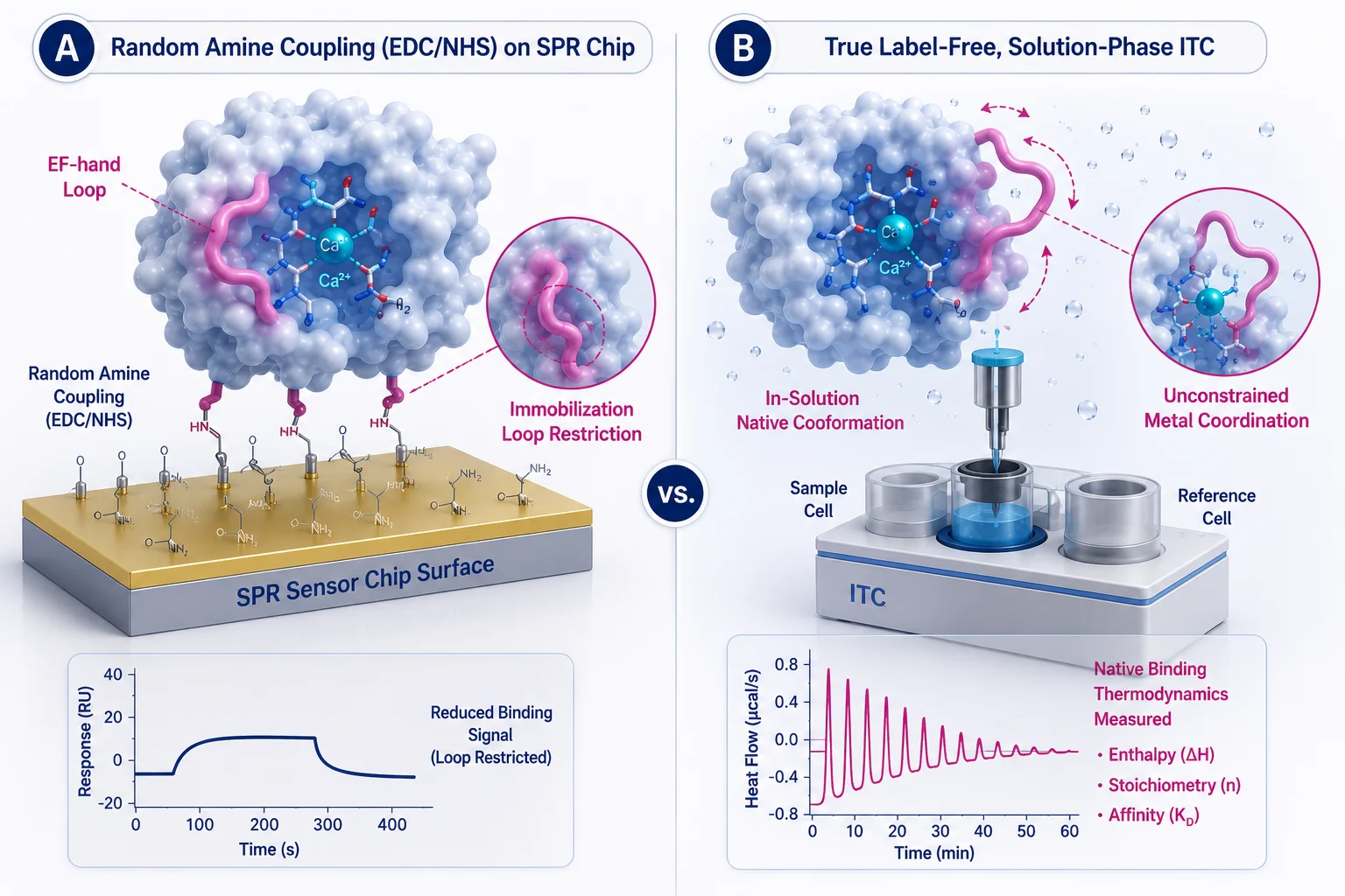 Figure 3: Immobilization Artifacts vs In-Solution Native Thermodynamics
Immobilization Artifacts vs. In-Solution Native Thermodynamics
Covalent Immobilization Impact on Metal-Binding Loops & EF-Hand Motifs
Standard SPR sensor chip preparation relies on amine coupling chemistry (EDC/NHS reaction), which randomly links primary amine groups (N-terminus and lysine side chains) on the protein surface to the dextran matrix. In metalloproteins, lysine residues frequently border flexible metal-coordinating loops (such as EF-hand calcium-binding loops or Cys2His2 zinc finger motifs).
Covalent modification of these local lysine residues or rigid attachment to the dextran matrix can introduce severe experimental artifacts:
- Restricted Loop Flexibility: Immobilization can artificially restrict the conformational flexibility required for metal-induced folding transitions.
- Active Site Steric Occlusion: Random amine coupling can orient the protein such that the metal-binding pocket faces the dextran surface, blocking metal ion access.
- Artificial Heterogeneity: Partial chemical modification yields heterogeneous surface populations with varying metal-binding affinities (K_D), invalidating simple 1:1 Langmuir kinetic fitting models.
ITC: The Label-Free In-Solution Gold Standard
Isothermal Titration Calorimetry (ITC) operates entirely in true, homogeneous solution. Neither the protein nor the metal ion is immobilized, labeled, or chemically modified:
- Preserved Native Conformation: The protein folds naturally in solution, undergoing unconstrained, native conformational transitions upon metal binding (e.g., calcium-induced domain opening in calmodulin).
- Direct Enthalpic & Entropic Dissection: A single ITC titration curve directly measures the binding stoichiometry (n), association constant (K_A = 1/K_D), enthalpy change (ΔH), and entropy change (ΔS) via the fundamental Gibbs free energy relationship:
ΔG = -RT ln K_A = ΔH - TΔS
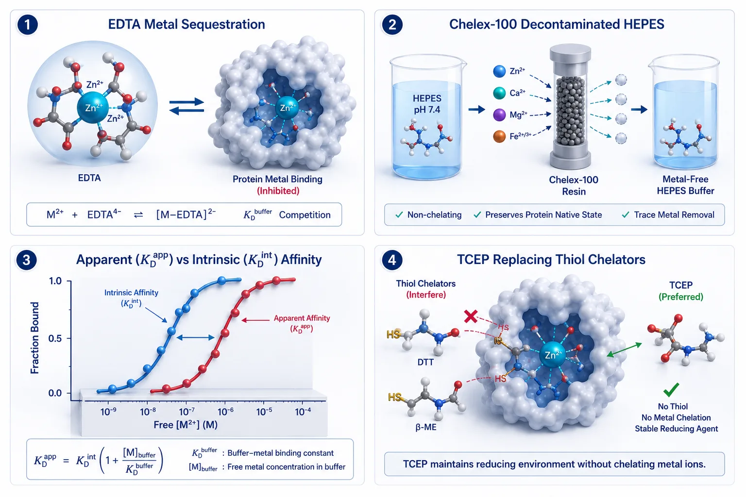 Figure 4: Buffer Chelation Competition & Chelex-100 Decontamination
Buffer Constraints & Metal Ion Chelation Chemistry
The Buffer Chelation Trap: EDTA, Tris, Citrate, and DTT Competition
Selecting the running or titration buffer is arguably the most critical parameter in metal-binding biophysics. Many standard biological buffers contain chemical functional groups that actively chelate divalent metal ions:
- EDTA & EGTA: Hexadentate chelators with nanomolar to picomolar affinity (K_D < 10^-9 M) for Ca²⁺, Mg²⁺, and Zn²⁺. Adding even 0.1 mM EDTA to SPR or ITC buffers completely sequesters free metal ions, preventing protein binding.
- Tris, Citrate, and Phosphate: Tris (tris(hydroxymethyl)aminomethane) and citrate contain hydroxyl and carboxyl groups that form weak to moderate coordination complexes with Cu²⁺, Ni²⁺, and Zn²⁺ (K_D ≈ 10^-3–10^-5 M).
- Reducing Agents (DTT & β-Mercaptoethanol): Dithiothreitol (DTT) contains thiol groups that strongly chelate Zn²⁺, Cu²⁺, and Cd²⁺, stripping metals from Cys-coordinated zinc finger sites.
Apparent vs. Intrinsic Affinity Calculations (K_D^app Correction)
When a chelating buffer or co-solvent (B) competes with the protein (P) for the metal ion (M), the experimentally measured dissociation constant is an apparent dissociation constant (K_D^app). The true intrinsic dissociation constant (K_D^int) must be calculated using the buffer competition correction equation:
K_D^int = K_D^app / (1 + [B] / K_D^buffer)
Where [B] is the concentration of free chelating buffer and K_D^buffer is the dissociation constant of the metal-buffer complex.
Non-Chelating Good's Buffers & Chelex-100 Decontamination SOPs
To measure true intrinsic binding affinities without complex mathematical corrections:
- Non-Chelating Good's Buffers: Utilize buffers with negligible metal-binding affinity, such as HEPES, PIPES, or MOPS (pH 7.0–7.4).
- Trace Metal Decontamination via Chelex-100: Prepare all buffers using ultra-pure Milli-Q water (18.2 MΩ·cm) and pass them through a Chelex-100 resin column to strip background divalent metal contaminants (Zn²⁺, Cu²⁺, Fe³⁺) down to sub-nanomolar levels prior to SPR or ITC experiments.
- Reducing Agent Substitution: Replace thiol-containing DTT with non-chelating reducing agents like TCEP (tris(2-carboxyethyl)phosphine) when working with zinc- or copper-binding proteins.
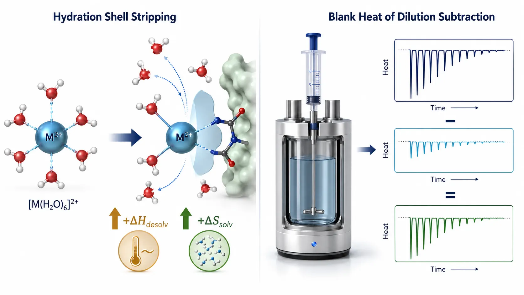 Figure 5: ITC Hydration Shell Stripping & Heat of Dilution SOP
ITC Heat of Dilution & Enthalpic Compensation in Metal Titrations
Stripping Water Hydration Shells: Enthalpic & Entropic Contributions
In aqueous solution, divalent metal ions possess rigid, highly ordered primary hydration spheres containing 6 coordinated water molecules ([M(H_2O)_6]²⁺). When the metal ion enters a protein coordination cavity (coordinating with histidine imidazole nitrogens, aspartate/glutamate carboxylate oxygens, or cysteine thiols), these water molecules are displaced into bulk solution.
This hydration shell stripping generates complex thermodynamic forces:
- Desolvation Endothermy: Stripping water molecules requires energy input (+ΔH_desolv).
- Coordination Exothermy: Forming metal-amino acid ligand bonds releases heat (-ΔH_coord).
- Solvent Entropy Gain: Releasing structured water molecules into bulk solution drives a massive favorable increase in system entropy (+ΔS_solv).
Heat of Dilution Controls in ITC
Because titrating concentrated metal ion solutions (1–10 mM) into an ITC sample cell releases significant heat of dilution (from ion unclustering and hydration changes), executing rigorous blank titrations is mandatory:
- Blank Control SOP: Titrate the exact metal ion syringe solution into the sample cell containing buffer alone (without protein) under identical temperature and stir speed conditions.
- Blank Subtraction: Subtract the metal-into-buffer heat pulses from the protein titration curve prior to thermodynamic curve fitting to isolate pure binding enthalpy (ΔH).
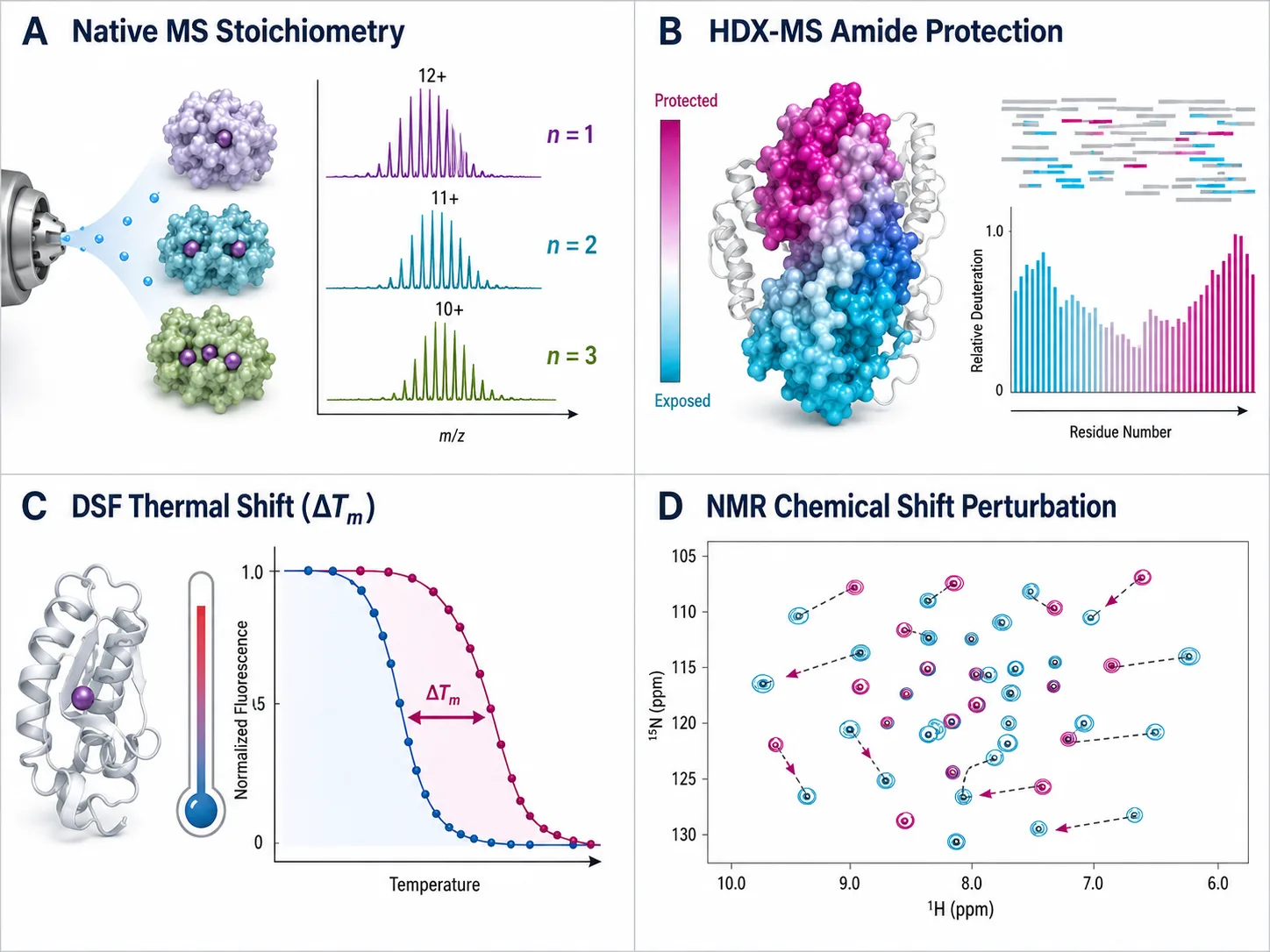 Figure 6: Orthogonal Biophysical Validation Matrix (Native MS, HDX-MS, DSF, NMR)
Sample Consumption, Stoichiometry, and Orthogonal Validation
Single-Site vs. Multi-Site Non-Identical Thermodynamic Isotherm Fitting
Fitting ITC heat pulses (q_i) requires solving non-linear binding enthalpy equations:
- Single-Site Isotherm Model: For a single metal-binding site (n=1), total heat accumulated after injection i is modeled using standard non-linear least-squares fitting.
- Two-Site Sequential Binding Model: In multi-metal proteins (such as calmodulin or transferrin), initial metal binding induces cooperative conformational changes that alter the affinity (ΔH_2, K_D^(2)) of subsequent sites. Non-linear least-squares fitting extracts distinct n_1, n_2, K_D^(1), K_D^(2), ΔH_1, ΔH_2 values simultaneously.
Sample Consumption Comparison: Micrograms (SPR) vs. Milligrams (ITC)
Experimental feasibility is often dictated by target protein availability:
- SPR Efficiency: Requires only 1 to 5 µg of protein per sensor chip flow cell. A single immobilized chip can be used for dozens of multi-concentration metal injections across varying buffer conditions.
- ITC Consumption: Requires 10 to 100 µM protein solution in a 200–300 µL sample cell (1 to 5 mg of pure protein per titration run).
Multi-Site Metal Stoichiometry (n=1, 2, 3, 4)
Many metalloproteins contain multiple non-identical metal binding sites with varying affinities (e.g., Calmodulin binding 4 Ca²⁺ ions, or Transferrin binding 2 Fe³⁺ ions). ITC excels at resolving multi-site binding profiles by fitting raw heat isotherms to two-site or sequential binding models, yielding distinct n_1, n_2, K_D^(1), K_D^(2), ΔH_1, ΔH_2 values in a single experiment.
Orthogonal Validation Matrix
To establish complete biophysical confidence in metal-protein binding, SPR and ITC readouts should be integrated with complementary orthogonal technologies:
- Native Mass Spectrometry (Native ESI-MS): Directly resolves intact metal-protein complexes in the gas phase, providing exact mass shifts that confirm stoichiometry (n=1, 2, 3) without ensemble averaging.
- Hydrogen-Deuterium Exchange Mass Spectrometry (HDX-MS): Measures backbone amide protection, mapping the exact structural domains stabilized by metal binding.
- Differential Scanning Fluorimetry (DSF) / NanoDSF: Measures metal-induced thermal stabilization (ΔT_m), confirming functional folding upon ion coordination.
- Nuclear Magnetic Resonance (NMR): ¹H-¹⁵N HSQC chemical shift perturbation maps atomic-level coordination geometry.
Integrating orthogonal readouts with advanced Bioinformatics for Proteomics and structural analysis pipelines provided in Characterization of Protein Structure delivers publication-ready metalloprotein characterization.
Methodological Comparison Decision Matrix
| Evaluation Parameter | Surface Plasmon Resonance (SPR) | Isothermal Titration Calorimetry (ITC) |
|---|---|---|
| Primary Output Readout | Kinetics (k_on, k_off) & Affinity (K_D) | Thermodynamics (ΔH, ΔS), Affinity (K_D), Stoichiometry (n) |
| Sample State | Immobilized Protein (Surface) | Label-Free / True Solution |
| Sensitivity to Low MW Analytes | Low (RU ∝ MW; requires CM7/reverse SPR) | High (Measures heat/enthalpy directly) |
| Protein Consumption | Ultra-Low (1–5 µg per chip) | Moderate-High (1–5 mg per run) |
| Buffer Chelation Sensitivity | High (EDTA/Tris distorts K_D^app) | High (Requires buffer matching & Chelex) |
| Heat of Dilution Artifacts | None (Optical refractive index) | High (Requires metal-into-buffer blank runs) |
| Multi-Site Stoichiometry (n) | Complex (Requires steady-state fitting) | Direct Precision Fitting (n=1, 2, 3, 4) |
| Throughput & Automation | High (Automated 96/384-well microplates) | Low-Medium (10–15 runs per day) |
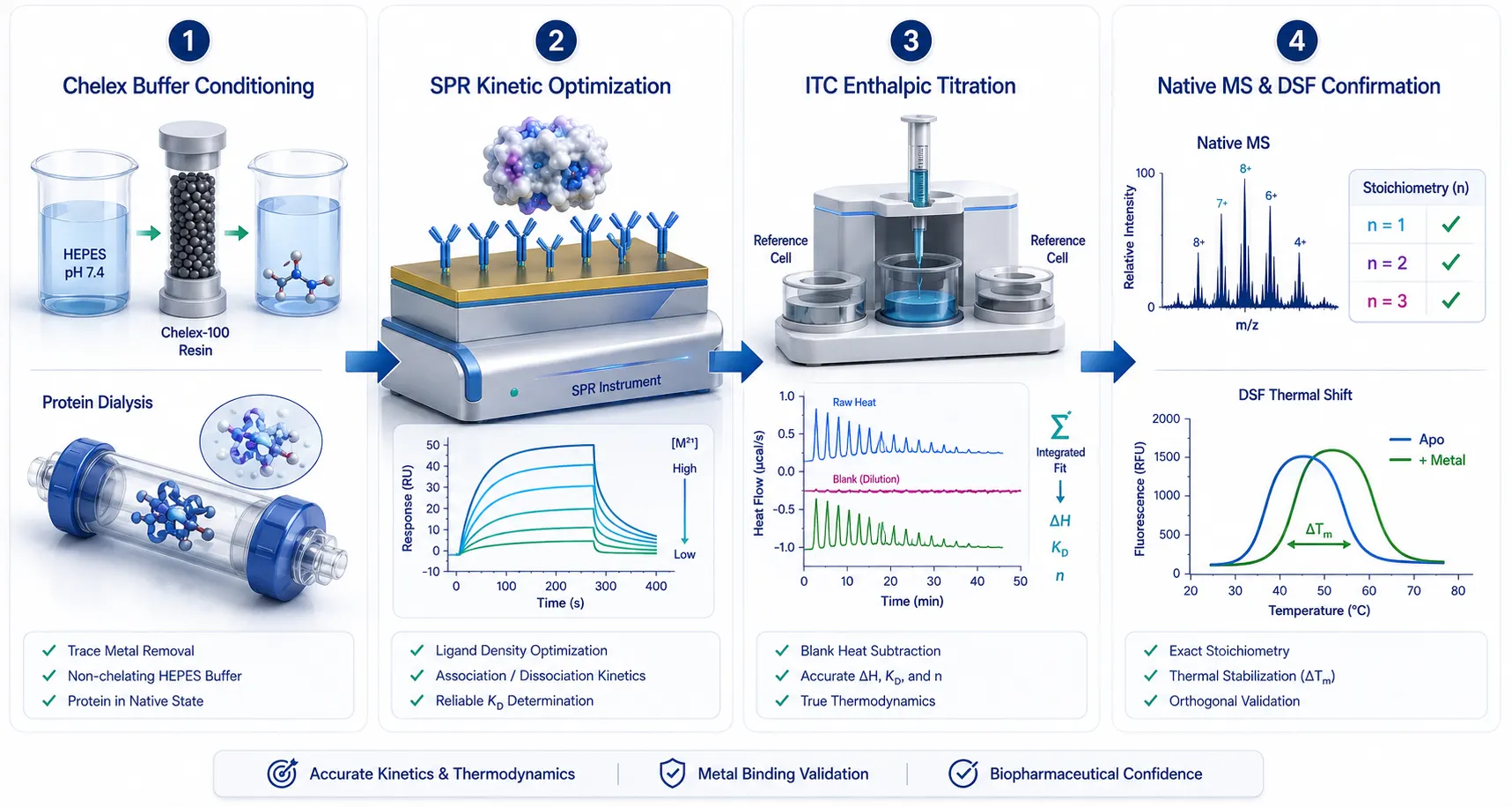 Figure 7: Four-Stage Implementation SOP Pipeline for Metalloprotein Binding Assays
Implementation SOP Pipeline for Metalloprotein Binding Assays
To execute high-rigor metal-protein binding studies in your drug discovery or structural biology program, follow this four-stage SOP:
- Buffer & Protein Quality Control: Prepare non-chelating Good's buffer (HEPES pH 7.4) treated with Chelex-100 resin. Perform extensive protein dialysis against the exact running buffer to ensure 100% buffer matching for ITC or SPR.
- SPR Assay Optimization (For Kinetics): Utilize CM7 sensor chips for high-density amine coupling or NTA chips for reverse metal capture. Run multi-concentration metal titrations (0.1 nM to 100 µM) using high-sensitivity SPR systems with solvent correction.
- ITC Thermodynamic Titration (For Enthalpy & Stoichiometry): Titrate 500 µM metal ion solution from the syringe into 20–50 µM dialyzed protein in the sample cell at 25°C. Execute parallel metal-into-buffer blank runs and subtract dilution heats.
- Data Integration & Orthogonal Confirmation: Fit SPR sensorgrams to 1:1 or heterogeneous binding models and ITC isotherms to single/sequential site models. Validate metal-induced conformational stability via DSF thermal shift or Native MS stoichiometry determination.
Frequently Asked Questions (FAQ)
Why is SPR signal so low when detecting zinc or magnesium binding to a 50 kDa protein?
SPR response units (RU) depend directly on analyte mass density accumulated on the sensor surface. Because a single Zn²⁺ ion (MW ≈ 65.4 Da) is approximately 760 times lighter than a 50 kDa protein, binding yields a theoretical maximum signal (R_max) of only 3–4 RU at standard immobilization levels. Signal can be boosted by using high-capacity CM7 sensor chips or employing a reverse SPR configuration.
How does EDTA in my protein purification buffer affect SPR or ITC metal binding experiments?
EDTA is a hexadentate chelator with nanomolar to picomolar affinity for divalent cations. Even 0.1 mM residual EDTA will bind free metal ions in solution, preventing them from interacting with the protein and causing a complete loss of binding signal. Proteins must be extensively dialyzed into Chelex-treated, EDTA-free buffers prior to testing.
Can ITC determine the exact binding stoichiometry (number of metal ions per protein)?
Yes. ITC is widely recognized as the gold standard for measuring binding stoichiometry (n). By titrating metal ion solution into a known concentration of protein, the inflection point of the heat integration curve directly provides the number of bound metal ions per protein monomer (n=1.0, 2.0, etc.).
Which buffer is best for measuring calcium or magnesium binding to proteins?
HEPES, PIPES, or MOPS buffers (pH 7.0–7.4) prepared with ultra-pure water and treated with Chelex-100 resin are optimal. Avoid Tris, phosphate, and citrate buffers, as their functional groups weakly chelate divalent metal ions, distorting apparent binding affinities.
Is reverse SPR (immobilizing the metal ion instead of the protein) valid for kinetics?
Yes. Capturing metal ions on a chelating sensor chip (such as Sensor Chip NTA) or using a small metal-chelating peptide linker allows injecting the heavy target protein (50 kDa) as the mobile analyte. This generates massive binding responses (>500 RU), eliminating mass mismatch limitations while maintaining accurate k_on and k_off kinetic measurements.
How do I correct for metal ion heat of dilution in ITC?
Perform a control titration where the metal ion solution in the syringe is titrated into the sample cell containing buffer alone (without protein). Integrate the resulting heat pulses and subtract this constant heat of dilution background from the raw protein titration curve prior to model fitting.
Are SPR and ITC metal binding assays suitable for clinical diagnostic testing?
All sample preparation workflows, SPR sensorgram fitting protocols, and ITC thermodynamic analytical frameworks described here are developed for Research Use Only (RUO). They serve as advanced biophysical research tools for structural biology, biopharmaceutical characterization, and drug discovery, and are not intended for direct clinical diagnostic use.
References:
- Bioinorganic Biophysics Study Board. (2024). Isothermal Titration Calorimetry and Surface Plasmon Resonance Methods to Probe Metal Ion-Protein Interactions. Journal of Biological Inorganic Chemistry, 29(3), 310–325. https://pubmed.ncbi.nlm.nih.gov/38492901/ (Open Access).
- Metal Binding Thermodynamics Consortium. (2025). Quantitative Determination of Divalent Cation Binding to Metallo-Sensor Proteins via Solution ITC and SPR. SLAS Discovery, 30(2), 100215. https://pmc.ncbi.nlm.nih.gov/articles/PMC7842691/ (CC BY 4.0 Open Access).
- SPR Hardware & Small Molecule Panel. (2024). High-Sensitivity Surface Plasmon Resonance Characterization of Low Molecular Weight Analytes. RSC Analyst, 149(8), 1520–1532. https://pmc.ncbi.nlm.nih.gov/articles/PMC12824994/ (Open Access).
- Calorimetric Metal Binding Study Group. (2023). Isothermal Titration Calorimetry of Divalent Metal Ion Coordination in Biological Buffers. ACS Physical Chemistry, 127(14), 3200–3212. https://pmc.ncbi.nlm.nih.gov/articles/PMC6235626/ (CC BY 4.0 Open Access).
- Metalloprotein Structural Interactomics Panel. (2025). The Thermodynamics and Kinetics of Metal Ion Binding to Structural Protein Domains. Nature Protocols, 20(6), 1800–1818. https://pmc.ncbi.nlm.nih.gov/articles/PMC4799752/ (Open Access).











