

Meta Intent: A practical research guide for designing cell-culture supernatant proteomics studies that distinguish bona fide secretion from serum background and intracellular leakage, while preserving enough material for defensible MS-based discovery and quantification.
Conditioned medium is often the most direct experimental window into what cultured cells release into their microenvironment. It can reveal soluble mediators, shed ectodomains, extracellular-matrix regulators, and vesicle-associated cargo that are not obvious from a cell-lysate experiment alone. It is also one of the easiest proteomics samples to misread. A protein list generated from cell-culture supernatant is not automatically a secretome: it may include residual medium proteins, material released by stressed or dying cells, and species introduced during processing.
A useful secretome study therefore begins with a classification problem, not an instrument choice: which proteins are actively released under the specified culture condition, which are vesicle-associated, which are soluble, and which are more plausibly explained by contamination or membrane damage? The answer depends on a connected design that links cell handling, collection-window controls, low-input recovery, matched cell material, and peptide-level analysis. A well-planned Protein Sample Preparation workflow protects that chain before LC-MS/MS acquisition begins.
Why Cell-Culture Supernatant Is Both Valuable and Vulnerable
The cell-conditioned secretome is biologically informative because it sits at the interface between a cell and its surroundings. Secreted growth factors, cytokines, proteases, matrix proteins, and cleaved receptor ectodomains can shape paracrine signaling, cell migration, matrix remodeling, and treatment responses. The secretome also contains material exported through multiple routes: conventional ER-Golgi secretion, non-classical release, surface shedding, and extracellular-vesicle pathways. MS-based secretomics can observe these routes without selecting only a predefined panel, but the same breadth creates an interpretation challenge.
Three issues dominate most discovery studies. First, serum-supplemented medium contains a complex protein background that is analytically much stronger than many proteins released by cultured cells. Even when cells are switched to a nominally serum-free collection medium, proteins retained on culture surfaces or in the extracellular matrix can persist. Second, secreted proteins are often dilute in a comparatively large volume of medium, so every transfer, filter, and concentration step can change recovery. Third, intracellular proteins enter conditioned medium when cells lose membrane integrity. A list enriched for cytoskeletal, ribosomal, mitochondrial, or abundant metabolic proteins may signal an interpretive problem rather than an unexpectedly broad secretome.
These risks do not mean that serum-free collection is always wrong or that intracellular proteins are never informative. They mean the collection condition must be treated as an experimental perturbation with a documented control window. Current secretomics literature consistently emphasizes that medium composition, collection duration, and cell viability can alter the apparent secretome; a short medium-renewal step can also help separate adaptation artifacts from accumulated released material.1
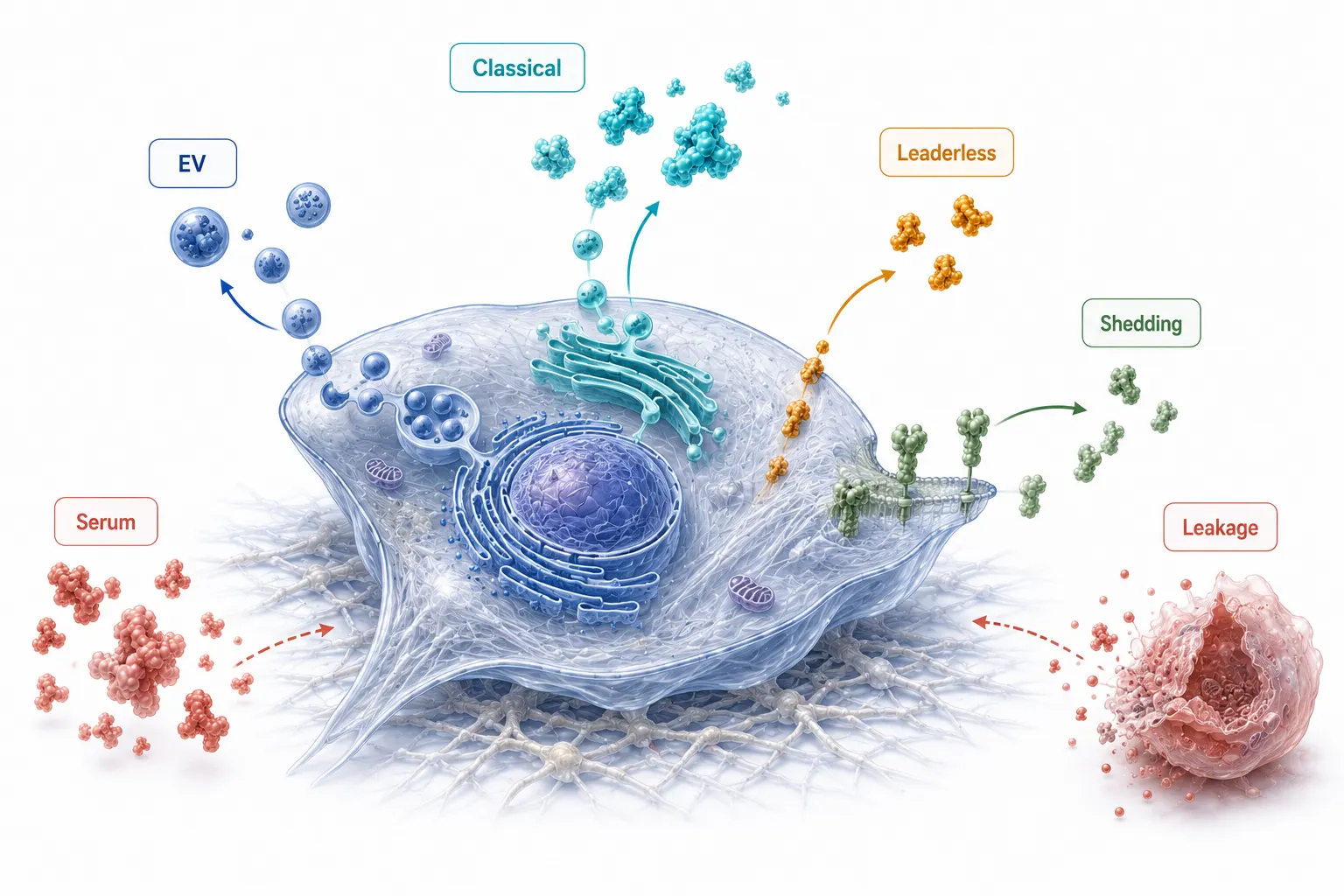 Figure 1. The cell-culture secretome includes soluble classical and non-classical exports, shed surface material, and vesicular cargo. Serum carryover and intracellular leakage are separate analytical sources that must not be conflated with secretion.
Define the Collection Window Before Removing Serum
Serum-free adaptation is a cell-model decision
Serum removal reduces a major background, but it can also change the biology being measured. Some cell types tolerate a short protein-free collection window with stable morphology and membrane integrity; others rapidly alter signaling, detach, or accumulate stress-related release. There is no universal collection duration, viability percentage, or lactate dehydrogenase threshold that proves every secretome is clean. The defensible approach is to run a small condition-finding experiment for the actual cell type, culture format, density, and treatment context.
Compare the intended collection medium and duration against the expansion condition using predeclared readouts: cell morphology, attached-cell area or count where relevant, a viable-cell measurement, a membrane-damage measurement such as LDH, and the total protein recovered from matched medium volumes. The purpose is not to optimize for the lowest possible LDH value in isolation. It is to identify a collection window that preserves the biology of interest while making release from damaged cells visible as a QC variable. If the intervention itself is cytotoxic, that fact must remain part of the design rather than being erased by a secretome filter.
Wash-out should be verified, not ritualized
Surface washing is needed to reduce serum carryover, but a fixed number of PBS washes is not a transferable SOP. Extra wash steps can detach sensitive cells or alter weakly adherent cultures; too few can leave substantial serum-derived peptide background. Use a gentle isotonic wash compatible with the model, document the number and timing of exchanges, and add a short pre-collection medium exchange when the adaptation experiment shows that it improves background control. The same procedure must be applied across biological groups so that a difference in washing does not become a difference in apparent secretion.
When cells cannot sustain the required serum-free interval, do not force the design into a nominally clean condition. Consider a serum-reduced or chemically defined system, metabolic-labeling strategy, or a focused capture method, then state the resulting analytical limitations. A Protein Identification Services project should record medium components and non-human protein databases up front, because contaminant annotation is easier before interpretation than after a candidate list has been generated.
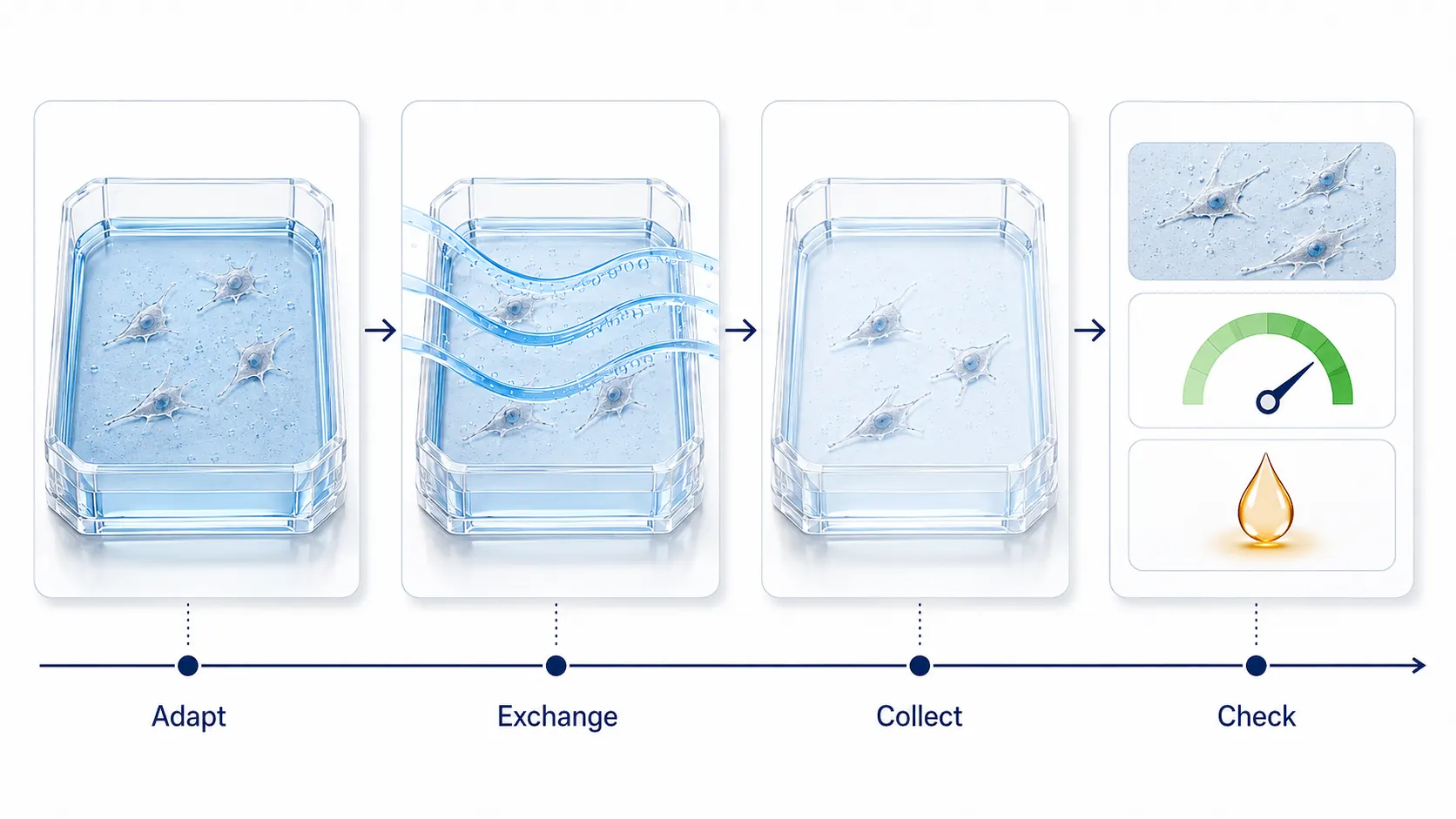 Figure 2. Serum-background control begins with a validated collection window. Adaptation, wash-out, morphology, viability, and membrane-integrity checks are interpreted together rather than as isolated pass/fail steps.
Process Conditioned Medium as a Low-Input Sample
Supernatant processing should remove cells and debris without turning a low-abundance sample into an uncontrolled fractionation experiment. Begin by documenting the starting volume, cell number or culture area, collection duration, and any visible signs of detachment. Clarify the medium using a sequence appropriate for the study question. Low-speed clearing removes cells; additional clearing can reduce larger debris. A low-protein-binding filter may be useful for soluble-secretome analysis, but it also changes what remains in the sample. If extracellular vesicles are a research question, do not apply a clarification or filter step that silently excludes the fraction you intend to measure.
Normalize the experimental design before normalization in software. Equal starting volume alone may not be meaningful if plates have different cell numbers, confluence, viability, or collection times. Choose a biological denominator that matches the question, such as viable-cell count at collection, a matched DNA measurement, culture surface area for a stable adherent model, or total cellular protein from a paired pellet. Record the denominator prospectively and do not select it only after seeing the MS results.
Include controls that locate the source of signal rather than merely adding more samples. A medium-only control identifies proteins contributed by the formulation or consumables. A processing blank can expose contaminants introduced during concentration and digestion. A cell-free control processed through the same concentration route can reveal whether low-binding plasticware, membrane filters, or precipitation chemistry produce a repeatable background. These controls should be interpreted as part of the experimental matrix, not removed as an afterthought once a differential-protein list is already assembled.
When the study compares treatments, randomize or balance processing order across groups where possible. A long collection day can otherwise align culture age, operator handling, and treatment group. Retain a pooled QC or a defined reference material when the acquisition strategy supports it, then inspect whether peptide retention, digestion behavior, and total signal drift with processing order. This will not make a poor collection window valid, but it makes technical variation easier to separate from an apparent secretion response.
Choose concentration chemistry by the failure you can tolerate
Centrifugal ultrafiltration is practical for modest volumes and offers a straightforward route to buffer exchange, but membrane adsorption and molecular-weight cutoffs can bias recovery. Organic-solvent or acid precipitation can be useful for larger volumes and can remove salts, yet pellets may be difficult to resolubilize and some protein classes may be recovered unevenly. Magnetic-bead capture methods such as SP3 are attractive when the material is dilute or low-input because capture, cleanup, and digestion occur in a compact workflow. They are not inherently lossless: bead-to-protein ratio, solvent composition, mixing, tube surfaces, and elution conditions still require a small recovery pilot.
The right comparison is not which method produces the largest total peptide count in one pooled sample. It is which method provides reproducible recovery of the protein classes that matter, produces compatible digestion chemistry, and preserves the intended comparison across replicates. For a quantitative study, an initial Precision Quantitative Proteomics Services discussion should define whether the priority is broad relative quantification, a smaller secreted-protein panel, or peptide-level confirmation of a few candidates.
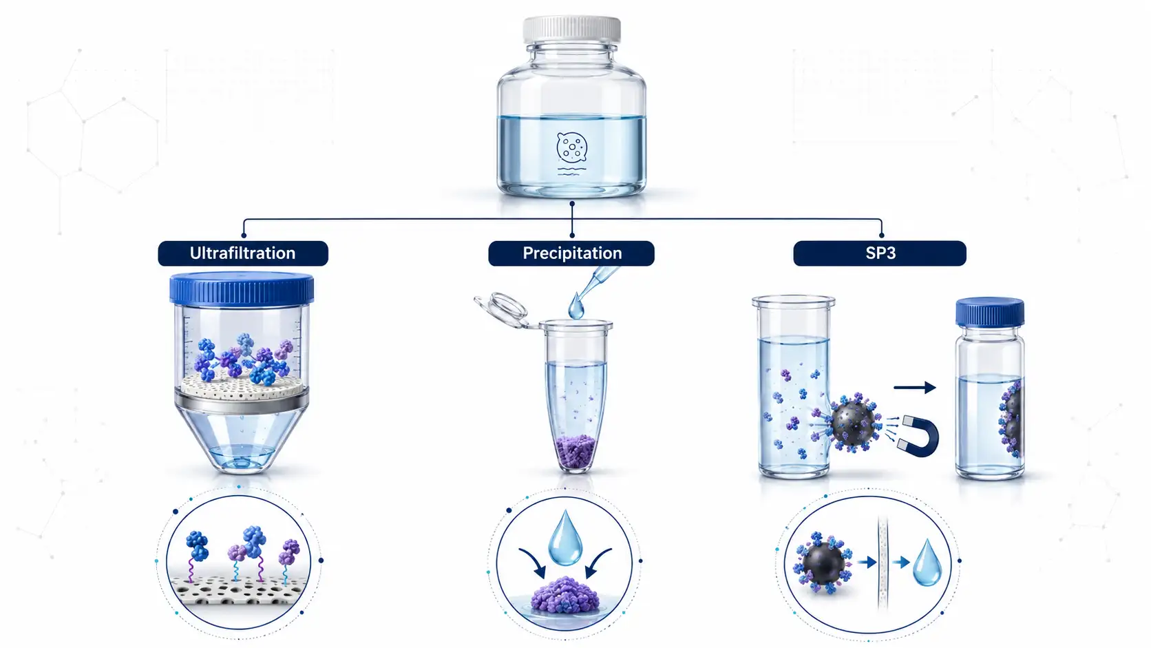 Figure 3. Concentration approaches solve different problems. Select ultrafiltration, precipitation, or SP3 on the basis of volume, expected input, recovery risk, and downstream measurement needs.
Classify Proteins With Evidence, Not a Single Prediction Tool
Signal-peptide prediction is valuable for identifying proteins compatible with classical secretion. Tools such as SignalP can provide a useful annotation layer, but a signal peptide is neither necessary for all extracellular proteins nor sufficient proof that a detected peptide arose from active secretion in the current experiment. Leaderless proteins may be released by unconventional pathways, while transmembrane proteins can appear through ectodomain shedding or vesicular export. Conversely, intracellular proteins can occur outside cells after membrane damage. Annotation is an evidence layer, not a verdict.
Peptide placement can sharpen that interpretation. For a membrane protein, peptides from an extracellular domain are more compatible with ectodomain shedding than peptides restricted to a cytosolic tail, although digestion and database assignment still limit certainty. For a processed precursor, a mature-chain peptide can provide different evidence from a propeptide. Glycosylation and other modifications can change peptide observability, so a missing unmodified tryptic peptide should not be treated as proof that the parent protein is absent. If route or proteoform is central to the biological question, define the expected peptide evidence before acquisition instead of relying only on protein-level roll-up.
Use a classification table that keeps each evidence source separate: signal-peptide prediction, transmembrane annotation, subcellular knowledgebase annotation, known extracellular-vesicle association, observed peptide locations, and abundance in matched cells. Prediction tools for leaderless or unconventional release should be treated as hypothesis generators because their performance and training assumptions vary. Comparative secretomics is more informative when it asks whether a protein is enriched in conditioned medium relative to the matched cellular compartment, while also considering known biology and experimental perturbations.1
Separate soluble secretome and vesicle-associated material intentionally
Conditioned medium can be analyzed as a total extracellular fraction, a soluble fraction, an extracellular-vesicle-enriched fraction, or paired fractions. These are different questions. Differential centrifugation, size-exclusion chromatography, and other separation methods enrich rather than absolutely purify a category of material, so describe the operational fraction that was collected rather than claiming that every identified protein is exclusively exosomal or soluble. Marker proteins, particle characterization, and process controls should match the study aim. A total-medium experiment is appropriate for a broad release phenotype; a fractionated design is appropriate when cargo route is the biological question.
For discovery datasets, a DIA Quantitative Proteomics Service can provide a consistent matrix across multiple conditions, while targeted follow-up is often the stronger choice once a small set of extracellular candidates has been prioritized. The acquisition strategy should follow the claim: broad discovery, differential secretion, route-specific fractionation, or confirmation of defined targets.
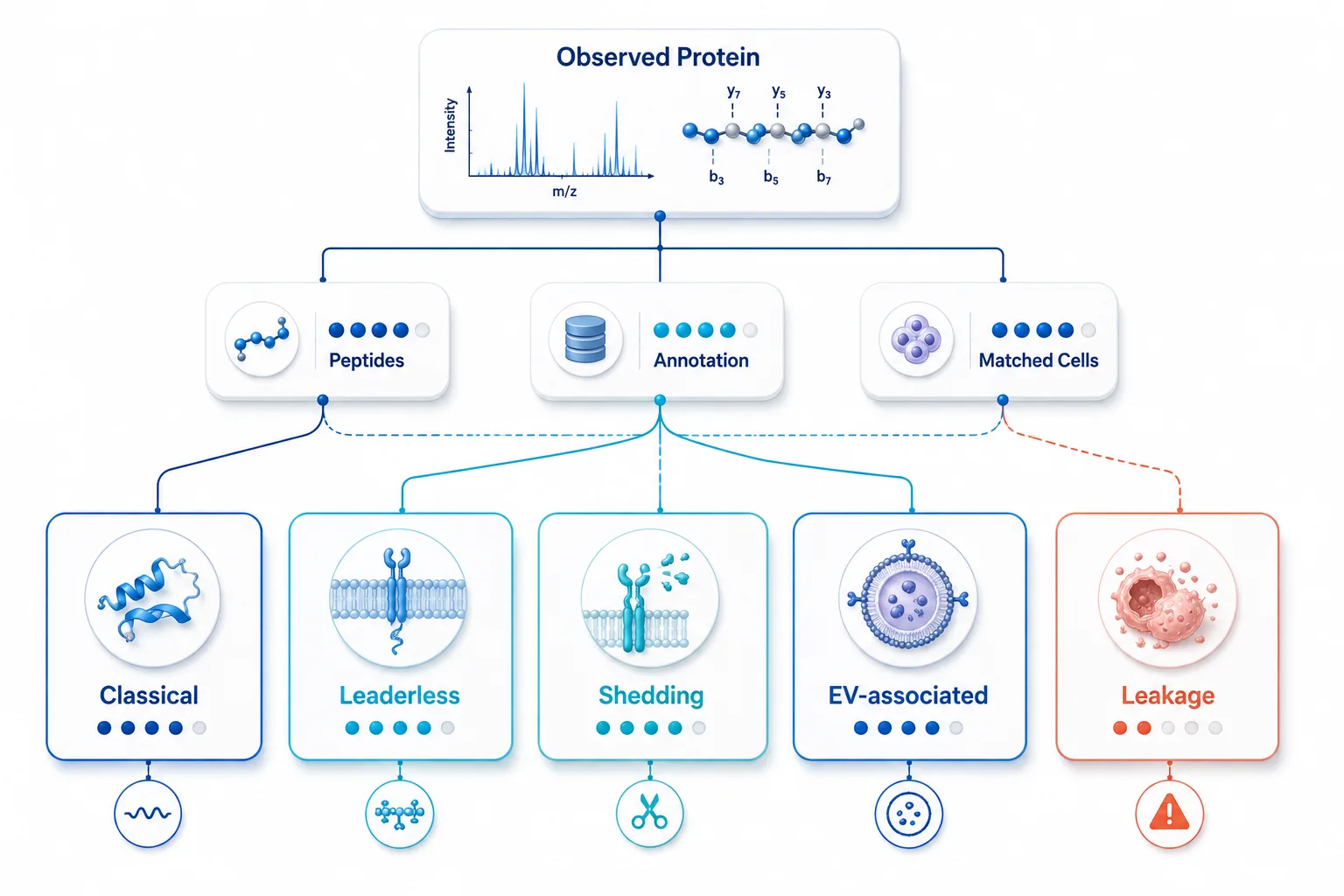 Figure 4. Secreted-versus-leaked classification requires converging evidence. Signal-peptide and unconventional-secretion predictions guide interpretation but cannot replace matched experimental controls.
Use Matched Cell Material to Test the Leakage Hypothesis
A matched cell pellet or cell-lysate sample collected from the same biological replicate is one of the most useful controls in secretomics. It does not make every intracellular protein a contaminant. Instead, it provides the context needed to ask whether a protein is disproportionately represented outside the cell relative to its cellular abundance. Comparative approaches have been shown to enrich for known secreted proteins while helping to expose candidates whose appearance in medium is more consistent with cellular leakage.
Do not convert this comparison into a universal CM-to-lysate cutoff. The ratio depends on how the two fractions were collected, total protein load, volume, cell number, digestion yield, normalization method, and protein turnover. A ratio can rank candidate behavior within a defined experiment; it does not establish a transferable boundary between "secreted" and "not secreted." Predefine how paired data will be scaled, retain peptide-level values where possible, and interpret the ratio alongside LDH, viability, protein annotation, and chromatographic evidence.
This control is especially valuable for treatments expected to alter adhesion, membrane permeability, or proliferation. If a treatment raises both LDH and the abundance of intracellular markers in medium, a difference in the supernatant may reflect damage-associated release. If a candidate is enriched in medium without a corresponding cell-lysis pattern and is supported by extracellular annotation or peptide location, it becomes a stronger secretion hypothesis. A Bioinformatics for Proteomics workflow should preserve these flags rather than collapsing all fractions into a single unannotated fold-change table.
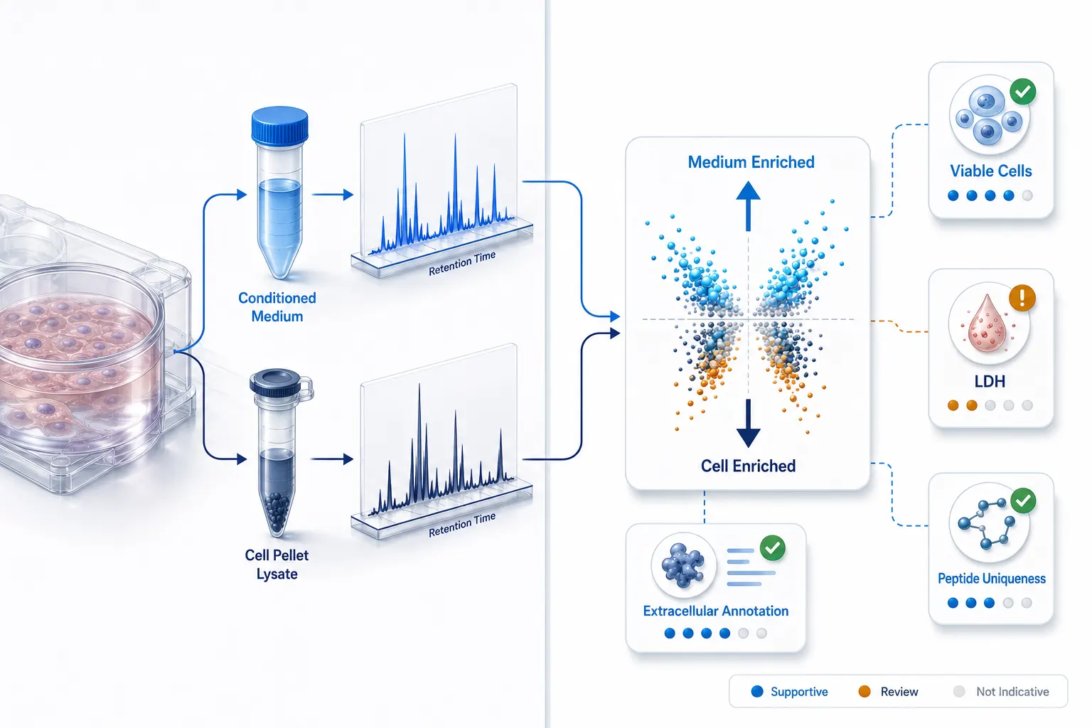 Figure 5. Matched cell material turns intracellular leakage into a testable explanation. The CM-to-lysate relationship is interpreted with QC and annotation evidence, not as a universal numeric cutoff.
Build the Method Around the Research Question
Secretome workflows are often overbuilt because the sample appears simple. Use the least complex design that can support the requested evidence. For a comparative screen of soluble proteins across many culture conditions, a validated serum-free window, clarified medium, low-input concentration, and DIA may be sufficient. For subtle treatment-driven changes, include enough biological replication and paired lysate controls to distinguish secretion from damage. For a narrow panel of candidates, a discovery run can nominate peptides, followed by a more focused Parallel Reaction Monitoring (PRM) or SRM & MRM assay.
Plan the comparison at the replicate level. Technical injections can reveal instrument or preparation repeatability, but they cannot substitute for independent cultures when the question is biological release. Keep the condition, collection time, initial cell state, and processing batch traceable for every replicate. If the experiment includes a pharmacological perturbation, include vehicle controls and decide before analysis whether the intended outcome is an extracellular abundance change, a change after normalization to viable cells, or a route-specific shift between soluble and vesicle-enriched fractions. Each definition can produce a different but valid answer when it has been specified in advance.
| Workflow question | Useful starting design | Key control | Do not infer automatically |
|---|---|---|---|
| Broad soluble release phenotype | Validated collection window, clarified medium, low-input DIA | Viability, LDH, medium-only and matched cells | That every detected protein is actively secreted |
| Low-input or dilute candidate discovery | Recovery pilot comparing ultrafiltration, precipitation, or SP3 | Recovery and reproducibility of relevant peptide classes | That the method with most IDs has the least bias |
| Vesicle versus soluble cargo | Predefined fractionation with fraction-specific MS analysis | Operational-fraction and marker characterization | That enrichment equals pure EV origin |
| Small, decision-driving target list | Discovery nomination followed by PRM or SRM/MRM | Unique peptides and fit-for-purpose confirmation | That discovery intensity is an absolute concentration |
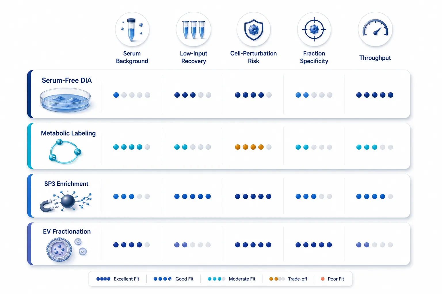 Figure 6. Method selection matrix. A workflow should be chosen for the bottleneck it resolves, not for the number of processing layers it adds.
Use This Four-Phase Secretome Implementation Plan
- Define the biological release question. Specify whether the study concerns total extracellular material, soluble proteins, vesicle-associated cargo, surface shedding, or candidate confirmation. Define the required evidence claim before the collection condition is selected.
- Validate the collection window. Test the relevant culture duration and medium condition with morphology, viable-cell, membrane-integrity, and medium-background readouts. Lock the selected procedure before comparing biological groups.
- Run a recovery-aware processing pilot. Compare a small number of appropriate concentration methods using representative material. Document starting volume, denominator, recovery behavior, and the steps that change the extracellular fraction.
- Interpret with paired controls. Analyze conditioned medium together with medium-only controls and matched cell material when possible. Combine peptide evidence, annotation, QC values, and fraction identity before calling a protein secreted or treatment-responsive.
This sequence makes secretomics a controlled inference workflow rather than a one-directional list-generation experiment. It also connects naturally to the other difficult-sample questions in this cluster: microproteins and sORF-encoded peptides in tissue proteomics require sequence-level evidence; database strategies for non-model organism proteomics determine what can be identified; and plasma DIA detectability depends on the relationship between target, matrix, depth, and missingness. In each case, the most reliable result comes from identifying the dominant source of ambiguity before scaling the method.
For studies that revisit existing files or require a harmonized comparison across runs, Bioinformatic Data Preprocess and Normalization Service can help make annotation, filtering, and statistical decisions explicit. It cannot recover material that was lost before acquisition, which is why collection-window validation remains the highest-leverage part of a supernatant study.
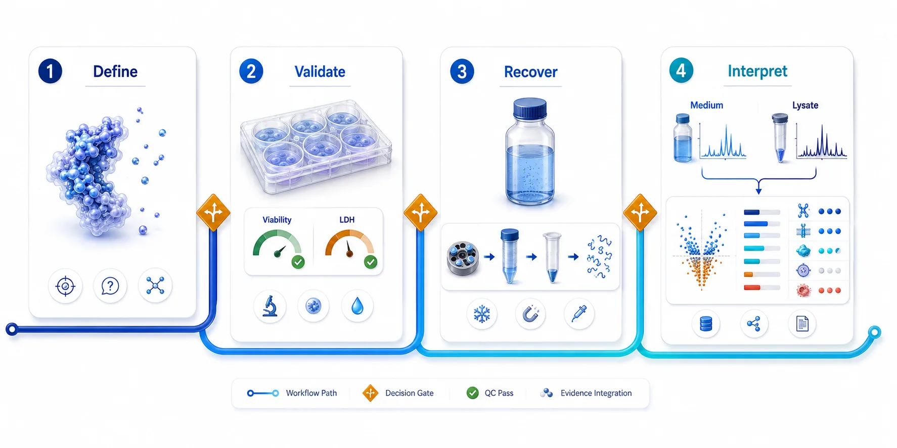 Figure 7. Four-phase secretome implementation plan. The workflow moves from biological question to collection QC, recovery-aware processing, and evidence-based classification.
Frequently Asked Questions
What if cells round up or detach during serum-free collection?
Treat this as a collection-window failure or a treatment-dependent phenotype that must be modeled, not as a condition to ignore. Shorten or redesign the collection interval, consider a defined medium, and document whether the observed release pattern tracks with cell damage.
Can residual bovine serum albumin simply be excluded during data analysis?
A contaminant database can flag expected bovine proteins, but computational exclusion cannot prove that all serum effects have disappeared. Reduce carryover experimentally and retain medium-only or wash-out controls when serum background is plausible.
Should adherent and suspension cultures use the same collection protocol?
No. Their handling risks differ. Adherent cultures need attention to detachment and surface-associated carryover; suspension cultures need a clear strategy for separating viable cells, debris, and released material without losing the intended extracellular fraction.
Does glycosylation prevent secreted proteins from being identified by bottom-up MS?
No. It can alter peptide observability and site assignment, but it does not make secreted proteins intrinsically inaccessible. If glycosylation is the question, specify a glycoproteomics or glycopeptide-enrichment design rather than assuming a standard digest will answer it.
Is a protein found in conditioned medium necessarily secreted?
No. It may originate from active secretion, vesicular export, surface shedding, serum background, or leakage from damaged cells. Matched cell material and QC evidence make these alternatives testable.
When should the study move from discovery DIA to a targeted assay?
Move when the candidate list is narrow and the study needs stronger peptide-level confirmation, optimized measurement of defined targets, or a focused follow-up across a larger sample set.
Related Articles in This Cluster
- Finding Microproteins and sORF-Encoded Peptides in Tissue Proteomics
- Database Strategies for Non-Model Organism Proteomics
- Will DIA Detect My Protein in Plasma?
References:
- Poschmann G, Bahr J, Schrader J, et al. Secretomics-A Key to a Comprehensive Picture of Unconventional Protein Secretion. Frontiers in Cell and Developmental Biology. 2022;10:878027. doi: 10.3389/fcell.2022.878027.
- Knecht S, Eberl HC, Kreisz N, et al. An Introduction to Analytical Challenges, Approaches, and Applications in Mass Spectrometry-Based Secretomics. Molecular & Cellular Proteomics. 2023;22(9):100636. doi: 10.1016/j.mcpro.2023.100636.
- Car I, Dittmann A, Klobucar M, et al. Secretome Screening of BRAFV600E-Mutated Colon Cancer Cells Resistant to Vemurafenib. Biology. 2023;12(4):608. doi: 10.3390/biology12040608.
- Rosa-Fernandes L, Rocha VB, Carregari VC, et al. A Perspective on Extracellular Vesicles Proteomics. Frontiers in Chemistry. 2017;5:102. doi: 10.3389/fchem.2017.00102.
- Mikulasek K, Konecna H, Potesil D, et al. SP3 Protocol for Proteomic Plant Sample Preparation Prior LC-MS/MS. Frontiers in Plant Science. 2021;12:635550. doi: 10.3389/fpls.2021.635550.














