

Meta Intent: A target-first decision guide for judging whether a protein of interest is likely to be detectable and reproducibly quantifiable by DIA-MS in plasma, and for choosing the least complex workflow that can produce defensible research evidence.
A plasma proteomics project often begins with a deceptively simple question: will data-independent acquisition, or DIA, detect my protein? The useful answer is rarely a binary yes or no. A protein can be biologically relevant, detectable in tissue, and even reported in plasma by another method, yet remain outside the measurable window of a particular plasma DIA workflow. Conversely, a protein observed once in a deep discovery run may not be quantified consistently enough for a comparative study. The practical task is to decide what level of evidence the project needs and whether the proposed workflow can deliver it.
This distinction matters because plasma is not a routine digest. A small set of highly abundant proteins dominates the ion population, while many proteins that motivate a research question occupy a much lower abundance region. Modern DIA acquisition improves consistency by sampling predefined mass ranges rather than stochastically selecting only the strongest precursors. It does not remove the physical limits imposed by co-elution, ion competition, peptide chemistry, sample preparation, and data-extraction thresholds. A well-scoped Plasma/Serum Proteomics Service therefore starts with the target and the decision needed from the data, not with an assumed protein count.
The framework in this guide is deliberately target-first: target biology -> plasma abundance window -> observable peptides -> workflow depth -> quantitative completeness. It helps investigators avoid two costly mistakes: applying a deep workflow to a target that has no suitable peptide evidence, and interpreting a missing DIA value as proof that the protein is absent.
"Detected" Is Not a Single Outcome
Before discussing depletion, instrument time, or software, define what the word "detected" must mean for this project. At least four outcomes are commonly collapsed into the same claim:
- Biological presence: the protein is produced in a relevant tissue, cell population, or model.
- Plasma presence: protein-derived material reaches plasma at a concentration and form that can survive collection and processing.
- Analytical identification: one or more credible peptide signals support a protein or protein-group assignment.
- Reproducible quantification: suitable peptides are measured across the planned samples with enough completeness and precision for the intended comparison.
These outcomes are progressively harder to establish. Tissue expression is often useful for hypothesis generation, but it does not establish that the protein enters plasma. A reported plasma identifier does not guarantee a unique peptide in the planned digestion. A unique peptide identified in a pooled discovery sample does not guarantee a stable quantitative trace in every individual sample. The correct opening question is therefore not "Can DIA find this protein?" It is "Which of these four outcomes must be demonstrated, in which sample set, and with what minimum evidence?"
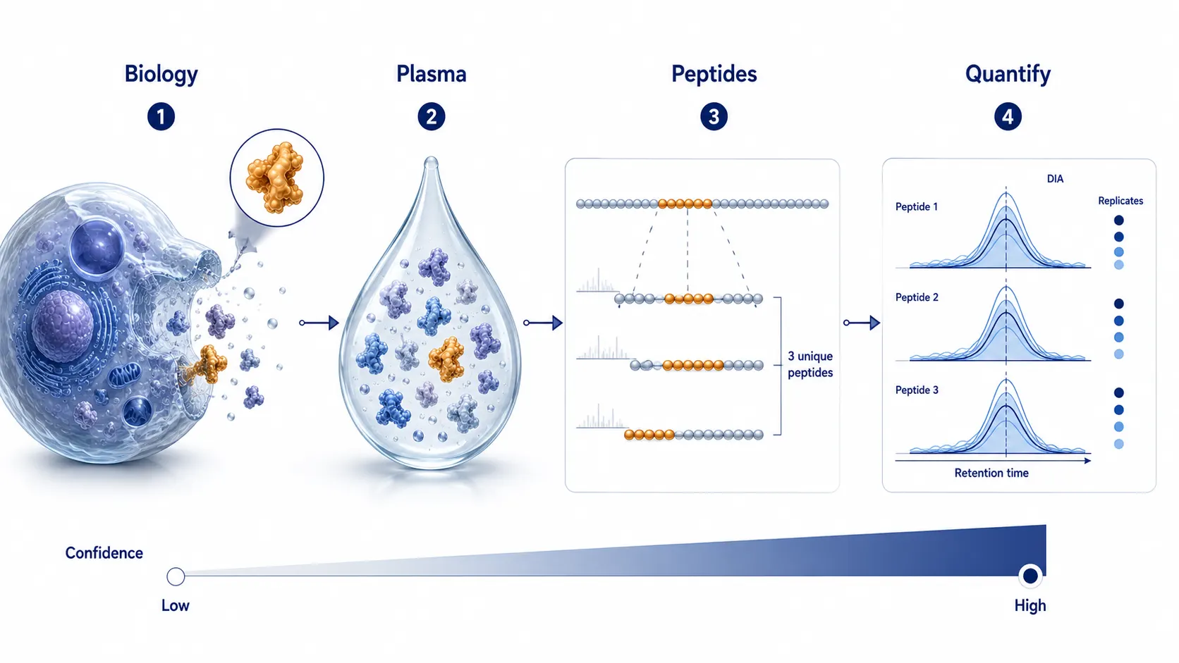 Figure 1. The target-to-measurement evidence chain separates biological relevance from plasma presence, peptide-level identification, and reproducible quantification.
Why Plasma Remains Difficult for DIA
Plasma combines extreme abundance differences with a large number of peptide species that can overlap in time and mass. Albumin, immunoglobulins, apolipoproteins, complement proteins, and coagulation-related proteins can occupy a large fraction of the measurable signal. Lower-abundance proteins may be present but contribute signals that are weak, obscured by neighboring precursors, or represented by peptides that are not ideal for a bottom-up assay. The core analytical challenge is not merely whether an instrument can fragment an ion. It is whether the relevant peptide can be isolated or deconvoluted with enough chromatographic points, fragment evidence, and signal-to-interference separation to satisfy the selected identification and quantification rules.
DIA changes the sampling problem. Instead of repeatedly choosing only the most intense precursor ions, DIA uses a defined window scheme to acquire fragment information across a selected mass range. That usually gives more consistent matrices than data-dependent acquisition in repeated plasma measurements. However, a wide window still contains multiple co-eluting precursors, and their fragments can interfere with one another. A weak target peptide can therefore fail at several points: it may never rise above the background, it may have no clean fragment trace, it may fall below a confidence threshold, or it may be identified but be too incomplete for the group-level comparison.
For this reason, an increase in total identified proteins should not be treated as proof that a specific target is now measurable. Recent multicenter benchmarking showed that current DIA workflows can improve coverage, reproducibility, and data completeness in plasma-like high-dynamic-range samples, while also showing that LC-MS configuration and data-processing choices still affect the result.1 The relevant output for a target-first project is not the largest possible protein list. It is a transparent answer to whether the target has enough peptide-level evidence in the actual workflow.
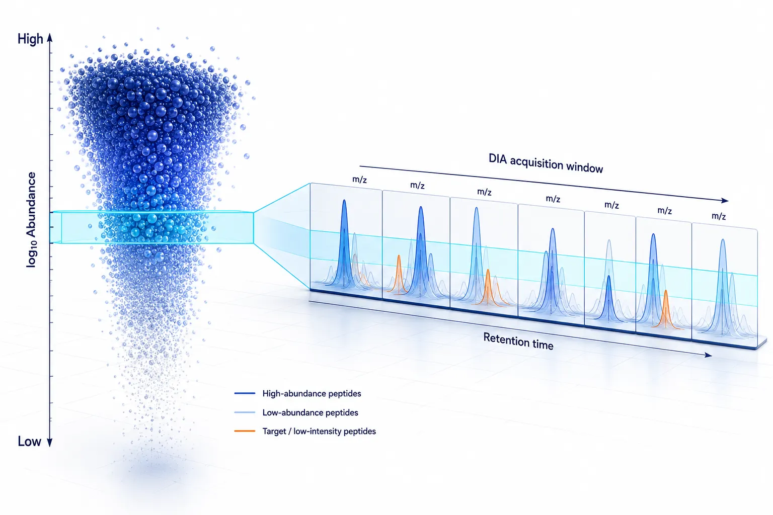 Figure 2. Plasma abundance distribution and the DIA observation window. DIA improves systematic sampling but does not eliminate ion competition or low-signal uncertainty.
Start With the Target, Not the Instrument
Ask whether the target is plausibly observable in plasma
A protein that is actively secreted, shed from a cell surface, released from a tissue, or carried in a stable complex has a different starting premise from a strictly intracellular protein. That does not make the first group automatically easy and the second group impossible. It means the project should state a mechanism of plasma presence before interpreting any detection result. Is the target expected to circulate freely, bind a carrier, associate with extracellular vesicles, appear only after cell damage, or fluctuate with collection conditions? If that logic is unknown, a plasma pilot should be framed as feasibility testing rather than as direct confirmation of a biological hypothesis.
Do not substitute transcript abundance or tissue proteomics for plasma evidence. These resources can make a target more or less plausible, but they cannot predict the final DIA trace by themselves. They do not account for secretion, clearance, binding partners, proteolysis, sample handling, or the proteotypic peptide landscape. A careful project brief records the predicted source of the protein, its likely plasma form, whether the desired conclusion concerns a protein family or exact isoform, and whether the study needs relative abundance or a more constrained quantitative endpoint.
Assess peptide observability before committing to depth
Bottom-up proteomics detects peptides, then infers proteins. A target may be difficult because it yields few tryptic peptides in a useful mass range, because its peptides are highly hydrophobic or poorly ionizing, because a sequence is shared among family members, or because a relevant site is frequently modified. A protein can also appear in a database with several predicted peptides but still lack a robust set of unique, consistently observed peptides under plasma conditions. The risk is especially high when the desired conclusion is isoform-specific or when homologous proteins share most of their sequence.
The most useful pre-study question is not simply whether a protein is listed in a reference database. It is whether the planned evidence can distinguish it from neighboring protein groups. For broad pathway exploration, protein-group-level evidence may be sufficient. For a named isoform or a target that will drive follow-up decisions, require stronger support: multiple credible peptides when feasible, explicit uniqueness assessment, coherent retention-time and fragment behavior, and consistent performance in a pilot. The same logic applies to a Protein Identification Services workflow: a long output table should not be allowed to hide weak target specificity.
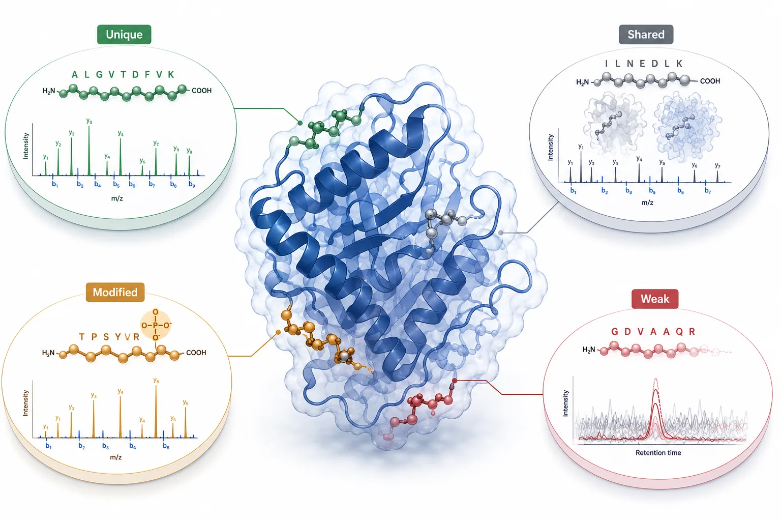 Figure 3. Peptide feasibility map. The ability to infer a target protein depends on the availability, uniqueness, and analytical behavior of its peptides.
Choose the Least Complex Workflow That Can Answer the Question
Neat plasma DIA: a strong starting point for many questions
Neat plasma DIA keeps the workflow simple. It minimizes additional manipulations, supports practical sample throughput, and preserves the original plasma matrix. It is often a sensible first choice when the target is expected to be among the more readily observed region of the plasma proteome, when the study is exploratory, or when the priority is a consistent comparative matrix across many samples. Good sample collection and controlled handling remain essential, so an early discussion of Protein Sample Preparation should include anticoagulant choice, time before processing, storage history, hemolysis risk, and freeze-thaw exposure.
The limitation is equally important: neat plasma does not offer a fixed low-abundance discovery guarantee. A target that is not observed in neat plasma may be below the workflow's practical observation window, represented by unsuitable peptides, or affected by pre-analytical and computational factors. It should not immediately be labeled biologically absent.
High-abundance depletion: use it as a different measurement lens
Depletion reduces the dominance of selected abundant proteins and can reveal a wider portion of the plasma proteome. This can be valuable when the target is plausibly present but likely masked by the upper abundance range. Yet depletion changes the sample. Proteins that bind, co-precipitate, or otherwise associate with removed components can be affected. The depleted and undepleted fractions should therefore not be treated as interchangeable versions of the same measurement. A finding from one fraction may need separate confirmation before it is used as a comparative conclusion.
Use depletion when the gain in target-relevant depth is more valuable than the increased workflow complexity and matrix alteration. A useful pilot compares target peptide observability, sample-to-sample completeness, and QC behavior across a small, representative subset. It does not rely only on an increase in aggregate protein IDs. This is the point at which a targeted DIA Quantitative Proteomics Service can be scoped around the target class and evidence threshold rather than around a generic request for maximum coverage.
Fractionation, enrichment, and targeted follow-up solve different problems
Peptide fractionation can reduce complexity further and deepen discovery coverage, but it adds analytical time and makes cross-sample design more demanding. Selective enrichment is appropriate when the biological question maps to a defined target class or modification. Neither option repairs poor peptide uniqueness. If the project already centers on a small number of well-defined proteins, moving to Parallel Reaction Monitoring (PRM) or SRM & MRM may be more defensible than repeatedly increasing discovery depth.
The escalation rule is simple: add complexity only when it addresses the demonstrated bottleneck. Use better separation when co-elution limits peptide clarity. Use depletion or enrichment when the target is plausibly present but masked by matrix dominance. Use targeted follow-up when the target list is already narrow and the project needs stronger peptide-level confirmation. Use a broader 4D-DIA Quantitative Proteomics Service when ion-mobility separation is relevant to resolving a complex peptide background, not merely because a higher-dimensional platform sounds more comprehensive.
This matrix-first logic is also useful for conditioned-medium studies, where serum carryover and intracellular leakage can be mistaken for extracellular biology. The corresponding design choices are covered in this guide to secretome proteomics from cell-culture supernatant.
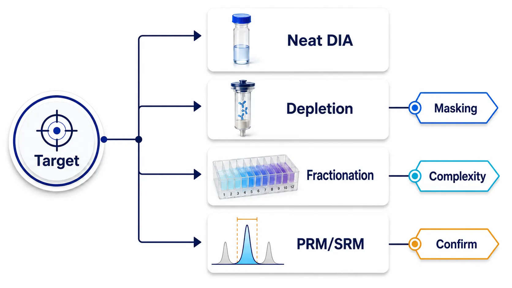 Figure 4. Workflow escalation decision tree. Each added layer should solve an observed bottleneck rather than serve as a default increase in complexity.
Depth Is a Design Variable, Not an Instrument Specification
Depth is often described as though it belongs to the mass spectrometer alone. In plasma DIA, it is an outcome of the complete method: sample matrix, amount loaded, chromatographic separation, peak width, acquisition window design, cycle time, spectral interpretation, and target-specific filtering. A longer gradient may create more separation between co-eluting peptides. Narrower or optimized windows may improve fragment selectivity in a relevant mass region. Better chromatographic stability can help align traces across a cohort. But these changes must be evaluated against the number of samples, the need for biological replicates, and the acceptable level of missingness.
For a large research cohort, a moderately deep workflow with stable retention times and predictable completeness may be more useful than an elaborate approach that achieves more identifications in a handful of samples but is difficult to apply consistently. For a small discovery pilot, more extensive separation may be justified if the purpose is to map candidate observability. The protocol should state which trade-off is being made. "Deep" without a defined target class, sample scale, or evidence criterion is not a meaningful method specification.
If the protein of interest is absent from the sequence resource, belongs to a rapidly evolving family, or requires non-canonical sequence interpretation, more depth will not repair the identification problem. In that situation, start with the evidence-controlled database choices described in this non-model organism proteomics database guide before increasing acquisition complexity.
This is also why a pilot should include at least two readouts. The first is coverage: were credible target peptides observed? The second is behavior: were those peptides sufficiently complete and stable across replicate injections or representative samples? A Bioinformatics for Proteomics plan should preserve this distinction rather than rolling every observation into one protein-level intensity table.
Read Missingness as Evidence, Not as a Spreadsheet Problem
A blank value is not a biological verdict. In plasma DIA, missingness can be informative when it is investigated at the level where it emerged. The first task is to determine whether the problem occurred before acquisition, during peptide observation, during identification, or during filtering and roll-up. Treating every blank as a value to be imputed too early can conceal the cause and create a misleading group comparison.
Four common causes of target missingness
- Signal below the observation window: the peptide may be present but too weak relative to background and interference.
- Peptide evidence is unsuitable: few unique peptides, inconsistent digestion, unstable modification states, or shared sequences weaken protein inference.
- Sample processing altered the target: handling, precipitation, depletion, digestion, or adsorption may reduce recovery or change comparability.
- Data extraction filtered the signal: confidence thresholds, library content, interference scoring, alignment, or protein roll-up rules may remove a weak but real trace.
The appropriate response depends on the pattern. If a peptide is absent in all samples and all replicates, assess whether the target belongs in the chosen workflow tier. If it appears in a subset with clear chromatographic traces, inspect sample covariates and pre-analytics before imputing. If precursor evidence exists but the protein group disappears after roll-up, review uniqueness and grouping rules. If the target is seen only after depletion, describe the result as depletion-dependent rather than as a universal plasma measurement. A Bioinformatic Data Preprocess and Normalization Service should document these choices so that missingness is traceable rather than silently overwritten.
Imputation can have a role in downstream modeling, but it should follow a declared detection and filtering strategy. It cannot create peptide evidence. Before applying a missing-value rule, record the proportion missing by peptide and protein, whether missingness differs by study group or batch, and whether the absent values are consistent with low-intensity censoring or with a handling-related pattern. For inferential work, the Statistical Analysis Service plan should be built around the observed data structure, not around the convenience of a complete matrix.
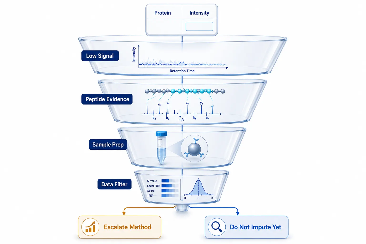 Figure 5. Missingness diagnostic funnel. Investigate the source of a blank value before applying a statistical replacement rule.
Build a Go/No-Go Pilot Around the Protein of Interest
A target-first pilot is not a miniature version of the full study. It is a decision experiment. Its job is to determine whether the target can support the intended evidence claim and which workflow tier is justified. The pilot should use representative samples whenever possible, because neat pooled plasma can make a target appear more stable than it will be across a biologically variable sample set.
Define the decision threshold before the first run. For an exploratory target, that may be reproducible detection of two or more target-specific peptides in a prespecified proportion of representative samples, with acceptable retention-time alignment and interference review. For a comparative study, it may also require that the target's peptide signals remain sufficiently complete to support the planned statistical model. Record both detected and non-detected observations; do not quietly replace the latter with zeros or a single imputed value during the feasibility stage. A pilot that shows the target only after aggressive searching is telling you that the workflow is fragile, not that the question is answered.
Use a small workflow matrix instead of a single yes-or-no experiment. The same representative material can be tested as neat plasma, depleted plasma, or a selected enrichment workflow, with the acquisition and analysis conditions held as comparable as possible. Review target peptide evidence, completeness across replicates, relative response to the intervention, and whether the intervention creates new analytical liabilities. This creates an auditable escalation decision: retain a simple DIA workflow, add complexity because it changes the target evidence materially, or redirect the target to a focused assay.
Use the following one-page brief before finalizing the acquisition plan:
- Target definition: protein group, family member, isoform, modified form, or candidate panel.
- Plasma rationale: expected source, circulating form, and key reasons that the target may or may not be observable.
- Peptide evidence: candidate unique peptides, potential sequence-sharing issues, and expected analytical vulnerabilities.
- Workflow tier: neat plasma, depletion, fractionation, enrichment, or targeted follow-up.
- Success rule: required peptide evidence, sample-level completeness, and reproducibility needed for the next decision.
- Escalation rule: the specific observation that would justify greater depth or a targeted assay.
A realistic success rule is not "find the protein." It may be "observe at least one unique peptide with coherent chromatographic and fragment evidence in most representative pilot samples," or "show that the target becomes consistently observable only after depletion." Either result is useful because it prevents the main study from being designed around an unsupported assumption. When the pilot identifies a credible candidate set, follow-up can move from broad discovery to targeted confirmation without pretending that the first DIA matrix answers every quantitative question.
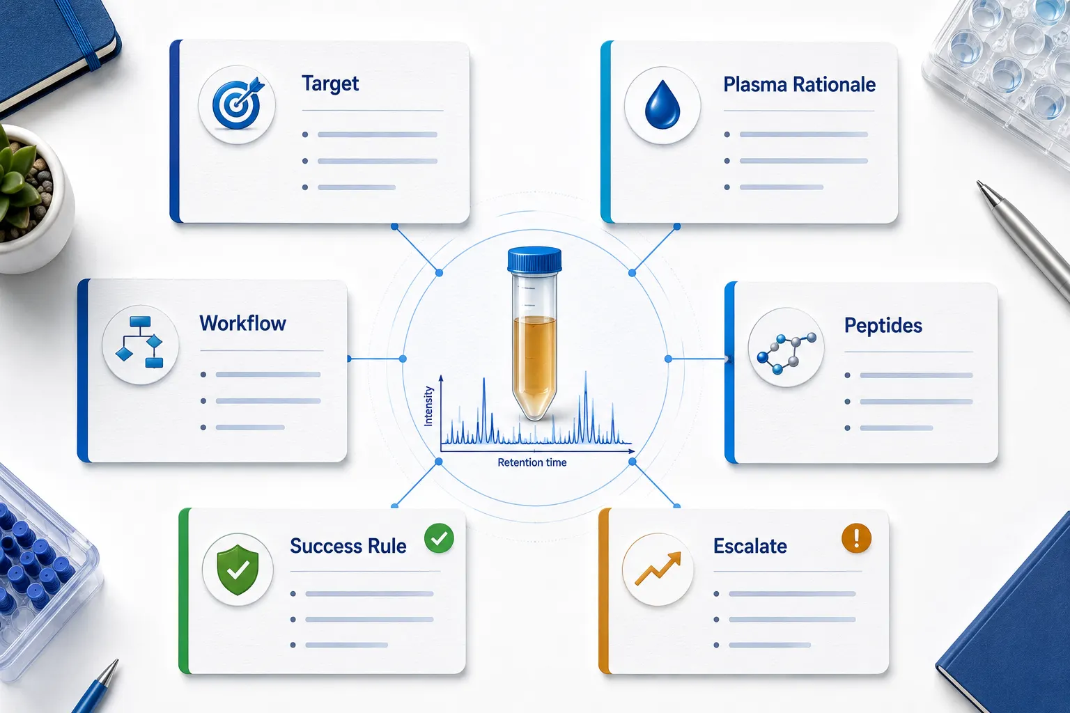 Figure 6. Plasma DIA feasibility worksheet. A concise project brief links the target hypothesis to a measurable pilot decision.
When DIA Is the Right First Choice - and When It Is Not
DIA is a strong first choice when the project needs an unbiased view of a discoverable region of the plasma proteome, expects to compare many samples, and needs lower stochastic missingness than a conventional data-dependent workflow. It is particularly useful when the target list is still open and the practical question is which proteins or pathways can be measured consistently enough to prioritize. It can also provide the peptide-level evidence needed to decide whether a more focused assay should be built.
DIA should not be positioned as a universal substitute for a targeted strategy. If the question concerns a very low-abundance protein, an exact isoform, a defined modification, or an absolute concentration, discovery DIA may be the wrong final endpoint even if it is an excellent feasibility screen. A low or inconsistent DIA signal is not failure. It is information about the distance between the target question and the current measurement window. The right next step may be a different plasma preparation, a target-enrichment method, or a focused PRM or SRM/MRM assay.
In short, ask DIA to do what it does well: map target observability and produce a reproducible discovery matrix. Do not ask it to prove that every biologically interesting plasma protein is measurable under every workflow. A disciplined workflow turns the answer into a practical decision: proceed with neat plasma, add a targeted depth strategy, or change the measurement approach before investing in the full cohort.
Frequently Asked Questions
Can DIA detect any protein that has been reported in plasma?
No. Reported plasma presence does not guarantee that a target has suitable peptides or falls within the observation window of a particular workflow.
Is depletion always required for low-abundance targets?
No. Depletion can improve depth, but it also changes the plasma matrix and may affect associated proteins. Use it when a pilot shows that matrix dominance is the relevant bottleneck.
Does a deeper protein list prove better target quantification?
No. Target quantification depends on peptide specificity, chromatographic behavior, interference control, and completeness across the intended samples.
What does a missing DIA value mean?
It may indicate low signal, unsuitable peptide evidence, a processing effect, or a data-extraction decision. It should not automatically be interpreted as biological absence.
When should a project move from DIA to PRM or SRM/MRM?
Move when the target list is narrow and the project needs more focused peptide-level confirmation or a method optimized around specific analytes.
Can a pilot decide the final plasma workflow?
Yes, when it is designed around predefined evidence and escalation rules rather than only around the total number of protein identifications.
References:
- Distler U, Yoo HB, Kardell O, et al. Multicenter evaluation of label-free quantification in human plasma on a high dynamic range benchmark set. Nature Communications. 2025;16:8774. doi: 10.1038/s41467-025-64501-z.
- Bennike TB. Advances in proteomics: characterization of the innate immune system after birth and during inflammation. Frontiers in Immunology. 2023;14:1254948. doi: 10.3389/fimmu.2023.1254948.
- Gegner HM, Naake T, Dugourd A, et al. Pre-analytical processing of plasma and serum samples for combined proteome and metabolome analysis. Frontiers in Molecular Biosciences. 2022;9:961448. doi: 10.3389/fmolb.2022.961448.
- Millioni R, Tolin S, Puricelli L, et al. High abundance proteins depletion vs low abundance proteins enrichment: comparison of methods to reduce the plasma proteome complexity. PLOS ONE. 2011;6(5):e19603. doi: 10.1371/journal.pone.0019603.
- Yadav AK, Bhardwaj G, Basak T, et al. A systematic analysis of eluted fraction of plasma post immunoaffinity depletion: implications in biomarker discovery. PLOS ONE. 2011;6(9):e24442. doi: 10.1371/journal.pone.0024442.












