


The Overlooked Small Proteome in Complex Tissues
Why Tissue Preparation Creates a Size-Selective Blind Spot
Database Design Determines What the Search Can See
Build a Microprotein-Specific Evidence Standard
Choose the Workflow That Matches the Question
Use Multi-Omics as a Constraint, Not a Candidate Multiplier
A Four-Phase SOP for Tissue Microprotein Discovery
Frequently Asked Questions
Meta Intent: A practical guide for tissue proteomics teams that need to decide whether a microprotein or sORF-encoded peptide is genuinely present, and how to build a defensible discovery-to-validation workflow rather than overinterpret a long candidate list.
The Overlooked Small Proteome in Complex Tissues
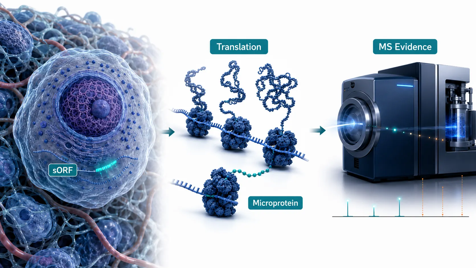
Many tissue proteomics studies begin with an understandable assumption: if a protein is expressed, a sensitive LC-MS/MS workflow should find it. That assumption works reasonably well for abundant, well-annotated proteins that generate several proteotypic tryptic peptides. It becomes unreliable for microproteins and sORF-encoded peptides (SEPs). These translation products are often shorter than 100 amino acids, may be below 10 kDa, and can arise from upstream, overlapping, alternative, or previously non-coding open reading frames. A conventional tissue workflow can therefore miss them before the mass spectrometer has even acquired a useful spectrum.
The problem is not simply instrument sensitivity. Tissue lysates contain a wide dynamic range of canonical proteins, lipids, salts, nucleic acids, and endogenous peptides. Small, soluble translation products can be lost during precipitation or filtration; hydrophobic species can be poorly recovered or retained; and a short sequence may yield only one observable peptide after digestion. If that peptide is absent from the database used for searching, no amount of downstream score optimization can recover the identification.
This is why the appropriate objective is not “find as many microproteins as possible.” It is to construct an evidence chain that survives three questions: Was the low-molecular-weight species retained during preparation? Was its sequence represented in a biologically plausible search library? Does the spectrum support one specific peptide better than every canonical alternative? Teams using broad Protein Identification Services can apply this framework before treating an unannotated hit as a biologically meaningful discovery.

Figure 1: The Microprotein Landscape: Genomic Origins, Biogenesis, and Proteomic Dark Matter
Why Tissue Preparation Creates a Size-Selective Blind Spot
Extraction is an analytical choice, not a neutral first step
Microproteins are vulnerable to losses that are easy to overlook when a protocol was designed for the bulk proteome. Organic-solvent precipitation, extensive cleanup, and high molecular-weight cutoff devices can improve a conventional sample by removing interference. They can also deplete exactly the small, soluble species under investigation. A tissue homogenate is especially challenging because proteolysis may begin rapidly after disruption, creating endogenous fragments that resemble authentic short proteins in both size and chromatographic behavior.
The practical response is to plan recovery and artifact control together. Keep tissue cold, limit the interval between disruption and denaturation, record protease-inhibitor conditions, and retain a small aliquot from every fractionation stage for troubleshooting. When the scientific question is explicitly about low-molecular-weight proteins, a pilot comparison between conventional extraction and an acid-compatible low-mass enrichment route is more informative than assuming a familiar lysis buffer is unbiased. Protein Sample Preparation should be treated as an experimental variable, with recovery checks designed around representative small and hydrophobic analytes rather than total protein yield alone.
Tricine-SDS-PAGE, low-mass solid-phase extraction, and size-aware fractionation can improve access to the target region, but each introduces a different bias. A gel fraction may enrich a small protein while reducing recovery of very hydrophobic species. A molecular-weight cutoff membrane may pass the analyte into a discarded fraction. A peptide-oriented cleanup can be useful for endogenous peptidomics but may not preserve an intact microprotein that requires a later digestion strategy. The right approach is therefore conditional: choose the enrichment route based on whether the immediate goal is intact-protein evidence, discovery peptide coverage, or targeted confirmation.
Trypsin can make a short protein analytically invisible
Bottom-up proteomics is optimized for proteins that produce multiple peptides of suitable length, charge, and retention. A 40- to 80-amino-acid microprotein may yield only one candidate tryptic peptide, several fragments below the useful mass range, or no suitable peptide if its sequence is highly hydrophobic. One missing cleavage site, one modification, or one poorly ionizing segment can remove the only usable analytical handle.
Do not assume that more trypsin resolves this problem. Over-digestion can increase the abundance of short, non-specific fragments without creating a unique proteotypic peptide. Instead, evaluate the predicted peptide map before committing scarce tissue. A complementary protease such as chymotrypsin, Asp-N, or Lys-C can create a second route to sequence coverage. A controlled comparison between digestion conditions is often more useful than a one-size-fits-all digestion protocol. Protein Digestion Service is particularly relevant when a candidate list contains sequences with few tryptic cleavage sites or strong membrane-associated regions.
For discovery questions centered on endogenous short products, a digestion-free or minimally processed route may also be appropriate. Overview of Peptidomics Services and Comprehensive Peptidomics Service can complement rather than replace a microprotein workflow: they help determine whether the observed signal behaves like an endogenous peptide product, while a proteogenomic experiment addresses its genomic origin and translation evidence.
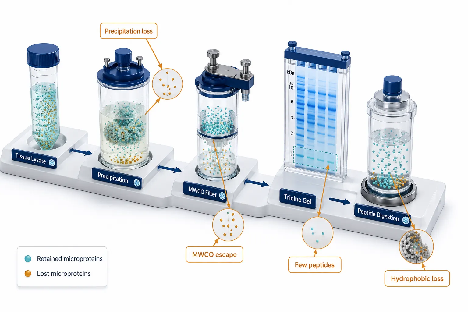
Figure 2: Biochemical Extraction and Enzymatic Digestion Bottlenecks for Low-Molecular-Weight Proteins
Database Design Determines What the Search Can See
A canonical database is necessary but insufficient
Swiss-Prot, RefSeq, and other canonical proteomes are essential controls. They are not comprehensive libraries of all translated tissue-specific ORFs. If a candidate sORF is not represented, its peptide spectrum can only be assigned to the closest available canonical sequence, remain unidentified, or be discarded during filtering. Conversely, adding every possible translation product to a database can create a search space so large that false matches become more difficult to estimate.
The useful contrast is not “canonical database versus six-frame translation.” It is “unconstrained candidate space versus evidence-constrained candidate space.” A tissue-specific custom library should start with the canonical proteome and known contaminants, then add non-canonical sequences supported by the relevant biological evidence. RNA-seq can capture expressed transcripts and sample-specific splice forms. Ribo-seq can further prioritize ORFs with translation evidence, including near-cognate initiation events. Public non-canonical ORF resources can provide additional candidates, but their entries should not be treated as equal evidence.
For every added sequence, record its source, coordinates, reading frame, transcript context, and reason for inclusion. That provenance becomes critical when a peptide matches multiple loci or resembles a canonical proteoform. Teams carrying out Bioinformatics for Proteomics should regard FASTA construction as part of the analytical method, not as a disposable preprocessing file.
These controls are the tissue-specific extension of a broader database strategy for non-model organism proteomics: in both cases, the useful database is the smallest evidence-supported search space that can still test the biological hypothesis.
Search-space inflation changes the meaning of a score
In a broad six-frame library, a spectrum can be compared with millions of additional peptide hypotheses. Some will match by chance. A score that looks convincing in a compact canonical search may be less specific after database expansion. Global target-decoy false discovery rate (FDR) control remains important, but it can be poorly calibrated for a small, low-abundance non-canonical subset that is overwhelmed by high-confidence canonical identifications.
The safer design is staged. Use a primary discovery search with a biologically constrained library, then subject candidate non-canonical peptides to separate checks: peptide-level uniqueness against the full canonical proteome and relevant proteoforms, independent decoy assessment for the non-canonical subset, and direct inspection of fragment-ion coverage. A reanalysis must also account for leucine/isoleucine ambiguity, sequence variants, homologs, and peptides that differ from known proteins by only one residue. De Novo Peptides/Proteins Sequencing Service can add value when a high-quality spectrum remains poorly explained by the constrained library, but de novo sequence output should still be reconciled with genomic context before an sORF claim is made.
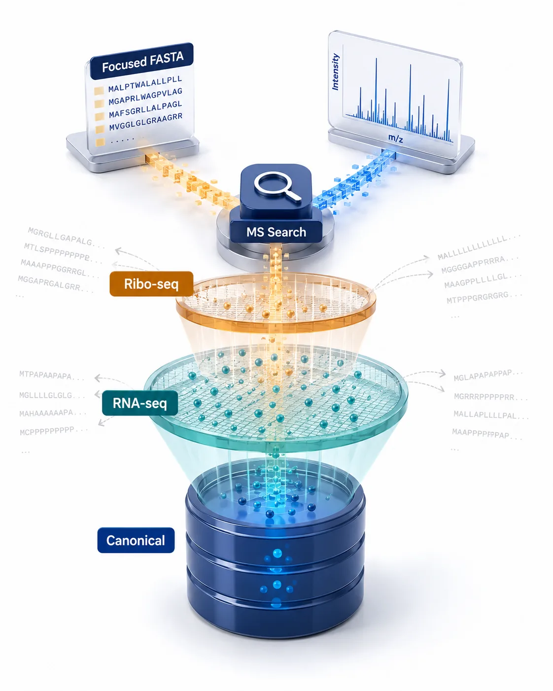
Figure 3: Database Construction Strategies: Canonical Proteome vs. Ribo-Seq-Guided Non-Canonical Libraries
Build a Microprotein-Specific Evidence Standard
One unique peptide can be informative, but it is not self-validating
Microproteins often cannot meet the familiar “two unique peptides per protein” convention because their sequence length makes two observable peptides unrealistic. Replacing that convention with a weaker standard is not the answer. Replace it with a more explicit standard.
First, the peptide should be strictly unique after comparison with canonical proteins, splice isoforms, pseudogene translations, common contaminants, and plausible single-amino-acid variants. Second, the observed MS/MS spectrum should contain an interpretable series of fragment ions rather than a score alone. Third, the signal should be supported by coherent precursor mass, retention time, charge state, and reproducibility information. The evidence should identify the peptide, not merely show that some low-abundance feature exists at the expected mass.
This standard matters most in tissue discovery, where a candidate can be confused with a proteolytic fragment of a high-abundance precursor. A peptide positioned internally within a large canonical protein, for example, does not become evidence of an independent microprotein because it appears in a low-mass fraction. The sequence must be assigned in the context of the full proteome and the experimental preparation. Peptide Mapping Service can help resolve whether a reported peptide belongs uniquely to the proposed sORF product or is more plausibly explained by an existing precursor.
Use tiered FDR and peptide-centric confirmation
Global 1% PSM FDR is a starting control, not the final answer for non-canonical claims. Report the search space, the number of non-canonical candidates, the number surviving each filter, and the subset subjected to manual spectral review. Where possible, compute or assess FDR separately for the non-canonical subset rather than allowing abundant canonical matches to dominate the error estimate.
Peptide-centric validation is a practical second line of defense. PepQuery-style analysis asks whether the candidate sequence explains an experimental spectrum better than alternative peptides in the reference proteome. It is especially useful after a discovery search because it turns a broad, database-dependent result into a focused competition among plausible explanations. For the highest-priority candidates, compare endogenous retention time and fragmentation with a stable-isotope-labeled synthetic peptide. Parallel Reaction Monitoring (PRM) Service offers a targeted route to test whether the proposed microprotein-derived peptide reproduces across tissue replicates under a pre-specified transition set.
The result should be communicated as an evidence tier. “Translation-supported candidate with one strictly unique peptide” is different from “synthetic-standard-confirmed tissue microprotein.” The distinction makes the work more useful for downstream biology because it tells the next investigator exactly what has been established and what remains to be tested.
Make negative results diagnostically useful
A negative result is only informative when the workflow makes clear what it was capable of detecting. “No microproteins were identified” can mean that the tissue contains none at the chosen condition, but it can also mean that the low-mass fraction was depleted, the candidate ORFs were absent from the FASTA library, or the digestion produced no observable peptide. These explanations require different follow-up experiments.
For each prioritized candidate, build a simple observability record before interpreting absence. Note predicted molecular mass, expected protease products, peptide uniqueness, hydrophobic segments, transcript abundance, translation support, and whether a heavy peptide would fall within the validated LC gradient. If a sequence has no suitable tryptic peptide, a negative bottom-up result should redirect the experiment toward an alternative protease, peptidomics, or intact-protein measurement. If a suitable unique peptide is predicted but never observed, assess recovery and ionization using a synthetic standard before concluding that the candidate is absent.
This diagnostic discipline prevents a common failure mode: repeating the same broad discovery workflow with a deeper injection and expecting the answer to change. Greater depth can help when ion statistics are limiting. It cannot correct a missing sequence library, a lost low-mass fraction, or a peptide design that offers no selective analytical handle. The most efficient project therefore turns every negative result into a defined next decision, rather than treating it as proof against translation.
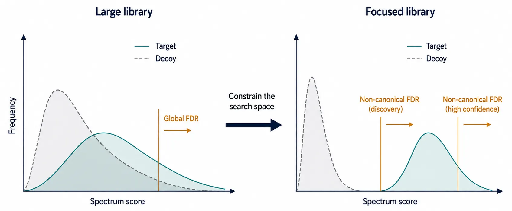
Figure 4: Search-Space Inflation and Two-Tier FDR Control Mechanics
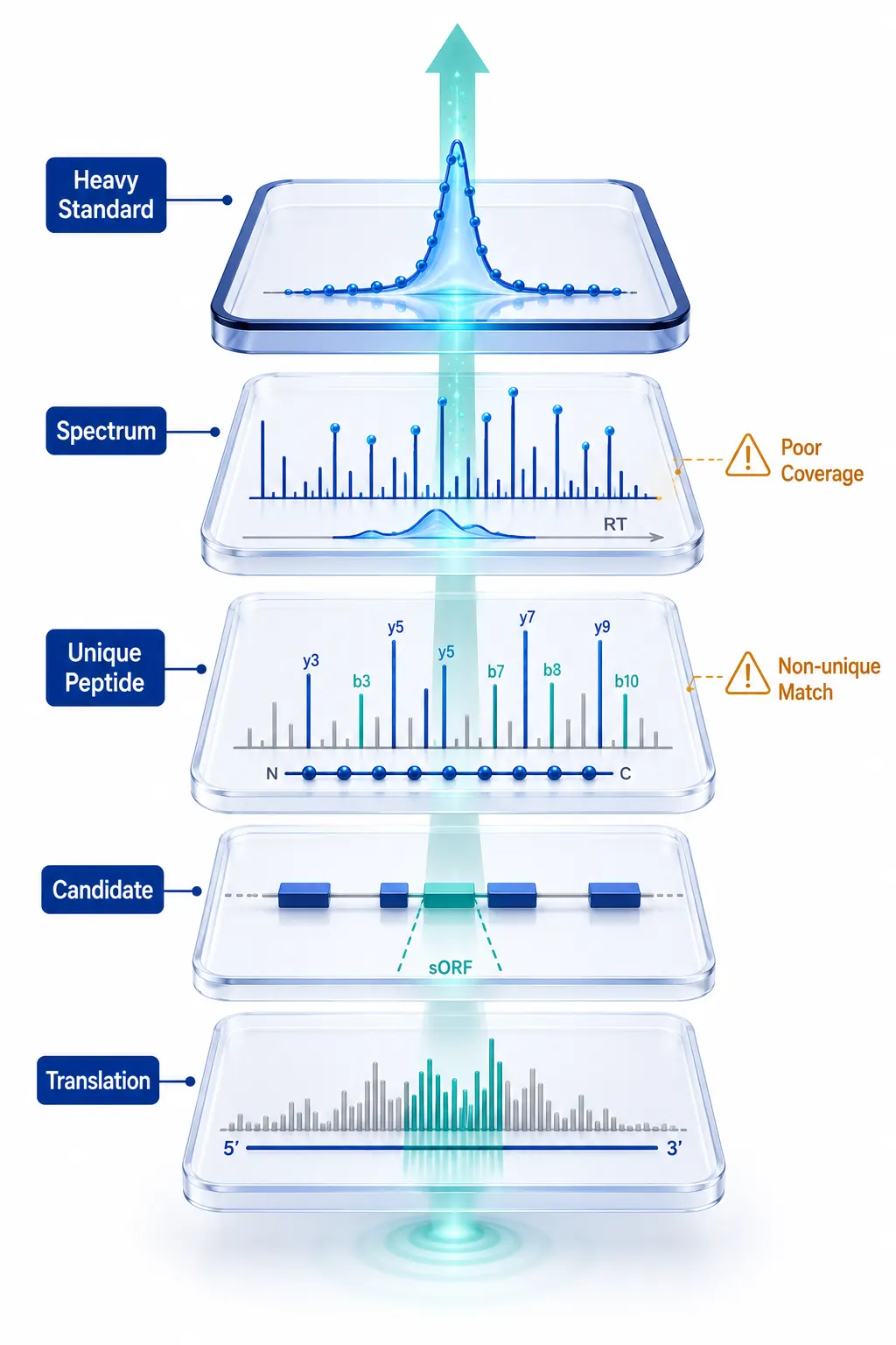
Figure 5: Proteogenomic Evidence Hierarchy: From Unique Peptides to Synthetic Standard Spectral Matching
Choose the Workflow That Matches the Question
No single acquisition strategy is optimal for every tissue microprotein question. Standard bottom-up discovery is appropriate when the aim is a broad first pass and sufficient tissue is available, but it is vulnerable to peptide scarcity. Low-molecular-weight enrichment and peptidomics improve access to short products but do not by themselves prove independent translation. Top-down analysis can increase sequence coverage for intact small proteins and retain proteoform context, although sample complexity and throughput remain practical constraints. Targeted PRM or MRM is the strongest route for a defined shortlist, not for unconstrained discovery.
| Research decision | Best first route | Main strength | Critical limitation |
| Broad tissue candidate discovery | Constrained bottom-up proteogenomics | Scalable search across many samples | Often only one observable peptide |
| Recovery of short endogenous products | Low-mass enrichment plus peptidomics | Better access to small soluble species | Can confound cleavage products with translation products |
| Intact sequence and proteoform context | Top Down Proteomics Service | Longer sequence coverage | Higher sample-complexity burden |
| Confirmation of a prioritized candidate | PRM/MRM with a heavy standard | Specific, repeatable evidence | Requires a pre-defined target |
For a project that begins with archived or public tissue datasets, Whole Proteome Profiling (Shotgun Protein Identification) and a focused reanalysis can establish which candidates warrant new experimental material. The most efficient workflow is frequently sequential: discover broadly, reduce the list aggressively, then spend targeted assay capacity only on candidates with a unique and biologically plausible peptide.
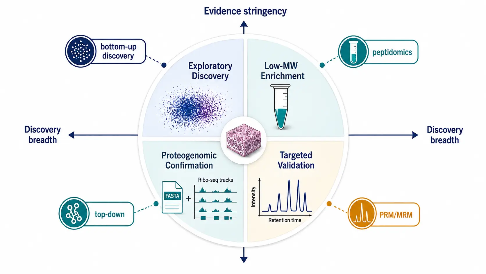
Figure 6: Methodological Comparison Matrix for Microprotein Discovery Workflows
Use Multi-Omics as a Constraint, Not a Candidate Multiplier
The most productive multi-omics integration does not simply add every RNA-seq and Ribo-seq ORF to the FASTA library. It asks whether independent data narrow the interpretation. Tissue RNA-seq indicates that the transcript is present. Ribo-seq suggests translation at a defined frame. LC-MS/MS provides direct peptide evidence. Targeted validation tests whether the signal is reproducible. Together, these layers can prioritize the small subset of candidates worth functional follow-up.
When the biological question moves from tissue translation products to proteins released by cultured cells, the evidence problem changes again: secretome proteomics from cell-culture supernatant requires controls for serum background and intracellular leakage in addition to a defensible sequence search.
This logic also keeps the current topic cluster distinct. Related proteomics articles may focus on database choice and data validation, DIA study design, or extracellular protein detection. Tissue microprotein discovery intersects with each of those decisions but has a separate endpoint: a low-mass, non-canonical translation product with a documented evidence tier. These are complementary decision guides, not substitutes for a tissue-specific microprotein workflow.
For related planning, see Proteomics Technology Selection, Database, and Data Validation, DIA Quantitative Proteomics, and Exosome Biomarkers and Advanced Detection Technologies. Each addresses a different analytical decision; none substitutes for a tissue-specific microprotein evidence workflow.
A Four-Phase SOP for Tissue Microprotein Discovery
Phase 1: Define the discovery claim before processing tissue
Specify the unit of evidence. Is the project trying to find translated candidate peptides, validate a small set of previously predicted ORFs, or quantify confirmed targets across tissue conditions? Establish this before choosing extraction and before generating a search library. Pre-register the minimum evidence needed for promotion from candidate to validated target, including the treatment of non-unique peptide matches.
Phase 2: Preserve and enrich the low-molecular-weight fraction
Process tissue under cold, time-controlled conditions. Compare a conventional protein extraction with one low-molecular-weight-aware route on a pilot subset. Use QC materials or representative peptides to monitor recovery through cleanup and fractionation. Document whether the workflow retains intact low-mass proteins, endogenous peptides, or digested peptides; those are different analytical objects.
Phase 3: Build a constrained search and test discovery evidence
Construct a provenance-tracked library from the canonical proteome plus tissue-relevant non-canonical ORFs supported by transcript and translation evidence. Search with defined mass tolerances and modification settings, then apply a separate review path for the non-canonical subset. Remove peptides that map to known proteins, closely related proteoforms, or plausible variant explanations. Inspect the spectrum, not just the search-engine score.
Phase 4: Confirm a shortlist with orthogonal evidence
Use peptide-centric re-querying, replicate tissues, and targeted PRM where the result will guide a downstream decision. For the highest-priority candidate, compare endogenous and synthetic heavy-peptide retention time and fragmentation. If the evidence remains ambiguous, report the candidate as unresolved rather than converting uncertainty into a definitive microprotein call. This is the point at which deep coverage, careful Protein Identification Services, and targeted confirmation become a connected research strategy rather than separate service requests.
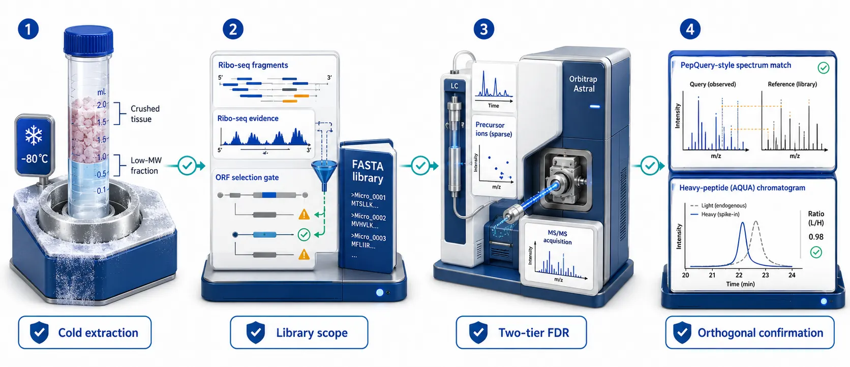
Figure 7: Four-Phase Implementation SOP Pipeline for Robust Tissue Microprotein Identification
Frequently Asked Questions
Can Ribo-seq evidence alone confirm a tissue microprotein?
No. Ribo-seq supports translation of an ORF, but it does not directly establish the stability, sequence, or tissue abundance of the resulting protein product. It is strongest when used to constrain the search library and paired with peptide-level evidence.
Should every sORF candidate have two unique peptides?
Not necessarily. Very short proteins may not yield two measurable, strictly unique peptides. In that situation, the evidence bar should shift toward superior spectral quality, strict uniqueness testing, replication, and orthogonal targeted validation.
How can a cleavage fragment be distinguished from an independent microprotein?
Search the peptide against the full canonical proteome and inspect its position within possible precursor proteins. A candidate also needs a defensible ORF and transcript context; low molecular weight alone is not sufficient.
Is six-frame translation the best default database strategy?
Usually no. It is useful for exploratory hypotheses but expands the search space sharply. A tissue-informed RNA-seq and Ribo-seq-constrained library is generally easier to interpret and validate.
When is top-down proteomics worth adding?
Consider it when intact sequence context or proteoform distinction is central to the question, especially after a bottom-up workflow produces ambiguous peptide evidence. It should be planned as a complementary measurement, not an automatic replacement for discovery proteomics.
What is the most convincing confirmation experiment?
For a prioritized peptide, agreement between endogenous and isotope-labeled synthetic standards in retention time and fragmentation, followed by targeted detection across independent tissue samples, provides a strong confirmation package.
References:
- Fijalkowski I, Peeters MKR, and Van Damme P. Small Protein Enrichment Improves Proteomics Detection of sORF Encoded Polypeptides. Frontiers in Genetics (2021).
- Olexiouk V, et al. Small Open Reading Frames, How to Find Them and Determine Their Function. Frontiers in Genetics (2021).
- Wang Y, et al. Mapping microproteins and ncRNA-encoded polypeptides in different mouse tissues. Frontiers in Cell and Developmental Biology (2021).
- Fabre B, et al. In Depth Exploration of the Alternative Proteome of Drosophila melanogaster. Frontiers in Cell and Developmental Biology (2022).
- Widespread stable noncanonical peptides identified by integrated analyses of ribosome profiling and ORF features. Nature Communications (2024).
More Articles
-
Proteomics Technology Selection, Database, and Data Validation
Compare technology selection, database design, and validation controls for defensible proteomics data. -
DIA Quantitative Proteomics
Explore the strengths and planning considerations of DIA-based quantitative proteomics workflows. -
Exosome Biomarkers and Advanced Detection Technologies
Review an extracellular-protein detection workflow where sample source and analytical sensitivity influence interpretation.