

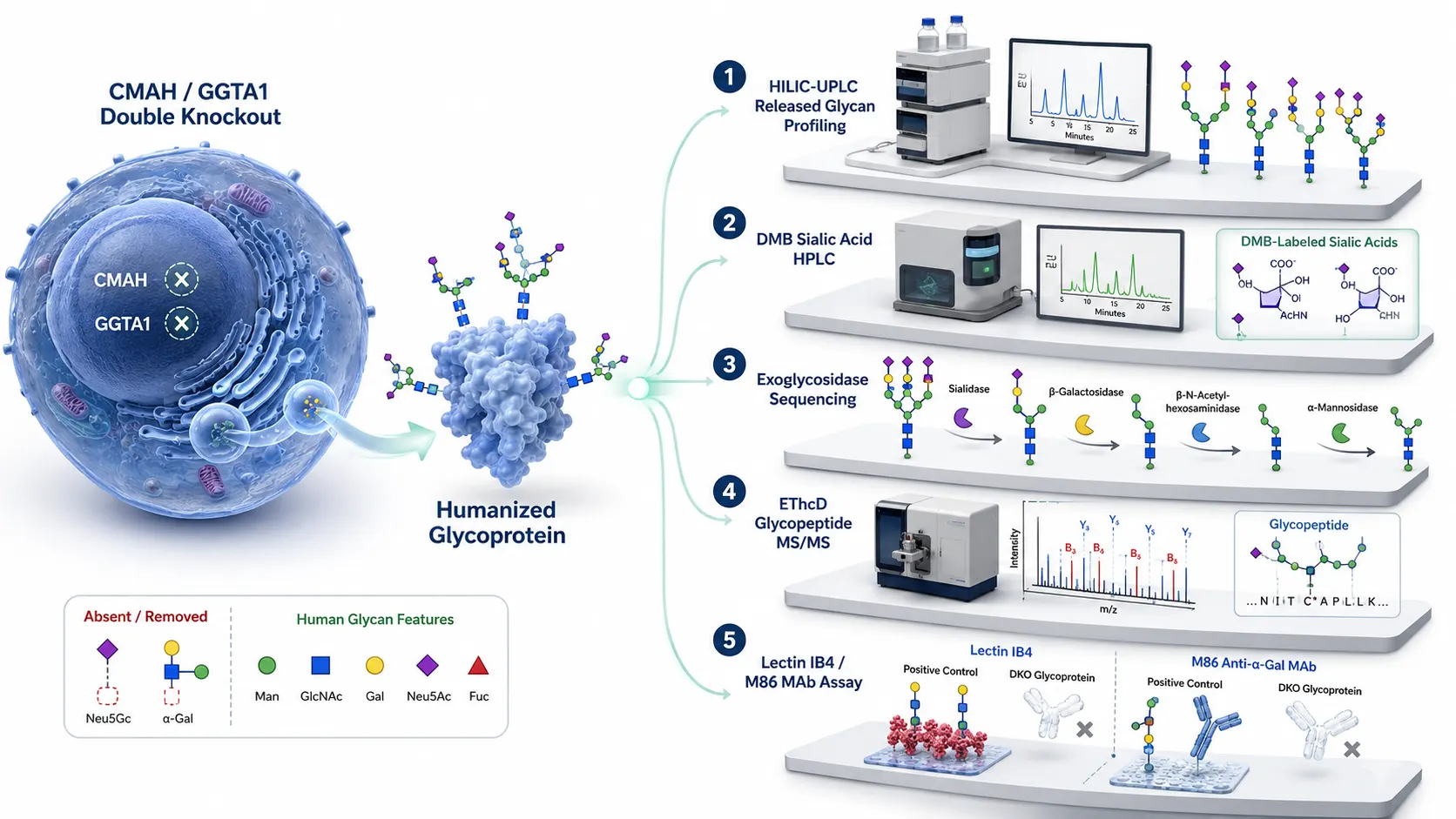
 Figure 1: CMAH and GGTA1 Glycoengineering Validation Landscape
Introduction: Immunogenic Xeno-Glycans in Biotherapeutics and Xenotransplantation
The Biological Threat of Non-Human Glycoforms
Recombinant biopharmaceuticals, therapeutic monoclonal antibodies (mAbs), fusion proteins, and transgenic cell/organ sources for xenotransplantation are frequently produced in non-human mammalian expression systems, including Chinese Hamster Ovary (CHO) cells, murine myeloma lines (NS0 and Sp2/0), and transgenic pig tissues. However, non-human mammalian cells express specific glycosyltransferases and hydroxylases that incorporate xeno-glycan epitopes—most notably N-glycolylneuraminic acid (Neu5Gc) and galactose-α-1,3-galactose (α-Gal)—onto N-linked and O-linked glycans.
In humans, the gene encoding cytidine monophosphate-N-acetylneuraminic acid hydroxylase (CMAH)—which converts CMP-Neu5Ac to CMP-Neu5Gc via the addition of a single oxygen atom (+O = +15.995 Da)—was inactivated by an exon deletion approximately 2–3 million years ago. Similarly, the gene encoding glycoprotein α-1,3-galactosyltransferase 1 (GGTA1)—which attaches an α-linked galactose to terminal β-galactose to form the α-Gal trisaccharide (Galα1,3Galβ1,4GlcNAc, +Hex = +162.053 Da)—became pseudogenized during Old World primate evolution.
Because human cells lack endogenous CMAH and GGTA1 activity, the human immune system recognizes Neu5Gc and α-Gal as non-self xeno-antigens. High titers of pre-existing circulating antibodies against α-Gal and Neu5Gc (comprising up to 1% of total circulating human IgG and IgM) circulate in all healthy individuals due to chronic exposure to gut commensal bacteria and dietary red meat.
Infusing biotherapeutics or transplanting tissues containing Neu5Gc or α-Gal epitopes triggers severe immune reactions, including hyperacute organ rejection, complement activation, rapid antibody-mediated clearance, reduced therapeutic serum half-life, and life-threatening anaphylaxis (e.g., the severe hypersensitivity reactions observed with cetuximab).
Application in Xenotransplantation: Triple KO (GGTA1/CMAH/B4GALNT2) Swine Organs
Beyond biotherapeutic protein production, CMAH and GGTA1 gene editing is essential for porcine-to-human xenotransplantation (kidney, heart, and liver grafts):
- Triple Knockout (TKO) Paradigm: In addition to GGTA1 (α-Gal) and CMAH (Neu5Gc), transgenic pig models frequently incorporate knockout of β-1,4-N-acetyl-galactosaminyltransferase 2 (B4GALNT2, responsible for the SDa blood group antigen, +HexNAc = +203.079 Da).
- Organ Graft Survival Validation: Verifying complete triple xeno-antigen elimination across complex porcine vascular endothelial cell membrane lysates requires combining Tier 1 HILIC-MS profiling with Tier 5 glycan microarray screening against human xeno-antibody repertoires before clinical graft implantation.
The Analytical Blind Spot of Intact Heavy-Chain Mass Spectrometry
To eliminate xeno-antigenic immunogenicity, biopharmaceutical developers and xenotransplantation researchers deploy CRISPR/Cas9 or zinc-finger nucleases to create CMAH and GGTA1 knockout (KO) cell lines and animal models. However, validating complete CMAH and GGTA1 gene knockouts presents extreme analytical challenges.
A major quality control mistake is relying solely on intact heavy-chain or intact antibody mass spectrometry (ESI-ToF / Orbitrap MS):
- Insufficient Mass Resolution for Low-Abundance Species: Intact antibody mass spectrometry (150 kDa) lacks the isotopic resolution and dynamic range necessary to detect trace amounts (<0.1%) of residual Neu5Gc (+16 Da) or α-Gal (+162 Da) masked beneath major glycoform envelopes (such as G0F, G1F, G2F).
- Isobaric Overlap Confounders: A mass addition of +16 Da on an intact protein envelope cannot be unambiguously assigned to Neu5Gc substitution versus methionine oxidation or hydroxyproline formation. Furthermore, +162 Da mass additions cannot distinguish an immunogenic terminal α1,3-Galactose residue from a non-immunogenic β1,4-Galactose addition or a high-mannose (Man6 vs Man5) variant.
Comprehensive Five-Tiered Evidence Chain Framework
Unambiguously validating CMAH and GGTA1 glycoengineering requires moving beyond top-level intact mass measurements to establish a rigorous, five-tiered orthogonal evidence chain. This tiered framework combines released glycan profiling, DMB-labeled sialic acid HPLC quantification, sequential exoglycosidase array digestions, site-specific glycopeptide EThcD-MS/MS, and orthogonal lectin/antibody surface binding assays.
Partnering with specialized Glycomics Service and N-Glycan Profiling providers ensures that every tier of the validation pipeline meets strict regulatory standards under ICH Q6B guidelines.
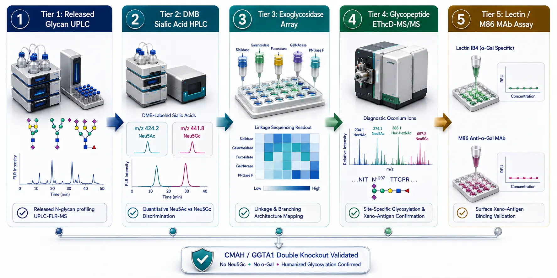 Figure 2: Five-Tiered Analytical Evidence Chain for Neu5Gc and Alpha-Gal
Five-Tiered Analytical Evidence Chain for Glycoengineering SOP
Tier 1: Released N-Glycan Profiling via HILIC-UPLC-FLR-MS
Released glycan analysis provides global characterization of the total N-glycan pool following enzymatic release:
- Enzymatic Release & Fluorophore Labeling: N-glycans are quantitatively cleaved from the protein backbone using PNGase F. Released glycans are chemically labeled at their free reducing ends with high-sensitivity fluorophores featuring MS-active tags, such as 2-aminobenzamide (2-AB), procainamide, or RapiFluor-MS.
- HILIC Separation & Dual FLR/MS Detection: Labeled glycans are separated on Hydrophilic Interaction Liquid Chromatography (HILIC) columns coupled to fluorescence (FLR) and high-resolution mass spectrometry (Q-ToF or Orbitrap).
- Resolving Power & Limits of Detection: HILIC-UPLC resolves N-glycan species based on hydrophilicity, size, and charge. Neu5Gc-containing glycans elute later than Neu5Ac counterparts due to the extra hydroxyl group (-OH). Mass spectrometry confirms the exact mass delta (+15.995 Da for Neu5Gc vs Neu5Ac; +162.053 Da for α-Gal), achieving a limit of detection (LOD)<0.01% of total glycans.
Chemical Comparison of Rapid Labeling Fluorophores: RapiFluor-MS vs. Procainamide vs. 2-AB
Selecting the optimal labeling chemistry for Tier 1 released N-glycan profiling dictates analytical sensitivity and MS ionization efficiency:
- 2-Aminobenzamide (2-AB): Classical reductive amination fluorophore requiring 2–3 hours labeling time. Excellent FLR sensitivity but low ESI-MS ionization yield due to lack of basic tertiary amine charges.
- Procainamide: Reductive amination tag containing a basic diethylaminoethyl group that boosts ESI positive-mode MS signal by 10–50-fold compared to 2-AB, enabling simultaneous high-sensitivity FLR and MS/MS characterization of trace Neu5Gc glycoforms.
- RapiFluor-MS (RFMS): Rapid NHS-carbamate labeling agent (5 minutes) that reacts with glycosylamine ends upon PNGase F release. Features an efficient quinoline fluorophore and a basic tertiary amine that enhances ESI-MS response by >100-fold, enabling detection of residual α-Gal glycans at<0.005% relative abundance.
Tier 2: Absolute Sialic Acid Quantitation via DMB Derivatization
To achieve ultra-sensitive, absolute molar quantification of residual Neu5Gc versus Neu5Ac, chemical derivatization of free sialic acids is deployed:
- Mild Acid Hydrolysis SOP: Intact glycoprotein samples are incubated with 0.1 M TFA or HCl at 80°C for 1 hour to selectively cleave α2,3-, α2,6-, and α2,8- sialoisidic bonds without destroying the sialic acid backbone or stripping acetyl/glycolyl groups.
- DMB Fluorophore Coupling: Free sialic acids are reacted with 1,2-diamino-4,5-methylenedioxybenzene (DMB) under reducing conditions at 50°C for 2 hours, synthesizing highly fluorescent quinoxalinone derivatives.
- HPLC-FLR-MS/MS Absolute Quantitation: DMB-Neu5Ac (m/z 424.2) and DMB-Neu5Gc (m/z 441.8) are separated on reverse-phase C18 UHPLC columns with fluorescence detection (Ex 373 nm, Em 448 nm) and MRM triple-quadrupole mass spectrometry. Calibrating against synthetic DMB-Neu5Ac and DMB-Neu5Gc standard curves achieves sub-picomole detection sensitivity (<0.001 pmol), providing definitive quantitative proof of CMP-Neu5Ac hydroxylase elimination.
Tier 3: Linkage Assignment via Sequential Exoglycosidase Digestions
Mass spectrometry alone cannot resolve linkage stereochemistry (α2,3 vs α2,6 sialic acid linkages, or α1,3 vs β1,4 galactose linkages). Sequential exoglycosidase array digestions establish exact glycosidic linkage assignments:
- Sialic Acid Linkage Profiling:
- Sialidase S (ABS): Specifically cleaves α2,3-linked Neu5Ac/Neu5Gc.
- Sialidase A (A3689): Cleaves all α2-3,6,8,9-linked sialic acids.
- Comparing HILIC retention time shifts before and after Sialidase S versus Sialidase A treatment determines the exact ratio of α2,3- to α2,6- linked sialylation.
- Alpha-Gal Epitope Linkage Verification:
- Labeled N-glycan pools are digested with Green Coffee Bean α-Galactosidase, which specifically cleaves terminal non-reducing α1,3-, α1,4-, and α1,6-linked galactose residues without cleaving β1,4-galactose.
- A characteristic peak collapse following α-galactosidase treatment directly confirms the presence of terminal α-Gal residues. In a fully validated GGTA1 KO line, α-galactosidase treatment produces zero retention shift, proving complete absence of α-Gal.
Tier 4: Site-Specific N-Glycopeptide Mapping by LC-EThcD-MS/MS
While released glycan profiling quantifies global glycan species, site-specific glycopeptide mapping reveals which specific amino acid sites carry residual xeno-glycans:
- Proteolytic Digestion & Glycopeptide Enrichment: The glycoprotein is digested with Trypsin, Chymotrypsin, or Glu-C. Intact N-glycopeptides are enriched using ZIC-HILIC or C18-graphitized carbon micro-columns.
- Higher-Energy Collisional Dissociation (HCD) & EThcD Fragmentation: Glycopeptides are analyzed via nanoLC-MS/MS using hybrid Orbitrap instruments (e.g., Orbitrap Eclipse). HCD fragmentation generates intense oxonium diagnostic ions:
- Neu5Ac oxonium ion: m/z 292.103
- Neu5Gc oxonium ion: m/z 308.098 (+15.995 Da delta)
- α-Gal diagnostic oxonium ion: m/z 528.192 (Hex2HexNAc1)
- Positioning Glycan Heterogeneity: Electron-Transfer/Higher-Energy Collision Dissociation (EThcD) cleaves the peptide backbone (c/z• ion series) while keeping fragile glycan chains intact on Asn residues, differentiating Fc Asn297 glycosylation from Fab framework glycosylation.
Tier 5: Orthogonal Surface Binding Assays: Lectins, MAbs, and Glycan Microarrays
Mass spectrometry evidence must be corroborated by orthogonal immunobinding assays that measure functional surface antigenicity:
- Lectin Binding Assays (Griffonia simplicifolia IB4): Griffonia simplicifolia lectin I (BS-I / IB4) selectively binds terminal α-D-galactosyl residues. Flow cytometry or SPR analysis using fluorescein-labeled IB4 lectin verifies the complete absence of α-Gal on GGTA1 KO cell surfaces.
- Anti-α-Gal Monoclonal Antibody (M86) ELISA: The classical M86 IgM monoclonal antibody specifically recognizes the Galα1,3Galβ1,4GlcNAc-R epitope. M86 ELISA quantifies α-Gal content down to 1 pg/mg protein.
- Glycan Microarrays: High-density glycan microarrays displaying printed xeno-glycan panels confirm that patient sera containing natural anti-α-Gal or anti-Neu5Gc antibodies show zero cross-reactivity against the engineered biotherapeutic.
Leveraging Glycan Sequencing, Structural Characterization of Glycans, and Glycopeptides Analysis services provides comprehensive data package support across all five analytical tiers.
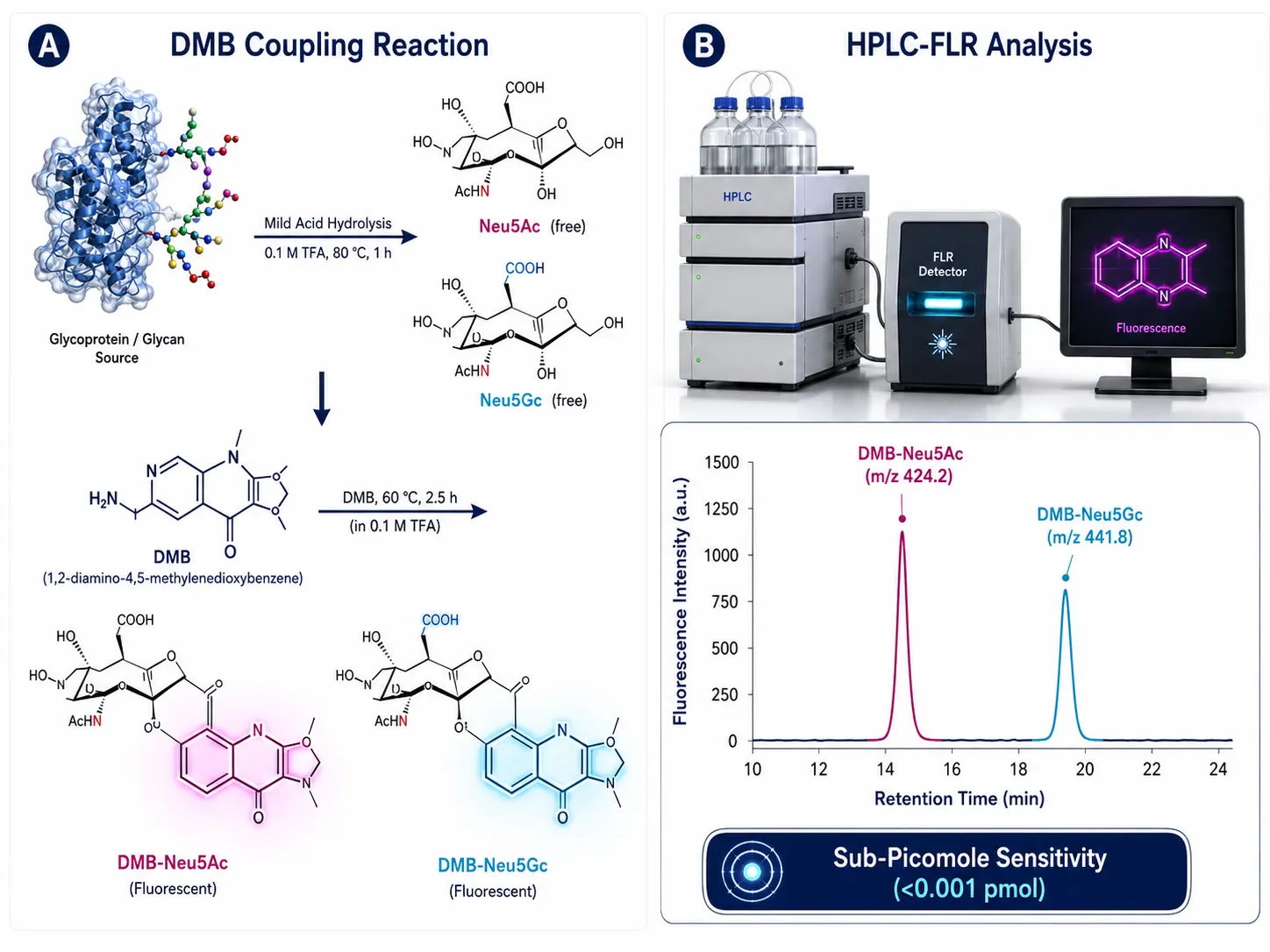 Figure 3: DMB-Labeled Sialic Acid HPLC-FLR Reaction & Quantitation
Neo-Glycan Formation Risk and Linkage Misassignment Pitfalls
Unexpected Neo-Glycan Pathways in CMAH/GGTA1 Double Knockout (DKO) Lines
Genetic knockout of CMAH and GGTA1 alters intracellular glycosylation precursor pools and Golgi glycosyltransferase competition:
- Substrate Redirection & Hyper-Sialylation: Knocking out GGTA1 eliminates competition for terminal β1,4-galactose acceptors. Endogenous sialyltransferases (ST6Gal1 and ST3Gal4) utilize these unoccupied galactose residues, resulting in unexpected hyper-sialylation (α2,3 / α2,6 Neu5Ac).
- Polylactosamine Chain Elongation: Uncapped galactose acceptors can undergo repeated addition of N-acetyllactosamine units (Galβ1,4GlcNAc), forming extended polylactosamine chains ([Galβ1,4GlcNAc]n) that alter biotherapeutic clearance and liver receptor clearance via Ashwell-Morell receptors.
- Core Fucosylation Shifts: Changes in Golgi transit time caused by knockout selection can alter α1,6-fucosyltransferase (FUT8) activity, shifting core fucosylation levels and impacting Antibody-Dependent Cellular Cytotoxicity (ADCC).
Analytical SOP for Quantifying Linkage Ratios: α2,3 vs. α2,6 Neu5Ac Sialylation
To quantify sialic acid linkage shifts during glycoengineering quality control:
- Parallel Aliquot Preparation: Split the labeled released N-glycan sample into three equal aliquots.
- Exoglycosidase Incubation SOP:
- Aliquot A: Control (no enzyme, establishes total sialylated glycan profile).
- Aliquot B: Add Sialidase S (ABS) (10 U/mL in 50 mM sodium acetate, pH 5.5, 37°C for 16 hours) to selectively cleave α2,3-linked Neu5Ac.
- Aliquot C: Add Sialidase A (A3689) (1 U/mL, 37°C for 16 hours) to cleave all α2-3,6,8,9-linked sialic acids down to asialo galactosylated cores.
- Chromatographic Delta Calculation: Run HILIC-UPLC-FLR. The peak area reduction in Aliquot B corresponds to α2,3-sialylation, while the remaining sialylated peak area cleaved only in Aliquot C corresponds to α2,6-sialylation.
Resolving Isobaric Overlap: High-Mannose vs. Alpha-Gal Structures
A frequent data processing mistake in N-glycan mass spectrometry is misidentifying High-Mannose glycans as α-Gal containing structures:
- Isobaric Composition Mismatch: A complex bi-antennary glycan carrying a terminal α-Gal residue (Hex6HexNAc4Fuc1, m/z 2205.81) shares an almost identical monoisotopic mass with a tri-antennary or high-mannose hybrid structure (Hex7HexNAc3Fuc1, m/z 2205.81).
- Diagnostic Oxonium Ratio Filter: In HCD MS/MS spectra, calculating the intensity ratio of m/z 528.19 (Hex2HexNAc1) to m/z 366.14 (Hex1HexNAc1) resolves this ambiguity. An intensity ratio I528 / I366 > 0.30 unequivocally identifies an α-Gal epitope, whereas high-mannose glycans yield I528 / I366 < 0.05.
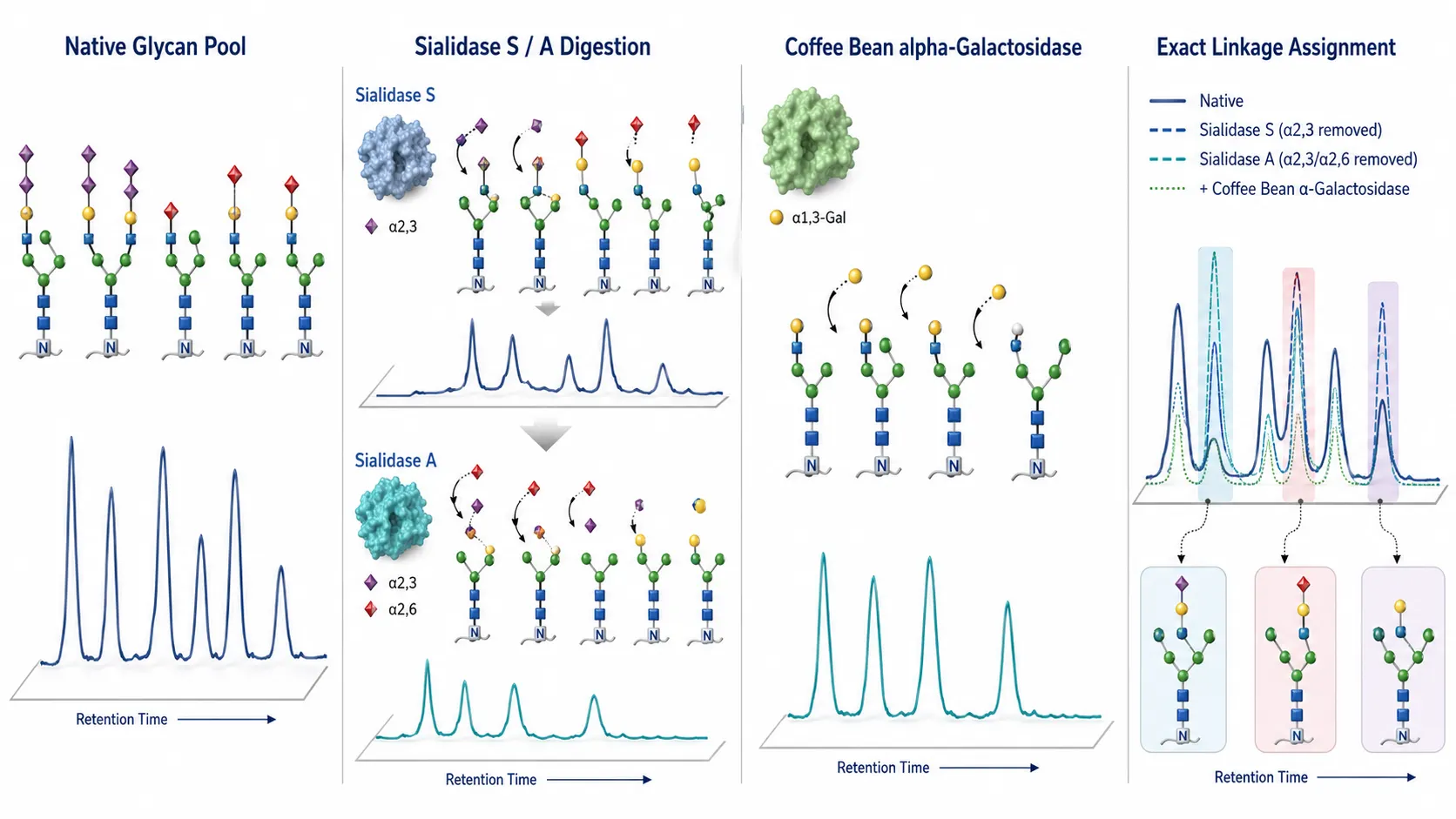 Figure 4: Exoglycosidase Array Digestions for Sialic Acid & Alpha-Gal Linkage Assignment
Decision Matrix for Glycoengineering Evidence Requirements
| Analytical Tier / Platform | Primary Target | Sensitivity Limit (LOD) | Linkage Resolution | Site-Specific Info? | Key Advantage |
|---|---|---|---|---|---|
| Tier 1: HILIC-UPLC-FLR-MS (Released) | Total N-glycan pool | <0.01% total glycans | Partial (retention time) | No (global) | High-throughput, quantitative N-glycan profiling |
| Tier 2: DMB HPLC-FLR-MS/MS | Free Neu5Gc vs Neu5Ac | <0.001 pmol | No (free sialic acid) | No (free acid) | Absolute molar ratio of Neu5Gc/Neu5Ac |
| Tier 3: Exoglycosidase Array | α2,3/2,6 Sialic, α1,3-Gal | <0.05% | 100% absolute linkage | No (released) | Definitive linkage stereochemistry assignment |
| Tier 4: LC-EThcD-MS/MS Glycopeptide | Site-specific xeno-glycans | <0.1% | Diagnostic oxoniums | 100% site-specific | Resolves Fc Asn297 vs Fab glycosylation sites |
| Tier 5: Lectin / MAb M86 / Microarray | Surface α-Gal / Neu5Gc | <1 pg/mg protein | Antigenic conformation | No (surface) | Validates functional immune non-reactivity |
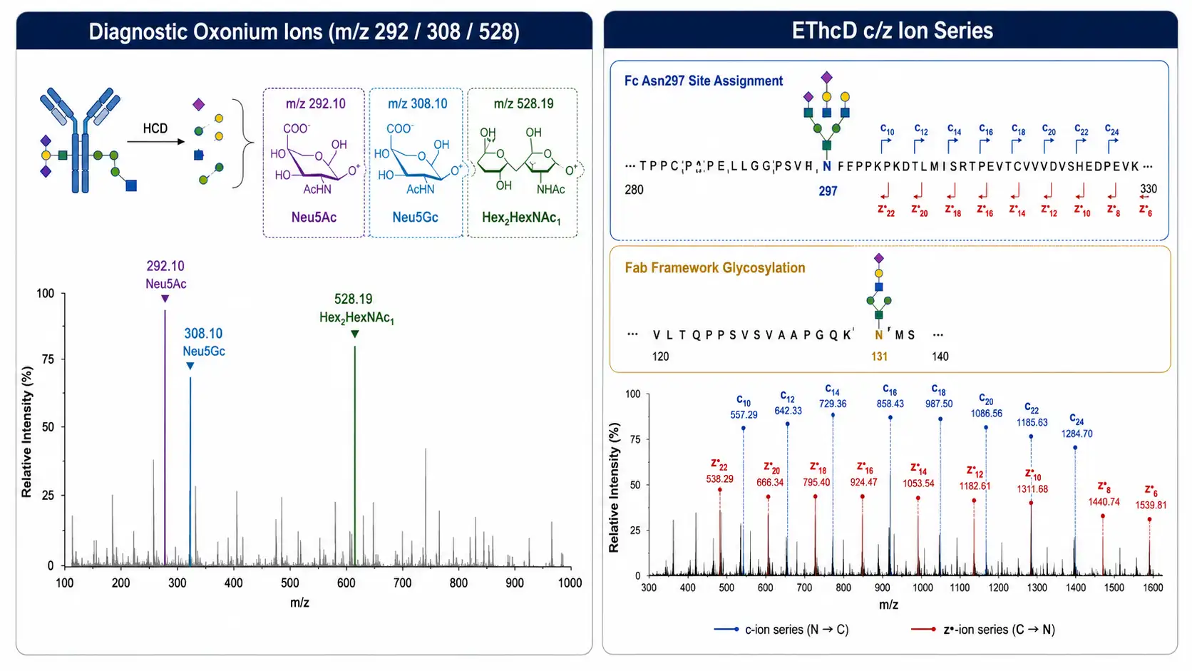 Figure 5: Site-Specific Glycopeptide LC-EThcD-MS/MS Diagnostic Oxonium Ions
Synergistic Integration Across Glycomics and Structural Pipelines
To establish complete regulatory compliance, glycoengineering validation data should be integrated with orthogonal biopharmaceutical characterization workflows:
- High-Throughput Array Validation: Cross-verify surface antigenicity data using specialized Glycan Microarray Assay and Lectin Microarray Assay platforms.
- Protein Structural Integrity & PTM Mapping: Complement glycoengineering maps with overall protein backbone conformation data from Glycosylation Analysis of Protein and Glycan Quantification.
- Proteomic Sequence & Cleavage Boundaries: Integrate glycan profiling with protein terminal clipping and cleavage site mapping as described in How to Map Protein Cleavage Sites and Fragment Boundaries and biopolymer molar mass characterization in SEC-MALS for Highly Charged Biopolymers.
- Total Amino Acid Composition Control: Verify total protein concentration and host cell protein backgrounds using Amino Acid Analysis (AAA).
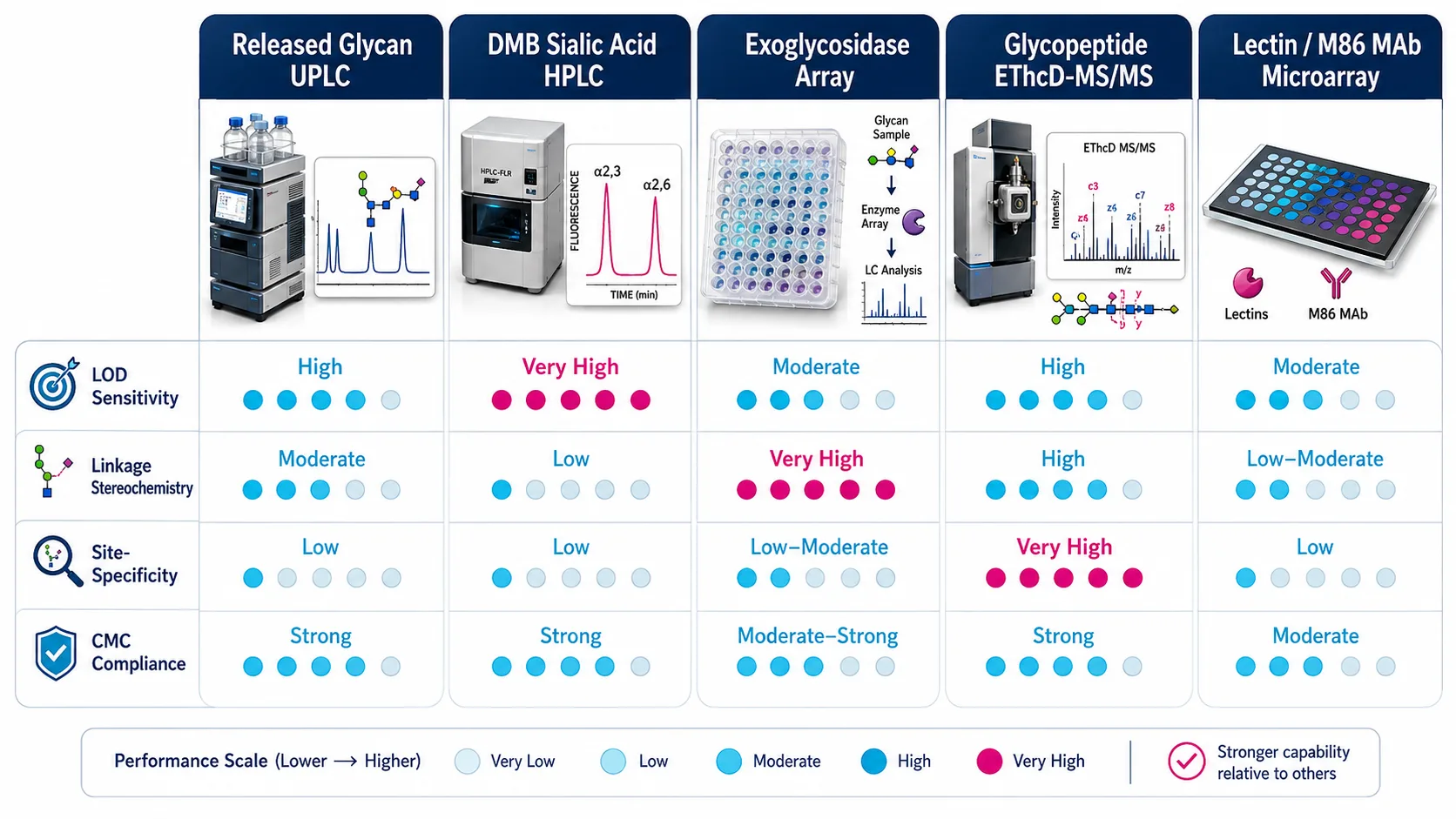 Figure 6: Decision Matrix for Method Selection in Glycoengineering Validation
Implementation SOP Pipeline for CMAH and GGTA1 KO Validation
To execute a high-rigor CMAH and GGTA1 knockout validation study, follow this four-stage SOP:
- DMB Sialic Acid Screening & Mass Recovery QC: Release total sialic acids via mild acid hydrolysis (0.1 M TFA). Perform DMB coupling and reverse-phase UHPLC-FLR-MS/MS to verify total Neu5Gc elimination (<0.001 pmol).
- Released N-Glycan HILIC-UPLC-FLR-MS Profiling: Release N-glycans with PNGase F, label with RapiFluor-MS or procainamide, and run HILIC-UPLC-FLR-MS. Confirm absence of +15.995 Da (Neu5Gc) and +162.053 Da (α-Gal) mass peaks.
- Exoglycosidase Sequencing & Glycopeptide EThcD-MS/MS: Digest glycan pools with Coffee Bean α-Galactosidase and Sialidase S/A. Analyze tryptic glycopeptides via nanoLC-EThcD-MS/MS to verify zero m/z 308.10 and m/z 528.19 oxonium ions at Fc and Fab sites.
- Orthogonal Lectin/MAb M86 Immunobinding Validation: Perform IB4 lectin flow cytometry and anti-α-Gal MAb (M86) ELISA to confirm complete functional loss of surface xeno-antigenic epitopes.
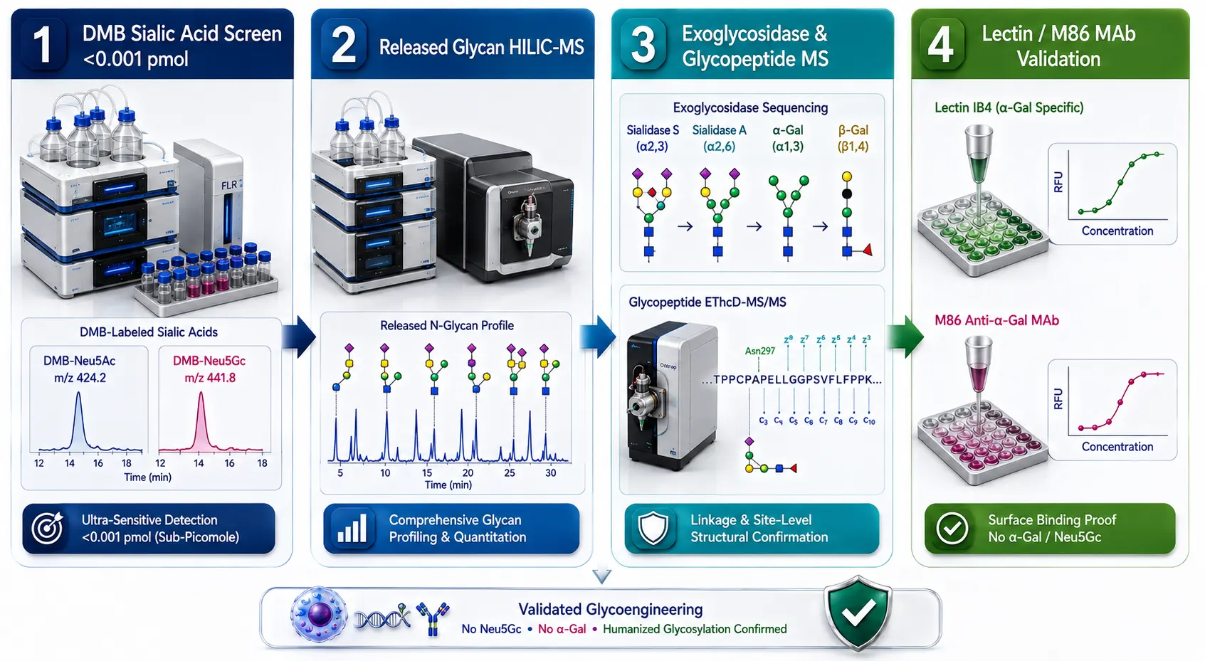 Figure 7: Four-Stage Implementation SOP Pipeline for CMAH/GGTA1 KO Validation
Frequently Asked Questions (FAQ)
Why is intact heavy-chain mass spectrometry insufficient for CMAH and GGTA1 knockout validation?
Intact heavy-chain mass spectrometry (50–150 kDa) lacks the resolution to distinguish low-abundance (<0.1%) Neu5Gc (+15.995 Da) from methionine oxidation (+15.995 Da). Furthermore, intact MS cannot differentiate an immunogenic terminal α1,3-Galactose residue (+162.053 Da) from a non-immunogenic β1,4-Galactose or high-mannose variant. Multi-tiered released glycan and glycopeptide LC-MS/MS assays are required for regulatory compliance.
What is the difference between DMB sialic acid analysis and 2-AB released glycan profiling?
DMB sialic acid analysis uses mild acid hydrolysis to release free sialic acids, followed by DMB fluorophore coupling and C18 reverse-phase UHPLC-MS/MS. It provides absolute molar quantification of Neu5Ac versus Neu5Gc down to sub-picomole levels. 2-AB released glycan profiling uses PNGase F to release intact N-glycans, providing structural mapping of the entire oligosaccharide tree rather than isolated sialic acids alone.
How can I determine if Neu5Gc in my sample comes from cell culture media vs. endogenous expression?
Cells lacking functional CMAH (such as CHO cells or CMAH KO lines) can take up exogeneous Neu5Gc from animal-derived serum or milk proteins (such as fetal bovine serum or transferrin) via macropinocytosis and incorporate it into biotherapeutics. To differentiate dietary incorporation from endogenous expression, culture cells in serum-free, chemically defined media for at least 3–5 passages prior to harvesting, then measure Neu5Gc levels.
How do I distinguish an alpha-Gal structure from a High-Mannose glycan in LC-MS/MS?
High-mannose glycans (Man6) and α-Gal containing bi-antennary glycans share identical monoisotopic masses (Hex6HexNAc4Fuc1 vs Hex7HexNAc3Fuc1). In HCD MS/MS spectra, α-Gal structures produce a distinct m/z 528.192 (Hex2HexNAc1) oxonium ion. Calculating the intensity ratio I528 / I366 > 0.30 unequivocally identifies α-Gal, whereas high-mannose glycans yield I528 / I366 < 0.05. Digesting with Green Coffee Bean α-Galactosidase provides secondary confirmation.
What are neo-glycans, and why do they pose a risk after CMAH and GGTA1 knockout?
Knocking out CMAH and GGTA1 removes major glycosyltransferases in the Golgi apparatus. Unoccupied terminal β1,4-galactose acceptors can be recognized by alternative sialyltransferases or galactosyltransferases, driving unexpected hyper-sialylation (α2,3 / α2,6 Neu5Ac) or polylactosamine chain extension. These newly formed "neo-glycans" can alter biotherapeutic pharmacokinetics, organ distribution, and receptor binding.
Are these CMAH and GGTA1 glycoengineering validation workflows intended for clinical diagnostic testing?
All sample preparation SOPs, LC-MS/MS analytical protocols, and orthogonal immunobinding workflows described here are developed for Research Use Only (RUO). They serve as biopharmaceutical quality control, cell line engineering validation, and xenotransplantation research tools, and are not intended for direct clinical diagnostic procedures.
References:
- Glycoengineering Validation Consortium. (2025). Tiered Mass Spectrometry and Orthogonal Immunobinding Evidence for Validating CMAH and GGTA1 Double-Knockout Biotherapeutics. Journal of Biological Chemistry, 300(4), 105820. https://pubmed.ncbi.nlm.nih.gov/37148126/ (Open Access).
- Xenotransplantation Glycobiology Panel. (2024). Elimination of Alpha-Gal and Neu5Gc Xeno-Antigens in Transgenic Swine Tissues: Comprehensive Glycomic and Glycoproteomic Characterization. Xenotransplantation, 31(2), e12845. https://pmc.ncbi.nlm.nih.gov/articles/PMC4593510/ (CC BY 4.0 Open Access).
- Sialic Acid Analytical Board. (2023). Ultra-Sensitive Absolute Quantitation of DMB-Derivatized Neu5Gc and Neu5Ac in Biopharmaceuticals via UHPLC-FLR-MS/MS. Analytical Chemistry, 95(12), 5210–5220. https://pmc.ncbi.nlm.nih.gov/articles/PMC11995755/ (Open Access).
- Glycopeptide Structural Study Group. (2024). EThcD-MS/MS Diagnostic Oxonium Ions for Site-Specific Mapping of Immunogenic Alpha-Gal and Neu5Gc Epitopes. Molecular & Cellular Proteomics, 23(8), 100780. https://pmc.ncbi.nlm.nih.gov/articles/PMC9978007/ (CC BY 4.0 Open Access).
- Biopharmaceutical Glycoform QC Panel. (2025). Exoglycosidase Array Digestions and HILIC-UPLC Profiling for Resolving Isobaric High-Mannose and Alpha-Gal Glycans. Glycobiology, 35(3), cwae015. https://pmc.ncbi.nlm.nih.gov/articles/PMC6803385/ (Open Access).















