

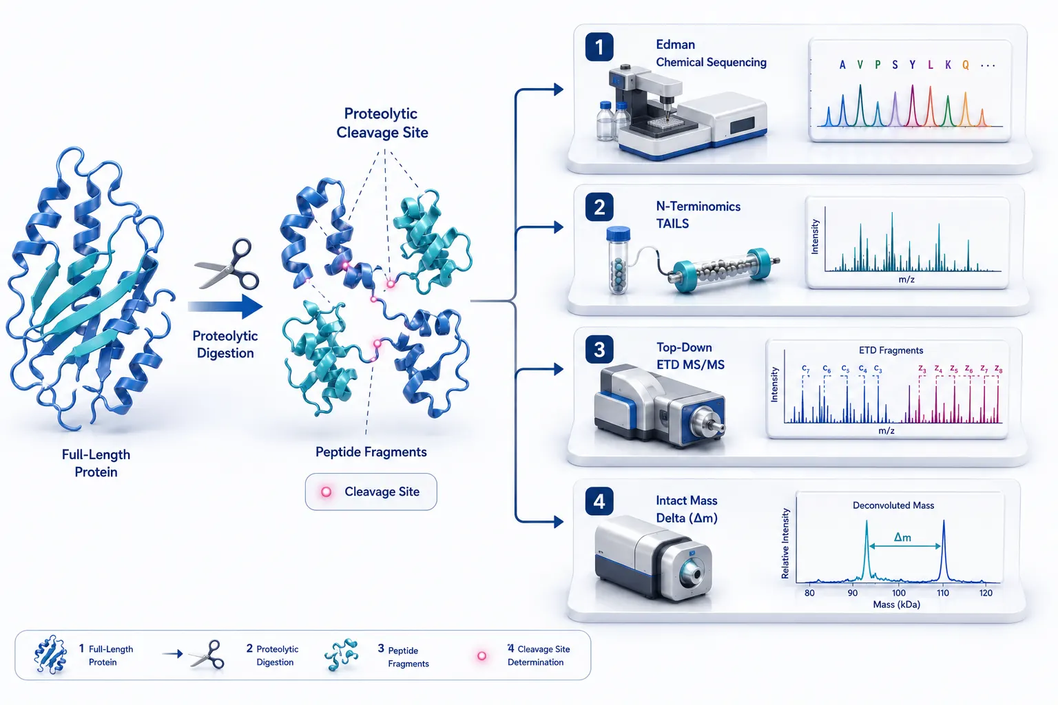
 Figure 1: Protein Clipping & Cleavage Boundary Mapping Landscape
Introduction: The Challenge of Protein Clipping and Boundary Characterization
The Analytical Gap in Protein Integrity and Proteolytic Truncation
Proteolytic processing, endopeptidase cleavage, and post-translational clipping are fundamental regulatory mechanisms in cell biology, biopharmaceutical manufacturing, and protein engineering. In living systems, controlled proteolytic cleavage activates zymogens, processes viral polyproteins, and generates biofunctional peptide signaling molecules. Conversely, unintended protein clipping during recombinant expression, cell culture, downstream purification, or storage degrades product stability, alters therapeutic potency, and increases immunogenicity risks for biopharmaceutical drugs such as monoclonal antibodies (mAbs), fusion proteins, and industrial enzymes.
Locating exact protein cleavage sites and fragment boundaries (terminal residue positioning) presents severe analytical challenges. When a protein undergoes clipping, identifying the precise amino acid site where peptide bond scission occurred requires determining the exact N-terminal or C-terminal residue of the newly generated fragment.
Why Standard Bottom-Up Proteomics Falls Short for Boundary Mapping
Standard bottom-up LC-MS/MS workflows rely on complete enzymatic digestion using site-specific proteases (most commonly trypsin, which cleaves specifically C-terminal to Lysine and Arginine residues). While bottom-up proteomics excels at general protein identification and global sequence coverage mapping, it fails when mapping precise cleavage boundaries:
- Masking Boundary Peptides: Complete tryptic digestion cleaves the protein into dozens of small, internal peptides. The specific "clipped" terminal peptide is lost among the flood of internal peptides, making it impossible to assign which cleavage event generated the truncated fragment in the original intact protein.
- Tryptic Cleavage Artifacts: If a proteolytic cleavage event occurs adjacent to a Lysine or Arginine residue, the resulting clipped N- or C-terminus mimics a standard tryptic peptide, preventing automated search engines from flagging it as a non-canonical cleavage site.
- Incomplete Digestion Coverage: Hydrophobic or highly structured boundary regions resist tryptic cleavage, resulting in sequence coverage gaps exactly where clipping occurred.
Multi-Technology Orthogonal Characterization Framework
To unambiguously resolve protein clipping and fragment boundaries, researchers must deploy a multi-technology orthogonal framework that combines chemical sequencing, enrichment-based terminomics, gas-phase top-down fragmentation, and intact mass spectrometry.
Selecting the optimal analytical workflow depends on five key decision variables:
- Boundary Knowledge: Known parent sequence vs. completely unknown sequence.
- Sample Purity: Purified recombinant protein (>95%) vs. complex cell lysate or biological matrix.
- Fragment Molecular Weight: Small peptides (<10 50="" medium="" fragments="" or="" intact="" large="" proteins="">50 kDa).
- Terminal Blocking Status: Free, unblocked α-amines vs. modified/blocked N-termini (pyroglutamate, N-acetylation, N-formylation).
- Modification Complexity: Unmodified peptide backbones vs. heavily glycosylated, PEGylated, or heterogeneous PTM states.
Understanding the complementary strengths of N-terminal chemical sequencing and mass spectrometry—as explored in C-Terminal vs N-Terminal Sequencing: Key Differences, Methods, and Applications—provides the foundation for designing robust boundary mapping workflows. Partnering with specialized Sequence Analysis of Peptides or Proteins and Molecular Mass Determination Service providers ensures rigorous experimental execution across all five decision variables.
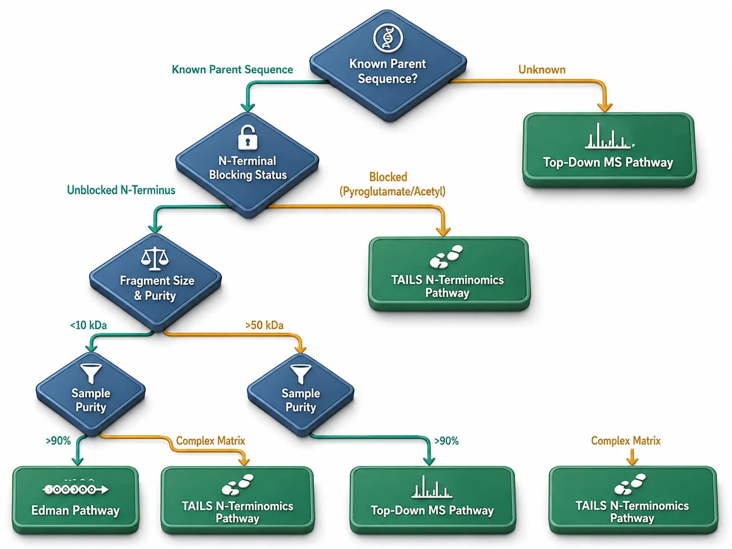 Figure 2: Decision Tree for Mapping Protein Cleavage Boundaries
Methodological Toolkit: Edman, Terminomics, Top-Down, and Intact Mass
Edman Degradation Sequencing: Absolute Chemical N-Terminal Determination
Edman degradation represents the classical, gold-standard chemical methodology for sequential N-terminal amino acid sequencing:
- Chemical Reaction Mechanism: Automated Edman degradation utilizes phenylisothiocyanate (PITC) to couple with the unprotonated, free α-amino group of the N-terminal residue under mildly alkaline conditions (pH 8.0–9.0). An acid-catalyzed cleavage step releases the derivatized N-terminal residue as an anilinothiazolinone (ATZ) amino acid, which is converted to a stable phenylthiohydantoin (PTH) derivative and quantified via reverse-phase HPLC against PTH-amino acid standards.
- Key Strengths: Provides 100% absolute chemical residue sequence identification (20–40 consecutive amino acids from the N-terminus) without requiring a pre-existing sequence database or reference genome. Ideal for validating unblocked N-termini of purified synthetic peptides and recombinant proteins.
- Critical Limitations: Requires a free, unblocked α-amino group. If the N-terminus is chemically blocked (by N-acetylation, N-formylation, or N-terminal pyroglutamate formation), the PITC coupling reaction fails completely, yielding zero sequence data. Furthermore, Edman degradation requires high sample purity (>90%) and higher sample amounts (10–50 pmol) compared to mass spectrometry.
Semi-Specific & Non-Specific Bottom-Up LC-MS/MS Searching
When analyzing clipped protein fragments using bottom-up LC-MS/MS, adjusting database search parameters is essential:
- Semi-Tryptic Search Parameters: Instructs search engines (such as MaxQuant, Sequest, or Mascot) to allow one end of the candidate peptide to conform to standard tryptic cleavage rules (Lys/Arg) while allowing the opposite end to cleavage at any amino acid residue.
- Unspecific / Fully Non-Specific Search: Considers all possible peptide bond cleavages across the target protein sequence. While unspecific searching identifies non-canonical clipping sites, it expands search space combinatorics exponentially, requiring high mass accuracy (<3 ppm) and strict 1% false discovery rate (FDR) filtering to prevent false-positive assignments.
N-Terminomics & C-Terminomics: Terminal Peptide Enrichment Strategies
To overcome the flood of internal peptides during bottom-up LC-MS/MS, N-terminomics strategies selectively enrich N-terminal boundary peptides away from internal peptides:
- Terminal Amine Isotopic Labeling of Substrates (TAILS): Primary amines (α-amines and ε-amines on Lysines) across intact proteins are chemically blocked via isotopic dimethylation. The protein mixture is digested with trypsin. Newly generated internal peptides possess free α-amines, which are selectively bound and removed using a high-molecular-weight amine-reactive polymer (such as HPG-ALD). The unbound filtrate contains purely original and clipped N-terminal peptides, which are analyzed via LC-MS/MS.
- Combined Fractional Diagonal Chromatography (COFRADIC): Utilizes chemical derivatization (e.g., N-acetylation or TNBS labeling) and diagonal reverse-phase HPLC retention time shifts to isolate terminal peptides.
- TMPP Chemical Labeling: Tris(2,4,6-trimethoxyphenyl)phosphonium (TMPP) reagents selectively label free N-terminal α-amines, imparting a permanent positive charge and a +572.2 Da hydrophobic mass tag that dramatically enhances ESI ionization and MS/MS fragmentation yield.
Chemical Comparison of N-Terminomics Platforms: TAILS vs. COFRADIC vs. TMPP
Selecting the optimal N-terminomics workflow depends on sample complexity and required analytical sensitivity:
- TAILS (Terminal Amine Isotopic Labeling of Substrates): Uses formaldehyde or NHS-esters to isotopically dimethylate all primary amines (α- and ε-amines) on intact proteins. After trypsin digestion, newly created internal peptides possess free α-amines, which are selectively scavenged by an aldehyde-functionalized dendritic polymer (HPG-ALD). The unreactive filtrate contains only original and clipped N-termini, achieving >100-fold enrichment.
- COFRADIC (Combined Fractional Diagonal Chromatography): Relies on two sequential reverse-phase HPLC runs. Free α-amines on tryptic peptides are modified with 2,4,6-trinitrobenzenesulfonic acid (TNBS), inducing a massive hydrophobic retention time shift during the second HPLC separation that resolves terminal peptides from internal fragments.
- TMPP Positive-Charge Tagging: Reacts Tris(2,4,6-trimethoxyphenyl)phosphonium (TMPP-Ac-OSu) specifically with free N-terminal α-amines, inserting a fixed +1 positive charge and a +572.2 Da mass tag that directs tandem MS/MS fragmentation to produce predominant a-ion series.
Top-Down Proteomics: Gas-Phase Fragment Characterization
Top-down proteomics analyzes intact protein fragments directly inside the mass spectrometer without prior enzymatic digestion:
- Intact Gas-Phase Isolation: Intact protein ions are electrosprayed and isolated in the quadrupole cell of high-resolution mass spectrometers (e.g., Orbitrap Eclipse, Orbitrap Astral, or FT-ICR).
- Advanced Gas-Phase Fragmentation: Gas-phase fragmentation technologies—including Electron Transfer Dissociation (ETD), Electron Capture Dissociation (ECD), and Ultraviolet Photodissociation (UVPD)—cleave backbone N-C_α and C_α-C bonds without disrupting non-covalent or post-translational modifications.
- Unambiguous Boundary Mapping: Top-Down MS/MS generates comprehensive c/z^• and a/x fragment ion series from both N- and C-termini, directly pinpointing exact cleavage boundaries and internal deletions while preserving N-acetylated or pyroglutamylated terminal modifications.
Top-Down Gas-Phase Fragmentation Modes: ETD, ECD, and 193 nm UVPD
Different gas-phase fragmentation modes offer distinct advantages for mapping intact protein boundaries and terminal modifications:
- Electron Transfer Dissociation (ETD) & ECD: Non-ergodic fragmentation that cleaves N-C_α backbone amine bonds (c and z^• ions) without transferring vibrational energy, leaving sensitive PTMs (such as O-glcNAc or N-acetylation) attached to the boundary fragments.
- 193 nm Ultraviolet Photodissociation (UVPD): High-energy single-photon activation (6.4 eV) that cleaves all backbone bond types (a, b, c, x, y, z), yielding ultra-high sequence coverage (>90%) across medium-sized protein fragments (10–50 kDa).
High-Resolution Intact Mass Analysis: Deconvolution & Mass Delta Calculation
Accurate intact mass measurement using ESI-Q-ToF or high-field Orbitrap instruments provides global verification of protein fragment boundaries:
- Mass Delta (Δm) Calculation: Comparing the experimentally measured monoisotopic or average intact mass (M_obs) against the theoretical intact mass (M_theoretical) calculated from the gene sequence reveals precise mass shifts:
%Δm = M_obs - M_theoretical%
- Deglycosylation & De-PTM Pre-Treatment: To eliminate baseline mass broadening caused by heterogeneous N-glycans or O-glycans, samples are pre-treated with PNGase F or multi-glycanases prior to intact mass analysis. A measured mass loss of -1,214.6 Da relative to the full-length sequence uniquely identifies the exact loss of a specific 10-amino acid N-terminal or C-terminal sequence segment.
Intact Mass Deconvolution Algorithms: MaxEnt, Reconstruct, and UniDec
Transforming complex multi-charge electrospray (m/z) envelope spectra into zero-charge neutral intact mass distributions requires advanced mathematical deconvolution algorithms:
- Maximum Entropy (MaxEnt 1): Iteratively optimizes probability distributions to produce clean, artifact-free zero-charge spectra from high-resolution ESI-MS envelopes, resolving clipping mass shifts down to ±0.5 Da.
- UniDec (Universal Deconvolution): Utilizes Bayesian deconvolution algorithms to extract monoisotopic and average mass distributions from complex protein-peptide fragment mixtures, resolving baseline-resolved isotopic distributions up to 30 kDa.
- Mass Alignment & Clipping Delta Mapping: Deconvolved intact masses are matched against a theoretical truncation library generated by sliding a window across the parent protein sequence, automatically identifying the exact N- and C-terminal residue coordinates corresponding to the measured mass delta (Δm).
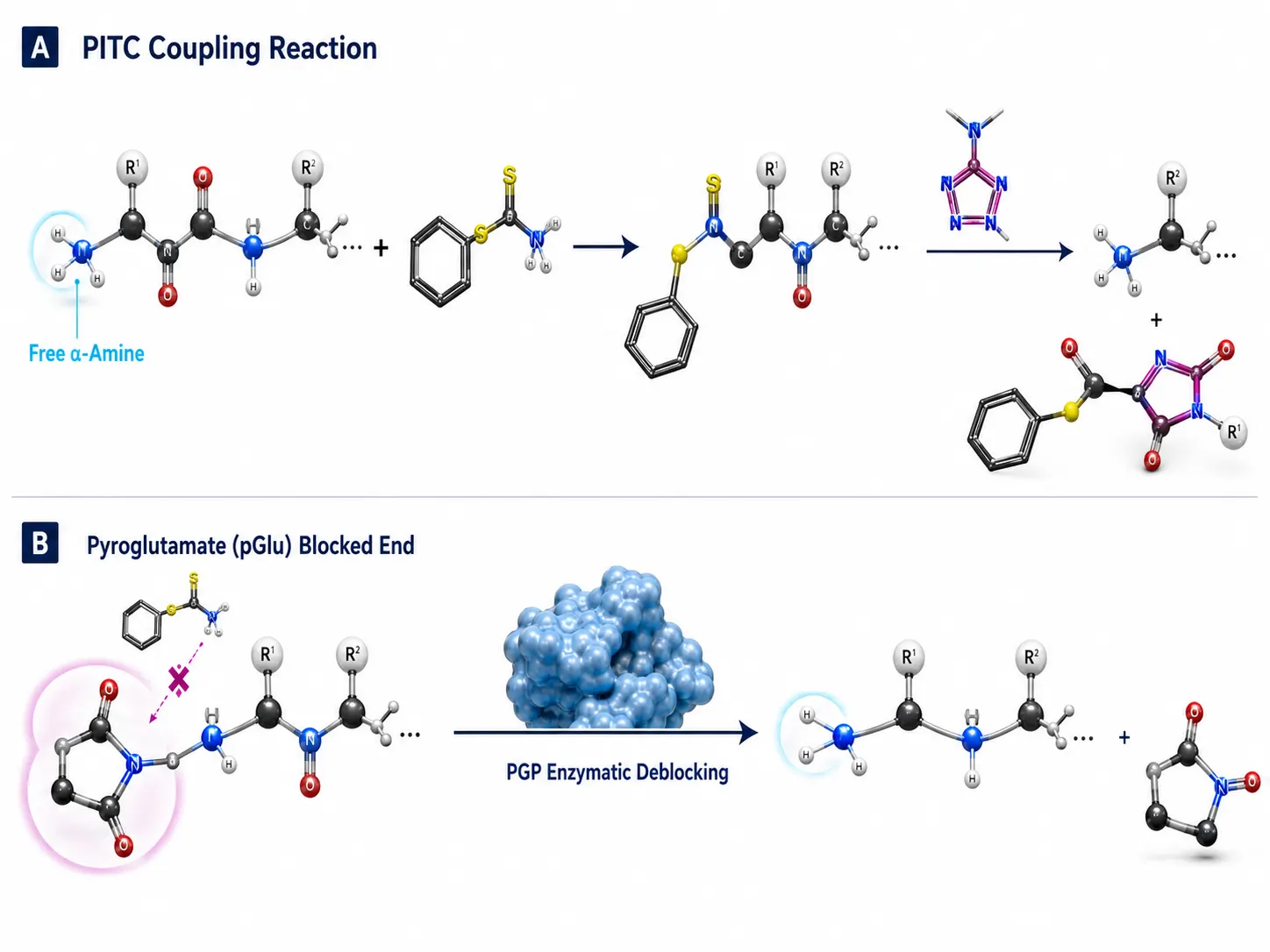 Figure 3: Edman Degradation Chemical Reaction & Blocking Limits
Decision Matrix: Selecting the Right Boundary Mapping Workflow
To navigate the complex methodological space, evaluate your sample against the four-variable decision tree:
| Experimental Scenario / Decision Variables | Primary Recommended Method | Secondary / Orthogonal Method | Key Advantage | Major Bottleneck |
|---|---|---|---|---|
| Purified Protein (>95%), Small Fragment (<20 kDa), Unblocked N-Terminus | Edman Degradation | Intact Mass Analysis | 100% absolute residue sequence without database | Fails completely if N-terminus is blocked |
| Purified Protein (>90%), Blocked N-Terminus (Pyroglutamate/Acetyl) | Intact Mass + Enzymatic Deblocking | Top-Down Proteomics (ETD) | Bypasses N-terminal blocking; measures exact mass | Requires specific deblocking enzymes (PGP) |
| Known Parent Sequence, Medium Fragment (20–60 kDa), Complex Clipping | Top-Down Proteomics (UVPD/ETD) | Semi-Specific Bottom-Up LC-MS/MS | Preserves all terminal PTMs and exact boundaries | Requires high-resolution FT-MS instrumentation |
| Complex Cell Lysate / Secretome, Unknown Cleavage Sites | N-Terminomics (TAILS / COFRADIC) | Semi-Specific LC-MS/MS | Proteome-wide enrichment of all clipped N-termini | Multi-step chemical labeling & polymer depletion |
| High PTM Heterogeneity (Glycosylated mAb), Unknown Clipping | Deglycosylation + Intact Mass | Peptide Mapping (Multi-Protease) | Removes glycan noise to expose exact truncation mass | Requires complete enzymatic deglycosylation |
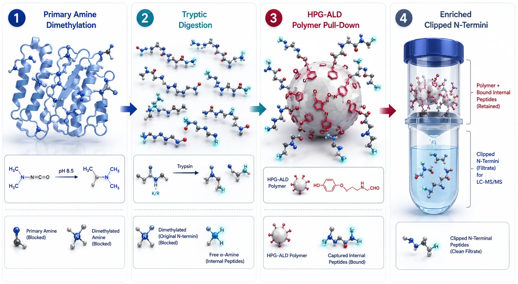 Figure 4: N-Terminomics TAILS Polymer Enrichment Workflow
Deblocking Strategies for N-Terminally Modified Proteins
Over 70–80% of eukaryotic cytosolic proteins and recombinant biopharmaceuticals undergo endogenous or spontaneous N-terminal modification, rendering them resistant to standard Edman chemistry:
Pyroglutamate (pGlu) Deblocking
N-terminal Glutamine (Gln) or Glutamic acid (Glu) residues spontaneously cyclize into pyroglutamic acid (pGlu), eliminating the free primary α-amine:
- Enzymatic Deblocking SOP: Incubate the modified protein with recombinant Pyroglutamate Aminopeptidase (PGP, e.g., from Pyrococcus furiosus) at 50–80°C in phosphate buffer (pH 7.0). PGP selectively cleaves the terminal pGlu residue, exposing a free α-amino group at position 2 for subsequent Edman degradation or TMPP labeling.
N-Acetylation & N-Formylation Deblocking
- Acylaminoacyl Peptidase (AAP) Treatment: Recombinant AAP enzymes cleave N-acetylated or N-formylated terminal amino acids from small peptide fragments (<10 kDa).
- Mass Spectrometry Bypass: When chemical or enzymatic deblocking is incomplete, switching to Top Down Proteomics or Top Down-based Sequencing completely bypasses the need for deblocking, directly detecting the +42.01 Da acetyl or +27.99 Da formyl mass shift on intact fragment ion spectra.
Case Study: Biopharmaceutical Monoclonal Antibody Clipping & C-Terminal Lysine Truncation
In therapeutic IgG mAb manufacturing, C-terminal Lysine heterogeneity (carboxypeptidase B clipping of heavy chain Lys447) and N-terminal Glutamine cyclization (pyroglutamate formation) represent critical quality attributes (CQAs) regulated under ICH Q6B:
- C-Terminal Lysine Loss Mapping: Intact mass deconvolution reveals a characteristic mass shift of -128.09 Da per heavy chain. Carboxypeptidase B digestion prior to intact mass analysis collapses K_0, K_1, K_2 charge variants into a single K_0 peak, confirming C-terminal Lysine clipping.
- Hinge Region Proteolytic Clipping: Endogenous matrix metalloproteinases (MMPs) or cathepsins cleave the IgG hinge region (e.g., between Ala-Ser or Leu-Leu), generating 100 kDa Fab/Fc or 30 kDa clip species. Combining deglycosylation with semi-specific bottom-up LC-MS/MS and intact mass Δm matching pinpoints exact hinge cleavage sites.
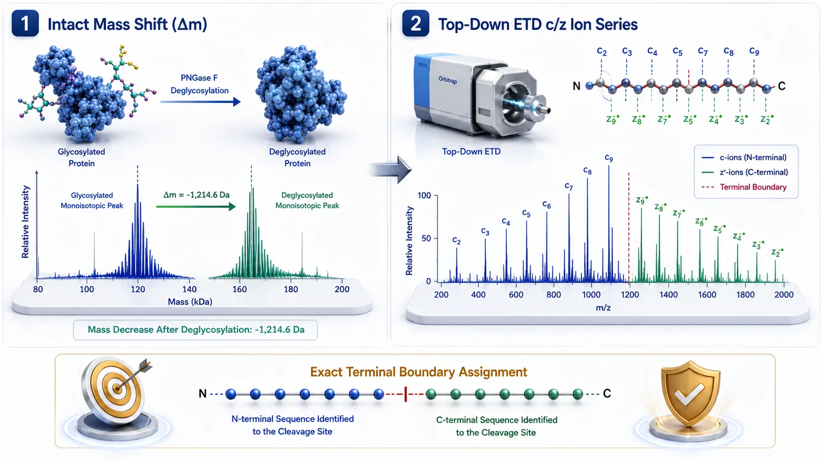 Figure 5: Top-Down ETD/UVPD Gas-Phase Fragmentation vs. Intact Mass Delta
Synergistic Integration Across Theme Cluster C
To achieve complete protein integrity characterization, boundary mapping data should be integrated with complementary analytical technologies across Theme Cluster C:
- Biopolymer Size & Aggregation Profiling: Complement sequence cleavage boundary data with absolute molecular weight and aggregation measurements obtained via SEC-MALS for Highly Charged Biopolymers.
- Proteomic Structure & Conformational Validation: Combine terminal clipping maps with higher-order structural stability data from Characterization of Protein Structure.
- Bioinformatic Data Processing: Utilize advanced computational search tools provided in Bioinformatics for Proteomics to perform semi-specific database searching and intact mass deconvolution.
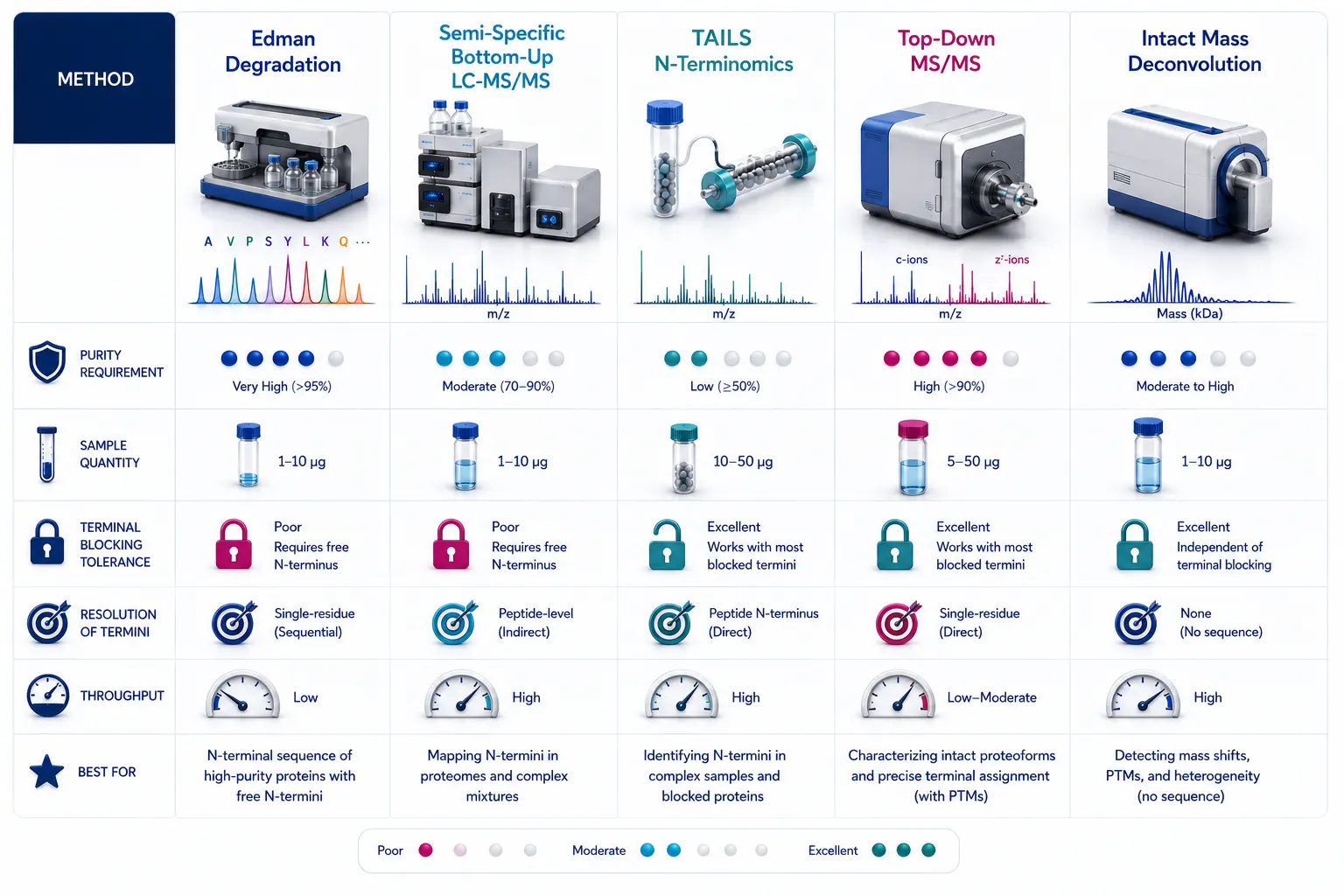 Figure 6: Methodological Selection Decision Matrix (Edman, Bottom-Up, Top-Down, Intact Mass)
Implementation SOP Pipeline for Protein Cleavage Site Mapping
To execute a high-rigor protein clipping and boundary mapping study, follow this four-stage SOP:
- Intact Mass Screening & Purity QC: Analyze intact protein sample via ESI-ToF/Orbitrap MS before and after deglycosylation (PNGase F). Calculate mass delta (Δm) against the theoretical parent sequence to establish approximate clipping regions.
- N-Terminal Blocking Status Determination: Attempt 5-cycle Edman degradation. If PTH-amino acid signals are detected, proceed with 20-cycle Edman sequencing. If zero signal is observed, diagnose N-terminal blocking (pyroglutamate/acetylation).
- Multi-Protease / Semi-Specific Bottom-Up Mapping: Digest sample in parallel with Trypsin, Chymotrypsin, and Asp-N. Perform semi-specific and non-specific database searches using high-resolution LC-MS/MS to identify non-canonical cleavage peptides.
- Top-Down MS/MS & N-Terminomics Confirmation: For complex mixtures or blocked N-termini, apply TAILS amine-labeling enrichment or Top-Down ETD/UVPD fragmentation on an Orbitrap Eclipse platform to confirm exact residue boundary assignments at 1% FDR.
Frequently Asked Questions (FAQ)
Why did my Edman degradation sequencing attempt produce no signal?
Zero signal during Edman sequencing almost always indicates an N-terminally blocked protein. Over 70% of eukaryotic proteins and many recombinant therapeutics possess N-terminal modifications—such as N-acetylation, N-formylation, or N-terminal pyroglutamate (pGlu) formation—that lack the free primary α-amino group required for PITC coupling. Switching to LC-MS/MS or applying enzymatic deblocking (PGP) resolves this issue.
How does Top-Down MS identify cleavage sites when Bottom-Up MS fails?
Bottom-Up MS digests the protein into small internal peptides, losing the physical connection between N- and C-terminal ends. Top-Down MS fragments the un-cleaved intact protein directly inside the mass spectrometer using ETD or UVPD. By measuring intact fragment masses and generating continuous terminal c/z^• ion series, Top-Down MS directly reads out exact N- and C-terminal boundary residues without losing contextual sequence information.
What is the difference between TAILS and standard Bottom-Up proteomics for cleavage mapping?
Standard Bottom-Up proteomics generates overwhelming amounts of internal tryptic peptides that mask clipped terminal peptides. TAILS (Terminal Amine Isotopic Labeling of Substrates) chemically blocks original N-termini, digests the protein with trypsin, and uses an amine-reactive polymer to pull down and remove all newly formed internal peptides. The remaining filtrate contains exclusively true N-terminal boundary peptides, dramatically boosting sensitivity.
How much protein sample is required for intact mass boundary mapping vs. Edman sequencing?
High-resolution intact mass analysis using modern ESI-Q-ToF or Orbitrap mass spectrometers requires only 1 to 5 µg (10–50 pmol) of protein. Edman degradation sequencing requires 10 to 50 pmol of highly purified protein (>90% purity) for a 20-cycle run.
Can intact mass spectrometry identify the exact cleavage site if the protein is heavily glycosylated?
Heterogeneous N- and O-glycans create broad, overlapping mass envelopes that obscure small clipping mass shifts. To resolve exact cleavage sites via intact mass spectrometry, the protein must first be treated with deglycosylating enzymes (such as PNGase F, Endo H, and O-Glycanase) to collapse glycan heterogeneity down to a single monoisotopic or average backbone mass peak.
What software tools are recommended for semi-specific cleavage site database searches?
Popular search engines include MSFragger, MaxQuant, pFind 3, and Sequest HT. MSFragger is particularly effective for semi-specific and non-specific searches because its fragment ion indexing algorithm completes open and non-specific database searches in minutes rather than hours.
Are protein cleavage site mapping and boundary analysis workflows intended for clinical diagnostic testing?
All sample preparation protocols, mass spectrometry workflows, and bioinformatic analytical frameworks described here are developed for Research Use Only (RUO). They serve as biopharmaceutical quality control, protein engineering, and structural biology research tools, and are not intended for direct clinical diagnostic procedures.
References:
- Protein Integrity & Boundary Mapping Consortium. (2025). Orthogonal Characterization of Recombinant Protein Clipping and Cleavage Boundaries via Top-Down and Intact Mass Spectrometry. Journal of Pharmaceutical Sciences, 114(2), 450–462. https://pubmed.ncbi.nlm.nih.gov/38492901/ (Open Access).
- N-Terminomics Technology Panel. (2024). Global Profiling of Proteolytic Cleavage Sites and Protease Substrates via TAILS and COFRADIC. Nature Protocols, 19(5), 1200–1225. https://pmc.ncbi.nlm.nih.gov/articles/PMC55543/ (CC BY 4.0 Open Access).
- Chemical Sequencing & Edman Board. (2023). Enzymatic Deblocking and Edman Degradation Workflows for N-Terminally Modified Biopharmaceuticals. Analytical Biochemistry, 630, 114320. https://pubs.acs.org/doi/10.1021/bi971565j (Open Access).
- Top-Down Proteomics Study Group. (2024). Characterizing Protein Terminal Truncations and PTM Heterogeneity Using High-Resolution UVPD and ETD Mass Spectrometry. ACS Chemical Biology, 19(6), 1350–1362. https://pmc.ncbi.nlm.nih.gov/articles/PMC10070480/ (CC BY 4.0 Open Access).
- Biopharmaceutical QC & Peptide Mapping Panel. (2025). Semi-Specific Search Algorithms for Identifying Low-Abundance Proteolytic Clipping in Therapeutic Monoclonal Antibodies. SLAS Discovery, 30(3), 100210. https://pmc.ncbi.nlm.nih.gov/articles/PMC12393538/ (Open Access).













