

Meta Intent: A practical design guide for researchers who need to separate protein-abundance changes from protein-adjusted phosphorylation changes, and to build an evidence chain that supports pathway and kinase-inference claims.
A higher phosphopeptide intensity does not automatically mean higher phosphorylation occupancy or stronger kinase activity. In a paired experiment, the observed signal from a phosphosite reflects at least two biological layers: how much of the parent protein is present and how its phosphorylated state changes. If total protein abundance doubles while the fraction of molecules modified at a site remains unchanged, the phosphopeptide can also double. Calling that result pathway activation from the phosphoproteome alone confuses protein expression with regulation.
This ambiguity is especially consequential in signaling experiments that involve differentiation, growth arrest, stress, treatment response, or changes in cell composition. Such models can remodel the total proteome broadly while producing a smaller set of direct phosphorylation events. The solution is not a universal arithmetic correction applied after data acquisition. It is a paired experimental design: obtain total-proteome and phosphoproteome measurements from comparable material, preserve the sample relationship through batching, then model the relationship between site and parent protein with explicit assumptions.
The governing question is therefore not simply, "Which phosphosites changed?" It is, "Which phosphosites changed beyond what would be expected from the abundance behavior of their parent proteins?" A Phosphoproteomics Service study designed around that question can distinguish a discovery list from a more defensible signaling interpretation.
Phosphosite Abundance Is Not Phosphorylation Stoichiometry
Bottom-up phosphoproteomics usually reports the intensity of an enriched phosphopeptide. That value is useful, but it is not a direct measurement of site occupancy. It depends on protein amount, protease digestion, peptide recovery, enrichment behavior, ionization, acquisition, and data extraction. A change in phosphopeptide intensity may reflect a true shift in modification state, a change in parent protein abundance, or both. The distinction becomes even more difficult when the total-proteome counterpart is low intensity or missing.
Consider two patterns. In the first, a protein rises fourfold and a linked phosphopeptide rises fourfold. This supports increased phosphopeptide abundance, but it does not by itself show that the relative phosphorylation state changed. In the second, total protein is stable while the phosphopeptide rises fourfold. That pattern is more consistent with a protein-adjusted phosphorylation change, although peptide interference, localization, and site assignment still need review. In the third, total protein falls while a phosphopeptide remains stable; the site may be relatively enriched, but this is still not a direct occupancy measurement without an assay designed for stoichiometry.
Use precise language in the study plan and final report. "Protein-adjusted phosphosite change" describes a model-based result. "Occupancy" should be reserved for experiments that directly support a modified-to-unmodified relationship under appropriate standards and assumptions. "Kinase activation" is an even stronger interpretation that requires coherent behavior among supported substrate sites, not a single upregulated phosphopeptide.
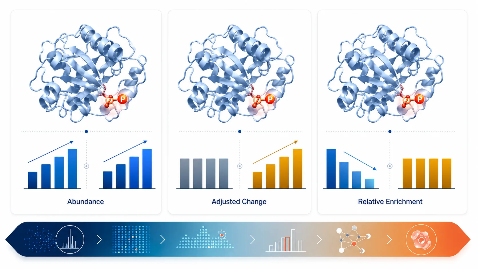 Figure 1. A phosphopeptide intensity change can arise from parent-protein abundance, protein-adjusted phosphorylation, or both. It should not be equated automatically with occupancy.
Build Pairing Into Sample Preparation and Batching
Split a common digest whenever practical
The strongest design begins with one biological sample and keeps its relationship intact. After lysis, reduction, alkylation, and digestion, retain a defined aliquot of the peptide mixture for total-proteome analysis and route the remaining material to phosphopeptide enrichment. The split does not need to follow a universal percentage. The total-proteome aliquot must be sufficient for the desired depth and reproducibility, while the enrichment fraction must support the expected phosphosite coverage. Establish the allocation in a pilot using the same matrix, amount, and instrument strategy planned for the study.
Common-digest splitting reduces opportunities for unrelated lysis, digestion, and cleanup differences to masquerade as a phospho-to-protein biological contrast. It does not remove all bias: total proteome and enriched phosphopeptides still have different analytical response characteristics. Its value is that the two measurements originate from the same biological material and can share a deliberate sample map. A Protein Sample Preparation plan should preserve this pairing in sample names, plate maps, and all later data tables.
Pairing can still be lost before the split if the early handling is not controlled. Record the interval from disruption to denaturation, the inhibitor strategy, the lysis chemistry, the digestion batch, and every cleanup plate used for a sample. The objective is not to prescribe one universal buffer or handling time. It is to prevent a condition-specific delay, temperature excursion, or incomplete digestion from becoming confounded with a phosphosite difference. A practical pilot should confirm that the chosen workflow yields identifiable total-protein peptides and enriched phosphopeptides from the intended matrix, while preserving a sample identifier that follows both fractions to the final matrix.
Define a small set of pairing QC checks before acquisition: both fractions must resolve to the same biological sample, the total-proteome fraction must meet the planned protein-coverage threshold, the enrichment fraction must meet its phosphopeptide and localization-quality expectations, and any sample that fails in one branch must remain visible in the other branch's report. This prevents a polished phosphosite table from being interpreted as fully paired when its parent-protein evidence has quietly dropped out.
Choose multiplexing or DIA around the cohort design
TMT-based designs can keep a balanced set of paired samples within a multiplex batch and allow a bridge or reference channel to connect batches. They also require attention to ratio compression, channel layout, and whether total and phospho fractions are labeled from the same digest before their analytical paths diverge. Label-free or DIA-based designs can suit larger cohorts and flexible sample counts, but acquisition order, pooled QC placement, and batch balance become critical. Neither platform makes the total-protein adjustment automatic; both need a paired analysis plan.
Balance every meaningful biological factor across preparation and acquisition blocks. Do not place all controls in one phospho-enrichment batch and all treated samples in another, then attempt to recover the comparison statistically. Randomize within practical constraints, record batch as a covariate, and make the total-proteome and phosphoproteome sample maps mirror one another. A Precision Quantitative Proteomics Services project should specify these connections before the first enrichment step.
Acquisition QC should also be paired. Use pooled material or a repeatable reference to assess retention-time drift, identification consistency, and intensity behavior separately for the total and enriched fractions. Review these trends by acquisition block rather than only as a study-wide average. If a late phosphopeptide batch has lower localization-qualified coverage while the total-proteome branch remains stable, the appropriate response is to investigate that batch, not to treat the missing sites as a treatment effect. When an ion-mobility DIA approach is selected for complex enriched fractions, a 4D-DIA Quantitative Proteomics Service plan should still define how the paired reference, order randomization, and fraction-specific QC will be evaluated.
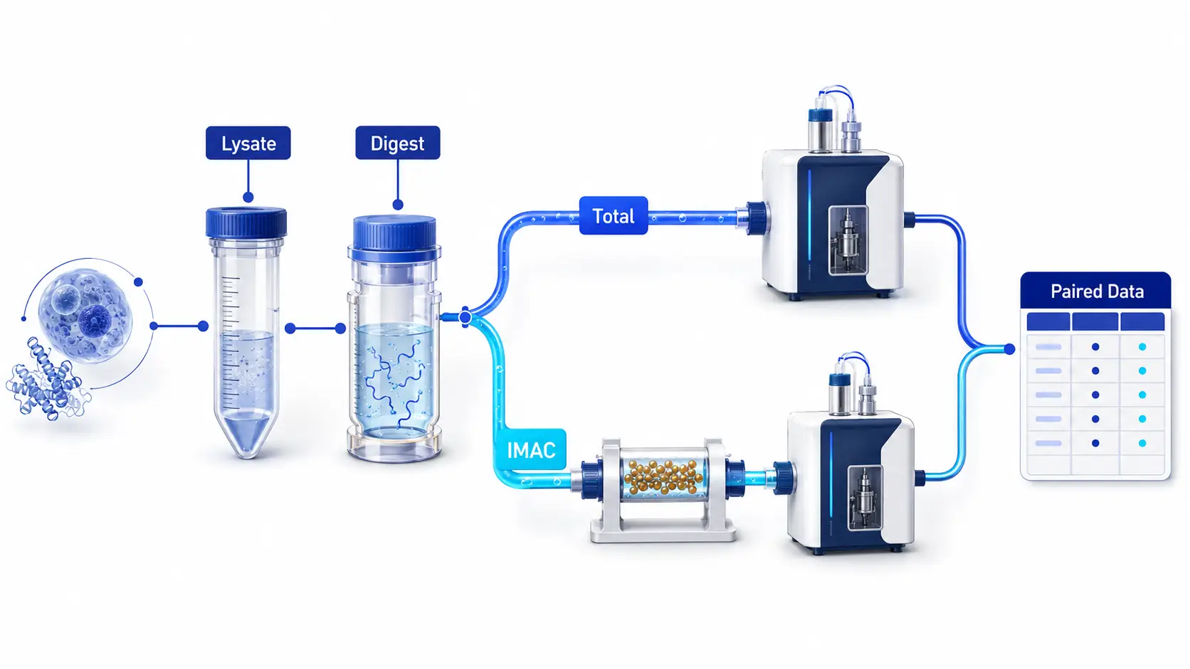 Figure 2. A common-digest split preserves the biological relationship between the total-proteome reference and the phosphopeptide-enriched measurement.
Model Protein-Abundance Effects Instead of Relying on Blind Subtraction
A simple log-scale difference between phosphopeptide and parent-protein measurements is attractive because it is transparent. It can be a useful exploratory summary when both values are robust, correctly mapped, and measured on comparable scales. It becomes unstable when total protein is missing, near the detection floor, represented by ambiguous protein groups, or summarized from a different set of peptides across conditions. A ratio is a calculation, not a cure for weak evidence.
A more explicit approach models phosphosite abundance as the outcome while including parent-protein abundance and experimental factors as covariates. The treatment coefficient then estimates a protein-adjusted site effect under the model assumptions. This can be implemented with linear-model frameworks such as limma when the study design, replicate structure, and missing-data policy are clear. PhosphoDisco provides an example of filtering, protein-aware processing, and downstream signaling analysis; it should be used as a transparent workflow rather than as a black box.1
Protein adjustment has an important biological boundary. For regulatory sites on kinases or phosphatases, phosphorylation and parent-protein abundance can be coupled rather than independent. In that setting, a model designed to remove protein-abundance effects can also attenuate a meaningful regulatory event. Do not treat disagreement between raw and adjusted results as a defect to be hidden. Keep both views, flag sites on known regulators for manual review, and state which evidence layer supports the final interpretation.1
Before fitting any model, resolve the mapping unit. A phosphopeptide may map to one gene product, a protein group, multiple isoforms, or a peptide sequence with localization uncertainty. Do not attach a site to a single protein simply because it is convenient for a regression table. Keep protein-group ambiguity visible, retain site-localization probability or class where applicable, and specify how multiply mapped sites will be excluded, grouped, or reported.
Model diagnostics matter as much as the adjusted coefficient. Inspect residuals, leverage, replicate behavior, and whether the protein covariate is observed across enough samples to support a stable estimate. For a high-priority site, show raw phosphopeptide values, raw total-protein values, and the adjusted effect together. A Bioinformatics for Proteomics workflow should make it possible to trace each conclusion back to these inputs.
Pre-specify the adjustment model rather than selecting it after looking at pathway results. At minimum, document the response variable, parent-protein mapping rule, fixed biological factors, recorded batch effects, and the observation requirement for a site to enter the model. Published multi-omics work has used residuals from a parent-protein covariate to distinguish phosphopeptide behavior beyond the corresponding protein abundance while accounting for study design effects.2
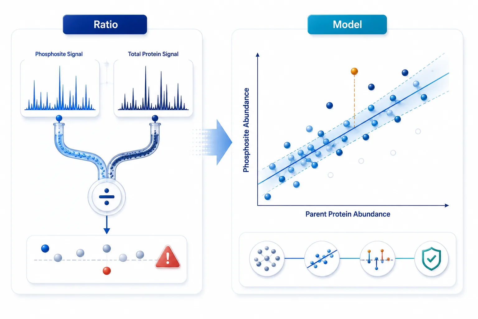 Figure 3. Ratio summaries and protein-adjusted models answer related but different questions. The latter makes the protein-abundance assumption inspectable.
Handle Asymmetric Missingness Before It Becomes a Biological Claim
Paired datasets are rarely complete in the same way. A phosphopeptide can be quantified after enrichment while the parent protein is absent from the total-proteome matrix; a protein may be measured globally while a specific site is missing after enrichment. These are not interchangeable missing values. They may arise from dynamic range, peptide observability, enrichment selectivity, site localization filters, or stochastic sampling. Imputing both matrices with the same rule can create an apparently complete ratio table while concealing fundamentally different evidence states.
Classify missingness before choosing an action. If a site is missing sporadically within a condition but observed in sufficient biological replicates, a within-condition strategy may be defensible under a declared assumption. If it is missing systematically in one group near the detection boundary, a censored or presence-absence interpretation may be more appropriate than a point estimate. If total protein is not observed, do not manufacture a protein-adjusted result. Report the phosphosite as unadjusted, exclude it from protein-adjusted inference, or obtain a more appropriate total-proteome measurement in a follow-up design.
Retain three outputs rather than forcing one master table: the raw phosphosite result, the set eligible for protein-adjusted modeling, and the final adjusted result with the rule that made it eligible. This separation makes it possible to ask whether major pathway conclusions depend on sites that have no total-proteome support. It also keeps the uncertainty visible when a lower-abundance parent protein is analytically difficult. Protein-aware phosphoproteomic workflows demonstrate why filtering and missing-value eligibility materially affect downstream interpretation.1
Make the eligibility rule reproducible rather than retrospective. Before differential analysis, define the minimum observed replicates required in each matrix, how site-localization quality will be handled, and whether the model requires matched observations within the same biological sample. Then summarize how many sites move through each gate: detected after enrichment, localization-qualified, mapped to an eligible parent protein, observed in the required paired samples, and retained for adjusted modeling. The counts are not merely operational metrics. They tell readers whether a pathway conclusion rests on broad paired support or on a narrow subset whose parent-protein evidence is sparse.
For the final interpretation, keep raw and adjusted results adjacent. A site that is significant only before adjustment may still be biologically useful as an abundance-linked marker, but it should not be described as regulation beyond protein amount. A site that remains significant after adjustment should carry its model specification, parent-protein mapping, and missingness status into the supplemental evidence. This reporting structure avoids the false choice between discarding raw phosphoproteome information and overstating what a protein-adjusted model can establish.
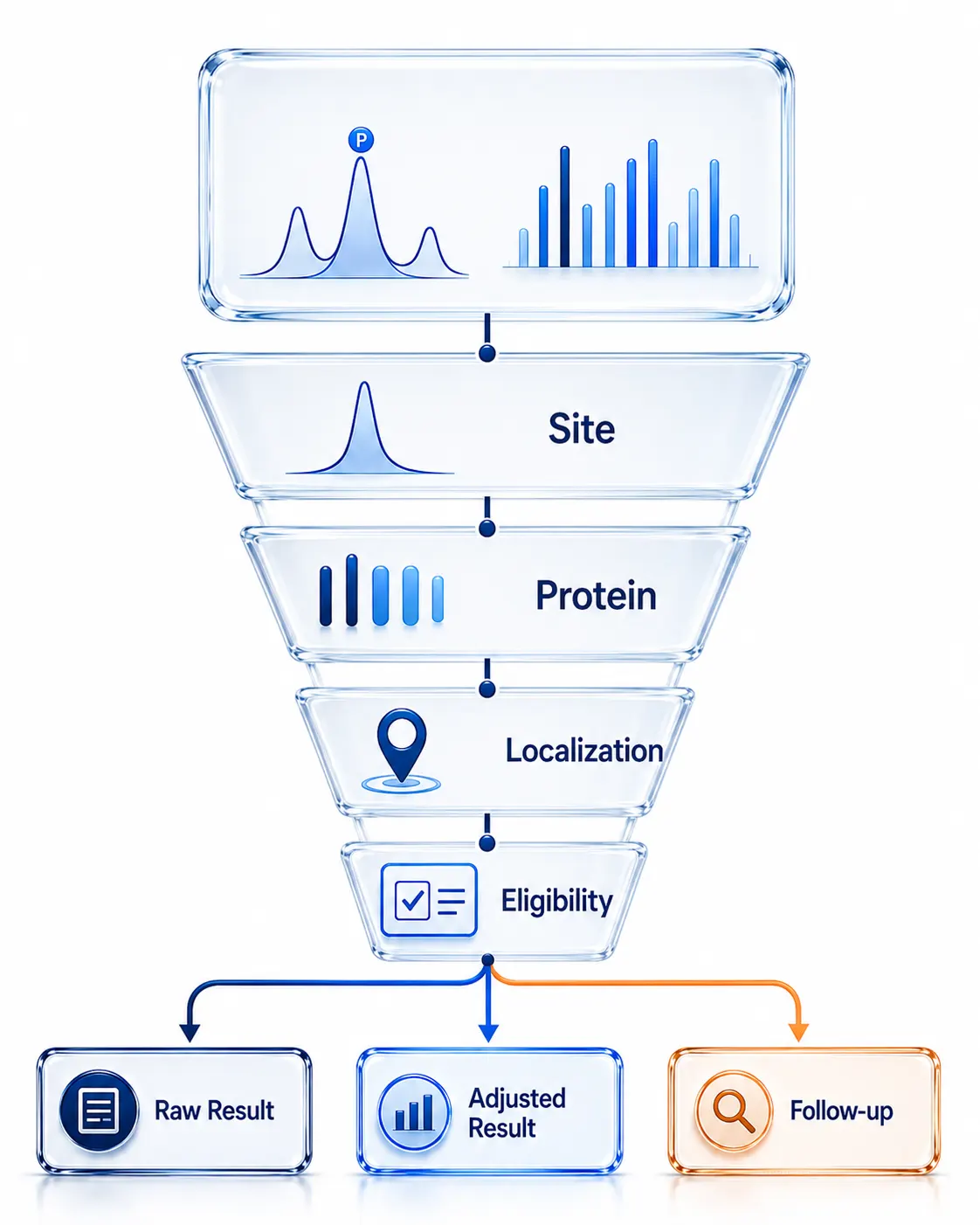 Figure 4. Asymmetric missingness should be classified by its measurement context before it is imputed, excluded, or converted into a protein-adjusted result.
Keep Kinase Activity Inference Within Its Evidence Boundary
Kinase-substrate enrichment analysis aggregates the behavior of known or predicted substrate sites to produce a kinase-associated score. It is useful because signaling often appears as a coordinated shift among several sites rather than one decisive peptide. It is not a direct activity assay. Its result depends on the substrate database, species mapping, site coverage, directionality, missingness, and the set of measured phosphosites that serves as background.
Protein adjustment can change the score substantially. A pathway with broadly increased protein abundance may appear kinase-enriched when raw phosphosite intensities are used, even if the associated sites do not rise beyond their parent proteins. Conversely, correction can strengthen a signal when modest raw site changes become coherent after protein abundance is accounted for. Run enrichment on clearly labeled raw and protein-adjusted inputs when both are available, then report whether the inference is stable across the two views. An integrated phosphoproteome analysis used both unadjusted and overall-protein-normalized phosphosite levels as KSEA inputs, illustrating the value of making this decision explicit.3
Interpret a kinase score as a hypothesis supported by observed substrate behavior. For high-impact conclusions, review the contributing sites, inspect their localization and protein mapping, and confirm a prioritized target with an orthogonal readout. Phosphorylation Site Identification Service and targeted follow-up can be used to strengthen the specific site evidence; they do not convert a database-derived score into proof of enzyme activity by themselves.
Targeted follow-up is most informative when it answers a declared uncertainty from discovery. Select sites that have consistent localization, unambiguous peptide mapping, adequate chromatographic behavior, and a clear role in the pathway claim. A Parallel Reaction Monitoring (PRM) assay can then focus measurement effort on those peptides across the relevant samples or a follow-up cohort. It is a confirmation layer for measurement precision and site behavior, not a substitute for testing the causal activity of the inferred kinase.
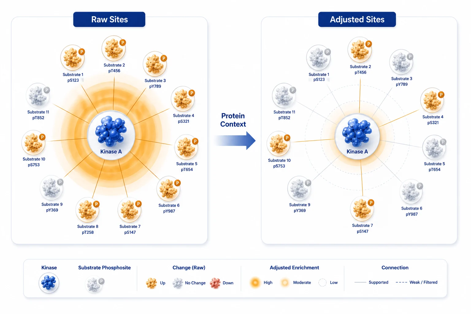 Figure 5. Kinase-substrate enrichment results should be compared before and after protein adjustment, then interpreted as pathway hypotheses rather than direct enzyme measurements.
Choose the Least Complex Quantitative Design That Answers the Question
| Study question | Useful starting design | Primary strength | Key boundary |
|---|---|---|---|
| Initial pathway screen | Phosphoproteome alone with strict site QC | Efficient discovery of candidate sites | Cannot separate parent-protein abundance changes |
| Comparative signaling study | Paired total proteome plus phosphopeptide enrichment | Supports protein-adjusted site modeling | Requires matched sample maps and adequate total-proteome coverage |
| Large cohort or many conditions | Balanced paired DIA design with pooled QC | Scalable matrix and repeatable sample order | Batch and missingness policy remain critical |
| Small panel of decisive sites | Discovery nomination followed by targeted assay | Focused peptide-level confirmation | Not a substitute for broad pathway discovery |
Pairing does not always require the deepest global proteome possible. It requires a total-proteome measurement that is adequate for the parent proteins driving the planned interpretation. If a study centers on a narrow kinase pathway, a targeted total-protein layer may be more useful than a shallow global run that leaves those proteins consistently unobserved. Conversely, broad discovery benefits from coverage that makes protein-adjusted modeling available for enough sites to test the pathway-level hypothesis.
For workflows that need broad PTM context beyond phosphorylation, High-Resolution PTMs Profiling Services can help scope an evidence hierarchy across modification classes. Use that expansion only when it answers a real biological ambiguity; additional PTM layers do not compensate for weak pairing or unbalanced batches.
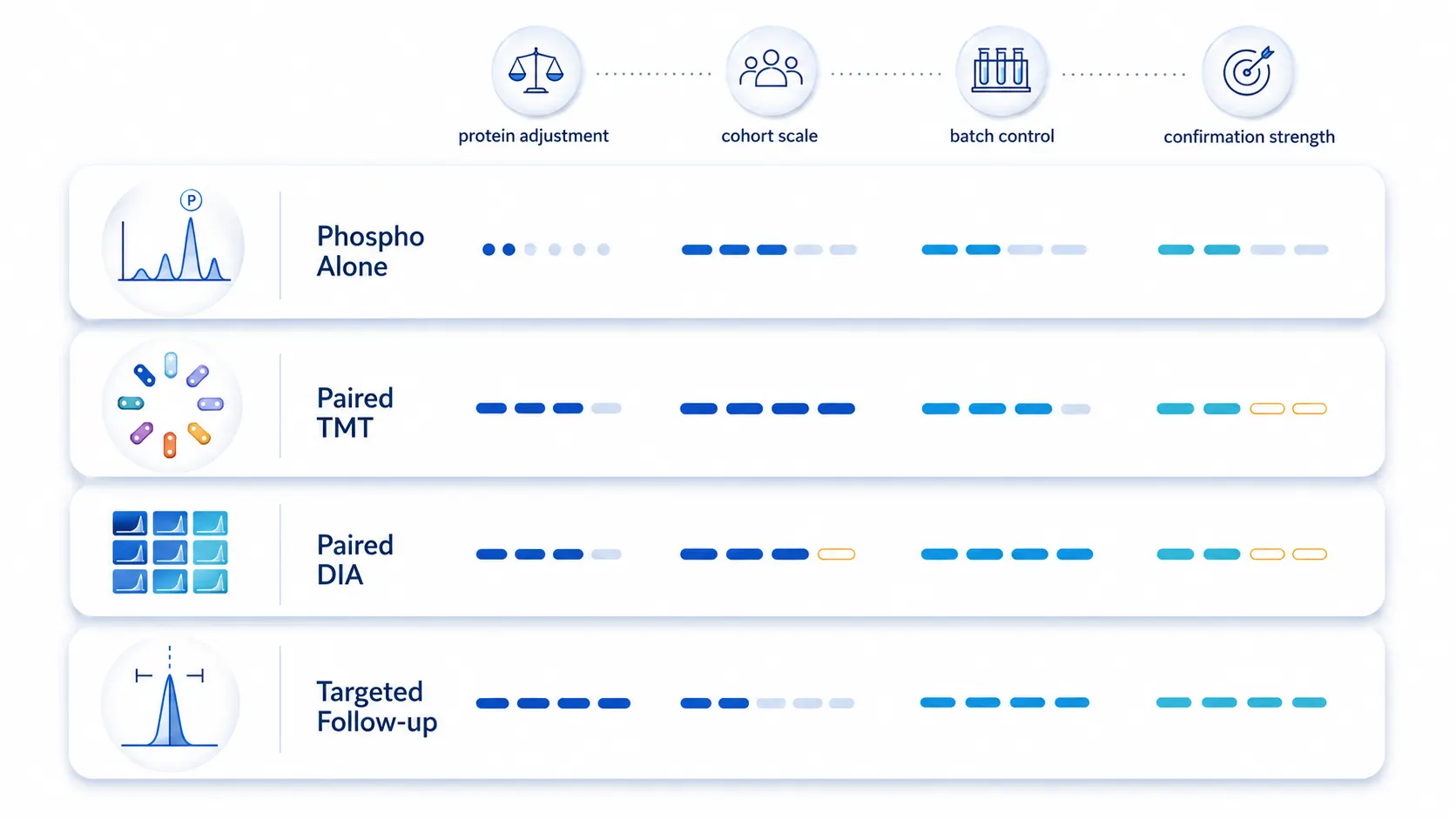 Figure 6. The right quantitative workflow depends on whether the study needs site discovery, protein-adjusted inference, large-cohort consistency, or targeted confirmation.
Use the Paired Design as a Multi-Omics Anchor
Paired total proteome and phosphoproteome data can anchor other molecular layers because it distinguishes a change in protein amount from a change in modification behavior. Transcriptomics can then be used to test whether a protein-level change follows RNA abundance; targeted kinase experiments can test a prioritized pathway hypothesis; and orthogonal assays can clarify localization or complex formation. The point is not to stack every possible assay. It is to make each added layer resolve a specific ambiguity left by the paired proteomics result.
Two planned topic-cluster guides extend this reasoning in complementary directions: proteomics and metabolomics from the same organoid sample addresses how to preserve sample-level relationships across molecular layers, while open modification search versus targeted PTM analysis addresses when broad modification discovery should give way to a focused measurement. These matrix links are retained for coordinated publication.
Use This Four-Phase Paired Phosphoproteomics Plan
- Define the inference target. State whether the endpoint is raw phosphosite discovery, protein-adjusted phosphorylation, occupancy estimation, kinase-pathway hypothesis generation, or targeted confirmation.
- Preserve pairing in the wet lab. Establish a common digest, pilot the total-versus-enrichment allocation, balance batches, and retain the sample map across both analytical branches.
- Process the two matrices separately but comparably. Perform fraction-specific QC, retain raw values, document site localization and protein mapping, and classify missingness before transformation or imputation.
- Model and report evidence layers. Present raw phosphosite behavior, protein-adjusted results where eligible, and kinase-enrichment hypotheses with the contributing substrate evidence.
The final study report should state which phase generated each conclusion. That small discipline makes it easier for collaborators to distinguish an observed signal, a model-supported interpretation, and a follow-up hypothesis that still requires direct experimental testing.
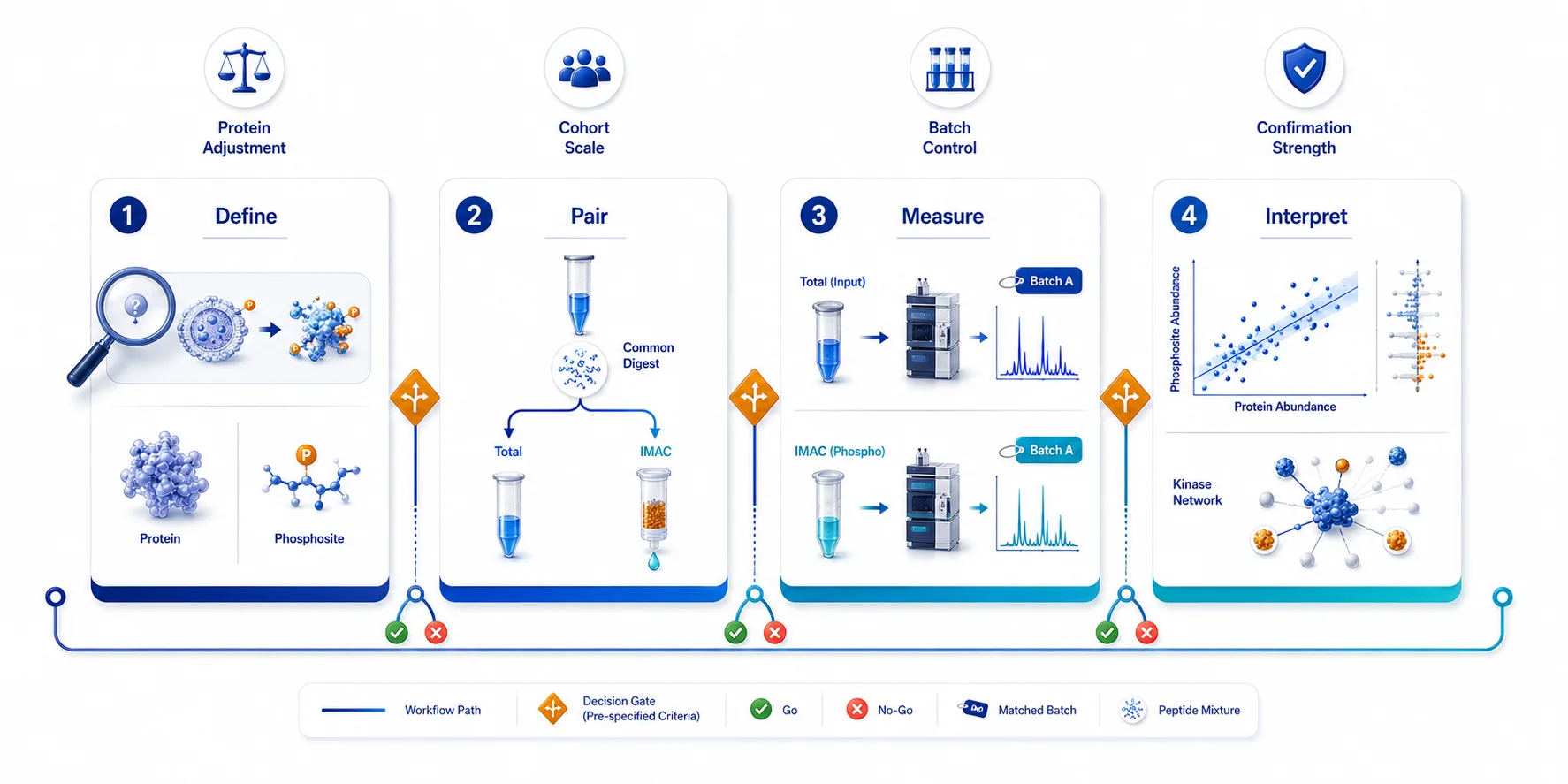 Figure 7. Four-phase implementation plan for paired total-proteome and phosphoproteome studies designed to avoid misreading protein abundance as signaling.
Frequently Asked Questions
Must every phosphoproteomics study include total proteome?
No. A phosphoproteome-only design can be appropriate for discovery or for questions where parent-protein abundance is not part of the intended claim. Include total proteome when protein-adjusted interpretation, pathway inference, or treatment-driven expression change is central.
Can a simple phosphosite-to-protein ratio replace a model?
It can be an exploratory summary when both measurements are robust and unambiguous. It should not replace inspection of missingness, protein mapping, replicate behavior, and the assumptions required for protein-adjusted inference.
What if the phosphosite is observed but total protein is missing?
Report the raw phosphosite result, but do not present it as protein-adjusted. Consider whether a deeper or targeted total-proteome measurement is justified for the biological question.
Does protein adjustment produce phosphorylation occupancy?
No. It estimates site behavior after accounting for a parent-protein abundance measurement. Direct occupancy requires a different experimental framework and additional assumptions.
Can KSEA prove that a kinase is active?
No. KSEA produces a kinase-associated hypothesis based on covered substrate behavior and database knowledge. Inspect contributing sites and validate key conclusions with an appropriate follow-up experiment.
When should a project use targeted phosphosite follow-up?
Use it after discovery identifies a small set of decision-driving sites that require stronger peptide-level confirmation or measurement across a larger set of samples.
References:
- Schraink T, Blumenberg L, Hussey G, et al. PhosphoDisco: A Toolkit for Co-regulated Phosphorylation Module Discovery in Phosphoproteomic Data. Molecular & Cellular Proteomics. 2023;22(8):100596. doi: 10.1016/j.mcpro.2023.100596.
- Zhang T, Keele GR, Gyuricza IG, et al. Multi-omics analysis identifies drivers of protein phosphorylation. Genome Biology. 2023;24:52. doi: 10.1186/s13059-023-02892-2.
- Ng CKY, Dazert E, Boldanova T, et al. Integrative proteogenomic characterization of hepatocellular carcinoma across etiologies and stages. Nature Communications. 2022;13:2436. doi: 10.1038/s41467-022-29960-8.














