

1. Introduction: The Nexus of Circulating Proteomics and Structural Tissue Imaging
1.1 Beyond Single-Organ Biomarkers: The Need for Whole-Body Systems Biology
Human aging and chronic non-communicable diseases are fundamentally multi-system, multi-organ processes. Pathological changes rarely remain isolated within a single anatomical compartment; instead, physiological stress, subclinical inflammation, and cellular senescence cascade across interconnected organ networks. Traditional clinical diagnostics rely heavily on single-organ biomarkers (such as serum creatinine for kidney function, cardiac troponin for myocardial injury, or alanine aminotransferase for liver health) or isolated organ imaging protocols. However, these localized metrics frequently fail to capture systemic inter-organ communication, endocrine signaling feedback loops, and early-stage multi-system organ decay.
Circulating blood plasma serves as the central communication highway of the human body, transporting thousands of secreted signaling proteins, hormones, extracellular vesicle surface markers, and inflammatory cytokines between anatomically distant tissues. Simultaneously, advanced non-invasive medical imaging modalities—such as magnetic resonance imaging (MRI) and computed tomography (CT)—provide high-resolution, three-dimensional quantitative assessments of organ volume, microstructural integrity, tissue composition, and functional connectivity. Bridging circulating blood plasma proteomics with multi-organ imaging-derived phenotypes (IDPs) establishes a revolutionary multi-scale systems biology framework, enabling researchers to map whole-body cross-talk, decipher organ-specific biological aging rates, and predict future multi-disease incidence decades before clinical symptom onset.
1.2 High-Throughput Plasma Proteomics Platforms (Mass Spectrometry, SomaScan)
Characterizing the vast, dynamic range of the circulating plasma proteome—spanning over ten orders of magnitude in protein concentration from high-abundance albumin to low-abundance interleukins—requires robust, high-sensitivity analytical platforms. Modern population-scale biobanks leverage three complementary high-throughput proteomic technologies:
- Mass Spectrometry-Based Proteomics: High-resolution liquid chromatography-tandem mass spectrometry (LC-MS/MS), utilizing Data-Independent Acquisition (DIA) and advanced antibody depletion protocols, provides untargeted, unbiased identification and absolute quantification of thousands of plasma proteins, capturing novel post-translational modifications (PTMs) and proteolytic cleavage fragments.
- Proximity Extension Assays: Utilizing pairs of antibodies conjugated to matching oligonucleotide tags, panels achieve ultra-high specificity and sensitivity through dual-recognition microfluidic qPCR or next-generation sequencing (NGS) readouts, quantifying over 3,000 to 5,400 plasma proteins simultaneously with minimal sample volume requirements.
- Aptamer-Based Arrays (SomaScan): Employing fluorophore-labeled, chemically modified single-stranded DNA aptamers (SOMAmers), this platform measures thousands of native protein analytes across wide dynamic ranges with high throughput.
Utilizing a specialized Quantitative Proteomics Service provides the rigorous analytical precision, dynamic range coverage, and batch-to-batch reproducibility needed to profile thousands of circulating plasma proteins across large clinical trial cohorts.
1.3 High-Resolution Imaging-Derived Phenotypes (IDPs) Across Multi-Organ Protocols
In parallel with proteomic advances, large-scale population biobanks (such as the UK Biobank) have standardized multi-organ imaging protocols. Quantitative image processing algorithms extract over 1,000 distinct Imaging-Derived Phenotypes (IDPs) representing organ morphology, tissue composition, and functional activity:
- Brain MRI IDPs: Regional gray matter volumes, white matter hyperintensity (WMH) volumes, hippocampal volume, cortical thickness, diffusion tensor imaging (DTI) fractional anisotropy, and resting-state functional connectivity networks.
- Cardiac MRI IDPs: Left and right ventricular end-diastolic volumes, myocardial mass, stroke volume, myocardial extracellular volume (fibrosis indices), and aortic compliance.
- Abdominal MRI/CT IDPs: Liver fat fraction (proton density fat fraction, PDFF), liver iron concentration, pancreatic volume, renal parenchymal fat, visceral vs. subcutaneous adipose tissue volumes, and kidney volume.
- Musculoskeletal IDPs: DEXA body composition, thigh muscle volume, and muscle fat infiltration (myosteatosis).
Integrating these quantitative 3D structural imaging traits with high-dimensional circulating proteomic profiles provides an unprecedented opportunity to decipher the molecular drivers of human organ health and multi-system pathology.
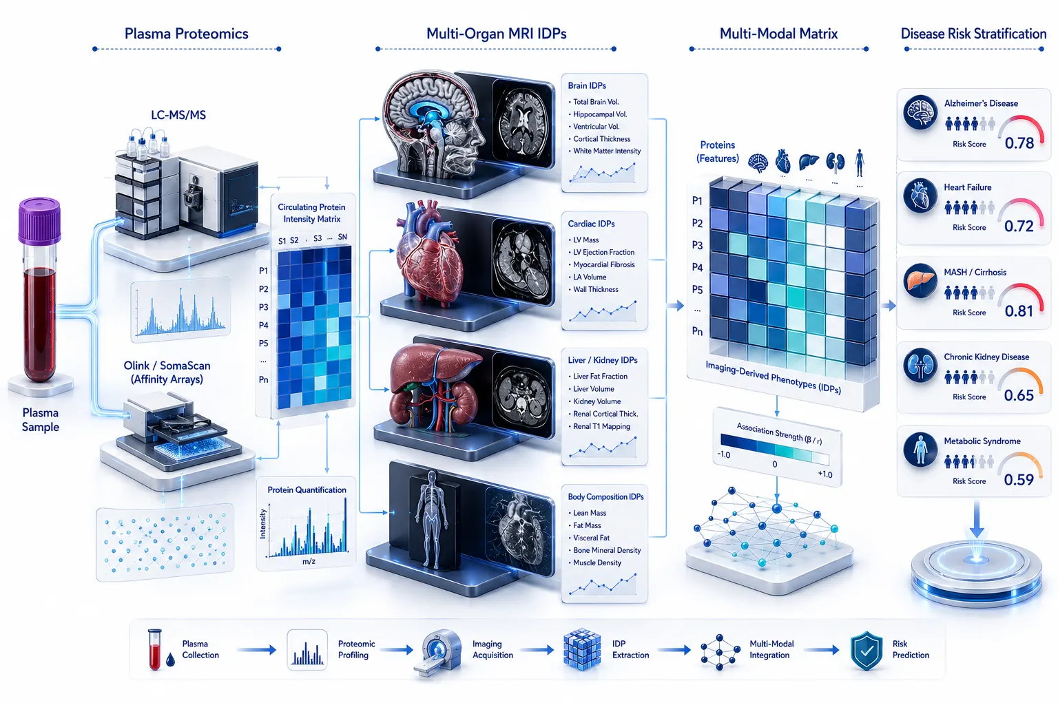 Figure 1: Plasma Proteomics & Multi-Organ IDP Integration
2. Mapping the Pan-Organ Proteomic-Imaging Landscape
2.1 Multi-Organ Phenotypic Associations: Pan-Organ vs. Organ-Specific Networks
Systematic multi-organ imaging-proteomics analyses reveal extensive, non-linear networks of association connecting circulating plasma proteins with quantitative tissue structures. Comprehensive biobank-scale analyses evaluating over 2,900 plasma proteins against 1,000+ multi-organ IDPs have uncovered more than 5,000 significant phenotypic protein-imaging associations.
These associations segregate into two distinct functional categories:
- Organ-Specific Proteomic Associations: Circulating proteins that exhibit highly specific correlations with structural traits of a single organ system (e.g., plasma Neurofilament Light Chain [NEFL] specifically correlating with brain ventricular enlargement and cortical thinning, or FABP4 correlating with abdominal fat accumulation).
- Pan-Organ Shared Proteomic Associations: Pleiotropic signaling proteins that correlate simultaneously across multiple organ imaging phenotypes (e.g., Growth Differentiation Factor 15 [GDF15] or Interleukin-6 [IL-6] correlating with reduced brain volume, increased myocardial fibrosis, elevated liver fat, and loss of skeletal muscle mass).
2.2 Distinguishing Tissue Secretion Origin: Organ-Derived Proteins vs. Remote Hepatic Mediators
A fundamental question in circulating biomarker discovery is determining where plasma proteins originate. By integrating external tissue-specific RNA expression datasets (such as GTEx and Human Protein Atlas) with plasma proteomic-imaging networks, researchers can determine protein secretion kinetics:
- Direct Organ-Derived Proteins: Plasma proteins associated with structural IDPs of the brain, lung, pancreas, or spleen are predominantly synthesized and directly shed by cells residing within those respective organs. Their circulating abundance directly reflects localized tissue cell turnover, cellular stress, or membrane damage.
- Remote Hepatic & Systemic Mediators: Conversely, plasma proteins correlating with structural traits of the heart, visceral body fat, and skeletal muscle are overwhelmingly synthesized in the liver. The liver acts as a central metabolic and endocrine processing hub, secreting systemic inflammatory, lipid-transport, and acute-phase proteins that remotely influence distant cardiovascular and musculoskeletal tissue architecture.
2.3 Resolving Organ-Shared Pathways and Protein-Protein Interaction (PPI) Networks
Enrichment analyses of organ-associated plasma proteins highlight convergent biological pathways. Proteins linked to multi-organ structural degradation are heavily enriched in extracellular matrix (ECM) organization, complement and coagulation cascades, TGF-beta signaling, insulin-like growth factor (IGF) regulation, and systemic senescence-associated secretory phenotype (SASP) pathways. Applying advanced Bioinformatics Analysis Service workflows enables constructing high-confidence protein-protein interaction (PPI) networks and functional modules that mediate cross-talk between anatomically separated organs.
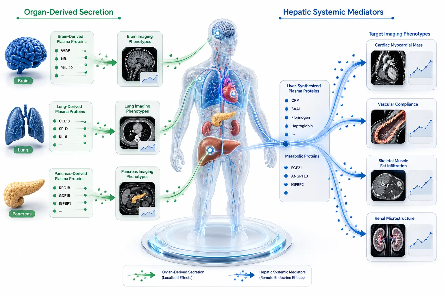 Figure 2: Pan-Organ Proteomic Secretion Origin Map
3. Organ-Specific Proteomic Aging Clocks and Asynchronous Biological Decay
3.1 Quantifying Biological Age Gap: Brain, Heart, Liver, Kidney, and Pancreatic Clocks
Chrono-age (calendar years) is an imprecise proxy for biological health. Individual organs within the same human body age at different velocities due to genetic predisposition, environmental exposures, and lifestyle factors. Using supervised machine learning algorithms (such as ElasticNet, Random Forest, or Gradient Boosting), researchers train organ-specific proteomic aging clocks using plasma proteins predominantly expressed or secreted by specific organs.
These organ aging clocks calculate organ-specific "Age Gaps"—the difference between an individual's predicted biological organ age and their chronological age:
- Brain Proteomic Age Gap: Driven by plasma proteins reflecting neuro-axonal integrity, glial activation, and neurovascular unit health.
- Heart Proteomic Age Gap: Driven by circulating markers of myocardial stretch, extracellular matrix turnover, and vascular calcification.
- Liver Proteomic Age Gap: Driven by plasma proteins reflecting hepatic lipid accumulation, extracellular fibrosis, and metabolic synthetic capacity.
- Kidney Proteomic Age Gap: Driven by circulating markers of glomerular filtration barrier health, tubular cell stress, and clearance capacity.
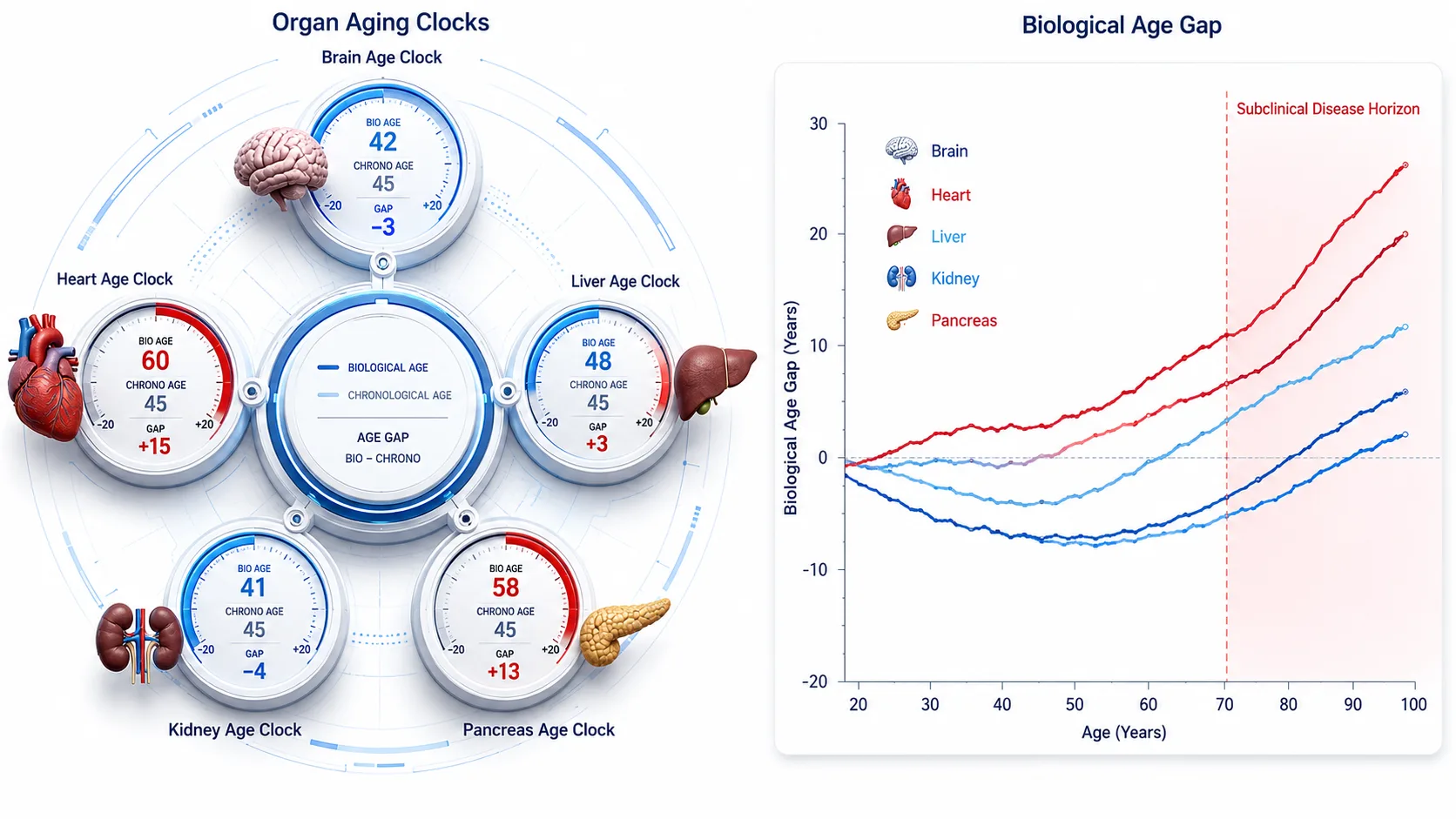 Figure 3: Multi-Organ Proteomic Biological Aging Clocks
3.2 Decades-Before-Onset Risk Prediction: Early Asymptomatic Disease Interception
A pivotal advantage of proteomic organ aging clocks is their ability to detect localized biological decay decades before clinical disease manifestation. Longitudinal biobank tracking demonstrates that an individual exhibiting a significantly elevated Brain Age Gap (e.g., biological brain age 10 years older than chronological age) has a dramatically increased risk of developing incident Alzheimer's disease or vascular dementia 10 to 20 years later, even when cognitive tests and standard laboratory metrics remain completely normal at baseline. Similarly, an elevated Liver Age Gap predicts future metabolic dysfunction-associated steatohepatitis (MASH), cirrhosis, and hepatocellular carcinoma years before clinical symptom onset.
3.3 Multi-Organ Agers vs. Single-Organ Agers: Systemic Inflammation & Longevity Metrics
Population stratification demonstrates that approximately 20% to 30% of individuals exhibit accelerated aging in at least one specific organ system ("Single-Organ Agers"), whereas approximately 1.5% to 3% exhibit accelerated aging across multiple organ systems simultaneously ("Multi-Organ Agers"). Multi-organ agers exhibit significantly higher systemic baseline inflammatory markers, accelerated multi-morbidity accumulation, and substantially higher all-cause mortality hazard ratios compared to normal agers, emphasizing the urgent need for multi-organ risk assessment.
4. Genetic Architecture and Causal Mechanisms (pQTLs & Mendelian Randomization)
4.1 Uncovering Genetic Roots: Identifying Causal Links Between Plasma Proteins and IDPs
Cross-sectional phenotypic correlations between plasma proteins and imaging-derived phenotypes do not inherently prove causality; they may reflect secondary reactive processes or shared environmental confounding. To uncover true causal mechanisms, researchers integrate genome-wide association studies (GWAS) with protein quantitative trait loci (pQTLs)—genetic variants that specifically regulate circulating plasma protein abundances. Combining pQTLs with multi-organ IDPs has identified over 8,000 genetic-root putative causal links between circulating proteins and structural tissue traits across the human body.
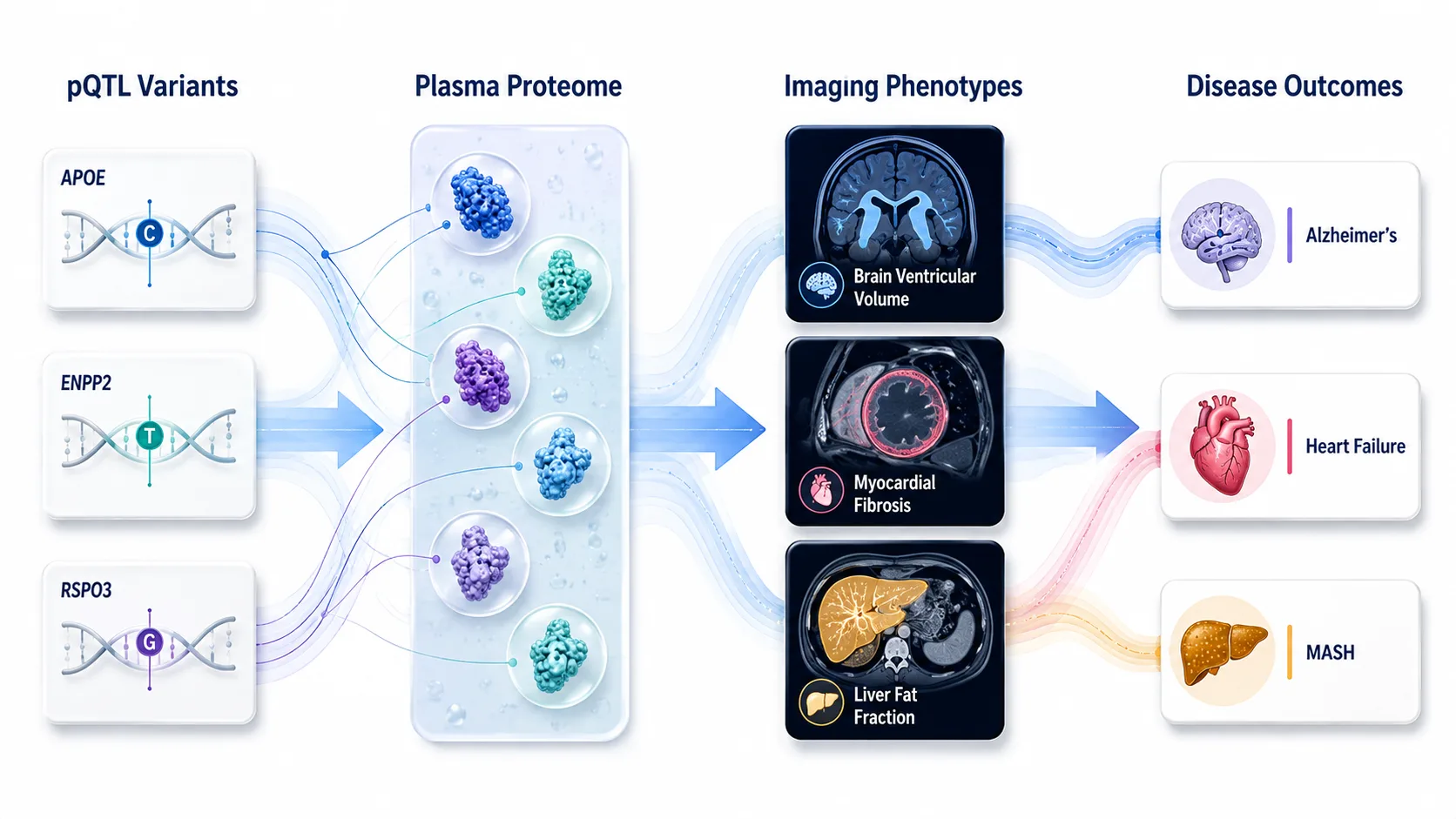 Figure 4: Genetic Causal Architecture (pQTL & MR)
4.2 Multivariable Mendelian Randomization (MR) for Drug Target Prioritization
Mendelian Randomization (MR) leverages the random assortment of genetic variants at conception as instrumental variables, mimicking a lifetime randomized controlled trial. Using multivariable MR approaches, researchers evaluate whether genetically predicted plasma protein levels exert direct causal effects on organ imaging traits and downstream disease outcomes:
- Neurodegenerative Targets: Mendelian randomization has identified circulating proteins (coding genes including APOE, ENPP2, RSPO3, MICB, and NSF) with direct causal effects on brain structural atrophy, ventricular enlargement, and incident Alzheimer's disease risk.
- Cardiometabolic Targets: Causal plasma protein drivers modulating left ventricular mass, arterial stiffness, and liver fat content provide validated therapeutic targets for drug discovery, minimizing clinical trial attrition.
Partnering with an experienced Biomarker Discovery Service provider allows biopharmaceutical teams to validate causal proteomic-imaging candidate targets and design targeted assays for drug mechanism-of-action (MoA) evaluation.
5. Translating Spatial Representations into Clinical Patient Stratification
5.1 Neurodegenerative Diseases: Brain MRI IDPs Aligned with Plasma Neuro-Proteomic Signatures
In neurodegenerative disease research, aligning non-invasive brain MRI phenotypes (hippocampal volume loss, ventricular expansion, white matter lesion load) with high-sensitivity plasma proteomics has transformed biomarker development. Ultra-sensitive assays quantifying plasma Neurofilament Light (NfL), phosphorylated Tau (p-Tau181, p-Tau217), Glial Fibrillary Acidic Protein (GFAP), and Amyloid-beta 42/40 ratios correlate precisely with MRI measures of cortical atrophy and neuro-inflammation, enabling early blood-based screening for preclinical Alzheimer's disease, Parkinson's disease, and vascular dementia.
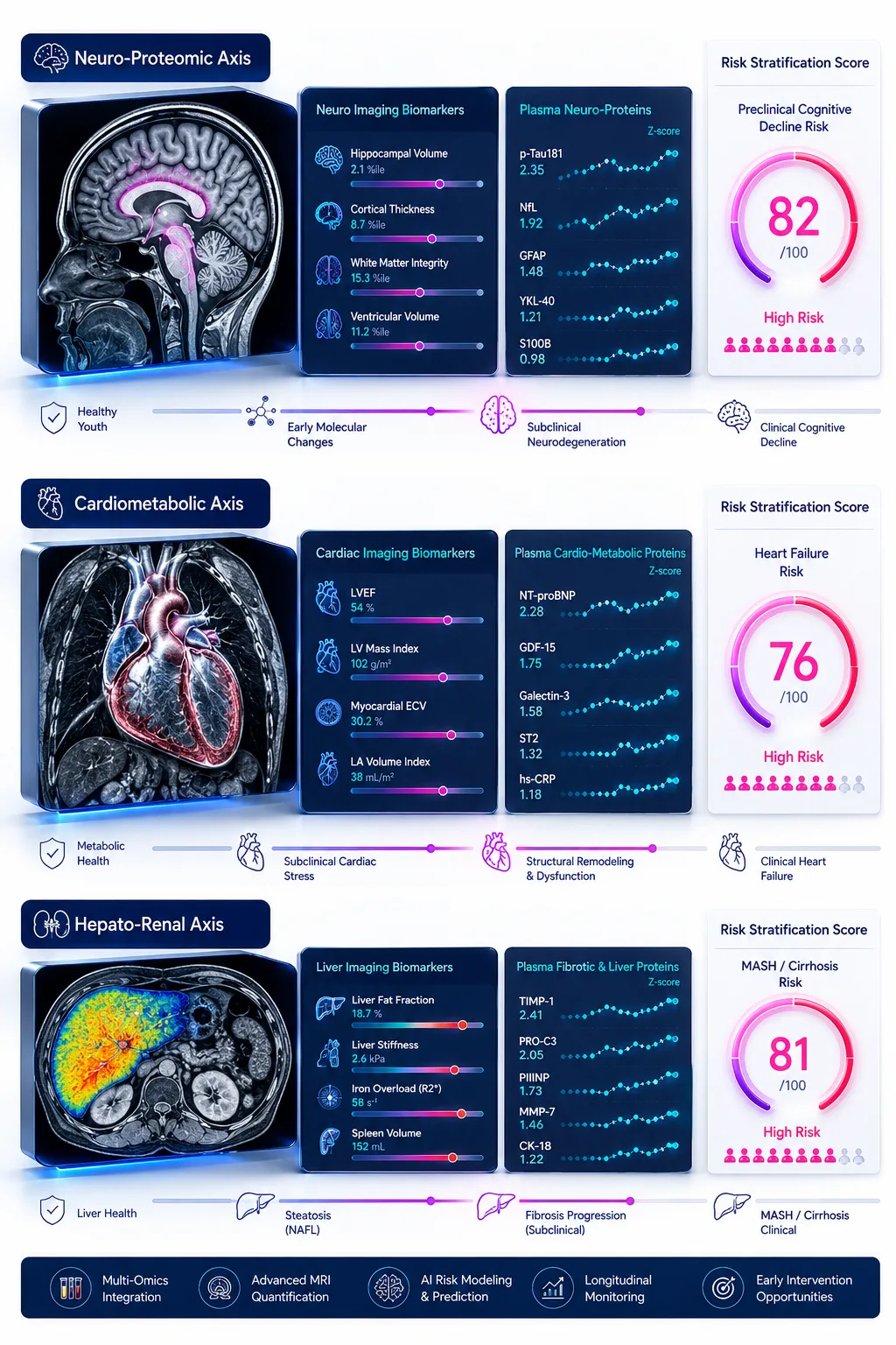 Figure 5: Multi-Disease Predictive Risk Stratification
5.2 Cardiometabolic Diseases: Cardiac/Abdominal IDPs Linked to Systemic Inflammation
Cardiovascular and metabolic diseases involve complex interactions between cardiac morphology, visceral fat deposition, and systemic inflammation. Plasma proteomic profiles correlated with cardiac MRI traits (such as N-terminal pro-B-type natriuretic peptide [NT-proBNP], troponins, growth factors, and matrix metalloproteinases) predict left ventricular remodeling, heart failure hospitalization, and coronary artery calcification. Simultaneously, plasma proteins reflecting liver fat fraction (PDFF) and visceral adipose tissue volume predict incident type 2 diabetes, metabolic syndrome, and major adverse cardiovascular events (MACE).
5.3 MASH & Chronic Kidney Disease: Structural Remodeling Reflected in Blood Omics
Metabolic dysfunction-associated steatohepatitis (MASH) and chronic kidney disease (CKD) are characterized by progressive tissue fibrosis and structural organ remodeling. Plasma proteomic signatures reflecting extracellular matrix turnover (such as TIMP-1, PRO-C3, and collagen cleavage products) correlate strongly with MRI-measured liver stiffness (MR elastography) and renal parenchymal fat infiltration, replacing invasive liver or kidney tissue biopsies with precise liquid biopsy panels. Furthermore, evaluating localized post-translational modifications via specialized PTM Analysis Service workflows reveals specific protein phosphorylation and glycosylation changes driving tissue fibrogenesis.
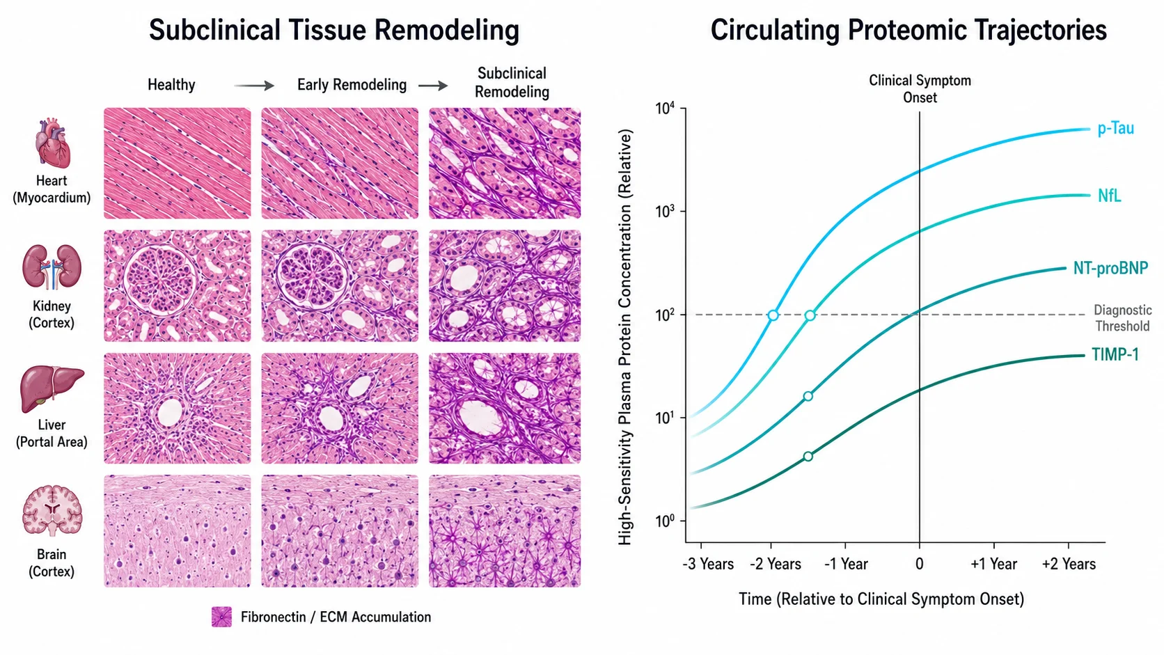 Figure 6: Asymptomatic Organ Remodeling vs. Proteomic Dynamic Gradients
6. Methodological Comparison: Traditional Clinical Phenotyping vs. Integrated Omics-Imaging
| Analytical Feature | Traditional Clinical Phenotyping | Integrated Plasma Proteomics & Multi-Organ Imaging |
|---|---|---|
| Primary Input Format | Single-organ biochemical markers & symptom assessment | Whole-body multi-organ 3D imaging & 3,000+ plasma proteins |
| Detection Window | Symptomatic or late-stage disease manifestation | Decades-before-onset preclinical subclinical risk detection |
| Organ Specificity | Low (Circulating markers often lack organ origin context) | High (Resolves organ-specific vs. remote hepatic secretion origins) |
| Causal Validation | Observational / Correlational only | Causal (Validated via pQTLs & Mendelian Randomization) |
| Biological Aging Tracking | Chronological age & generic blood chemistry | Quantifies organ-specific biological Age Gaps (Brain, Heart, Liver, Kidney) |
| Risk Prediction Accuracy | Moderate (Relies on static clinical risk factors) | High (Multi-scale multi-omics risk scores outperforming clinical models) |
| Scalability for Clinical Trials | High cost if relying exclusively on repeated MRI scans | High (Scalable non-invasive blood tests replacing repeated MRI) |
7. Translational Implementation Framework for Biopharmaceutical R&D
For translational research teams, biopharmaceutical developers, and contract research organizations (CROs) integrating plasma proteomics and multi-organ imaging into target discovery, mechanism-of-action (MoA) validation, and clinical trial stratification, we recommend a four-stage implementation framework:
- Multi-Modal Data Ingestion & Harmonization: Standardize plasma sample collection (EDTA plasma), high-throughput proteomic profiling (LC-MS/MS, or SomaScan), and quantitative 3D MRI image processing pipelines across multi-center cohorts.
- Organ-Derived Feature Extraction & Aging Clock Construction: Apply machine learning algorithms to isolate organ-derived proteomic signatures and compute organ-specific biological Age Gaps across target patient populations.
- Causal Target Prioritization via pQTLs & MR: Integrate genome-wide genetic data to perform multivariable Mendelian Randomization, prioritizing causal plasma protein drivers of structural tissue degradation over secondary reactive markers.
- Non-Invasive Blood Biomarker Panel Deployment: Train interpretable classifiers on clinical trial endpoints (e.g., organ disease progression, therapeutic response rate, or major clinical events), replacing costly multi-organ imaging protocols with scalable, non-invasive plasma biomarker panels for patient stratification.
For end-to-end experimental execution, leveraging an integrated Immunoproteomics Service and comprehensive Bioinformatics Analysis Service ensures robust antibody validation, high-sensitivity protein quantification, and rigorous multi-modal data integration.
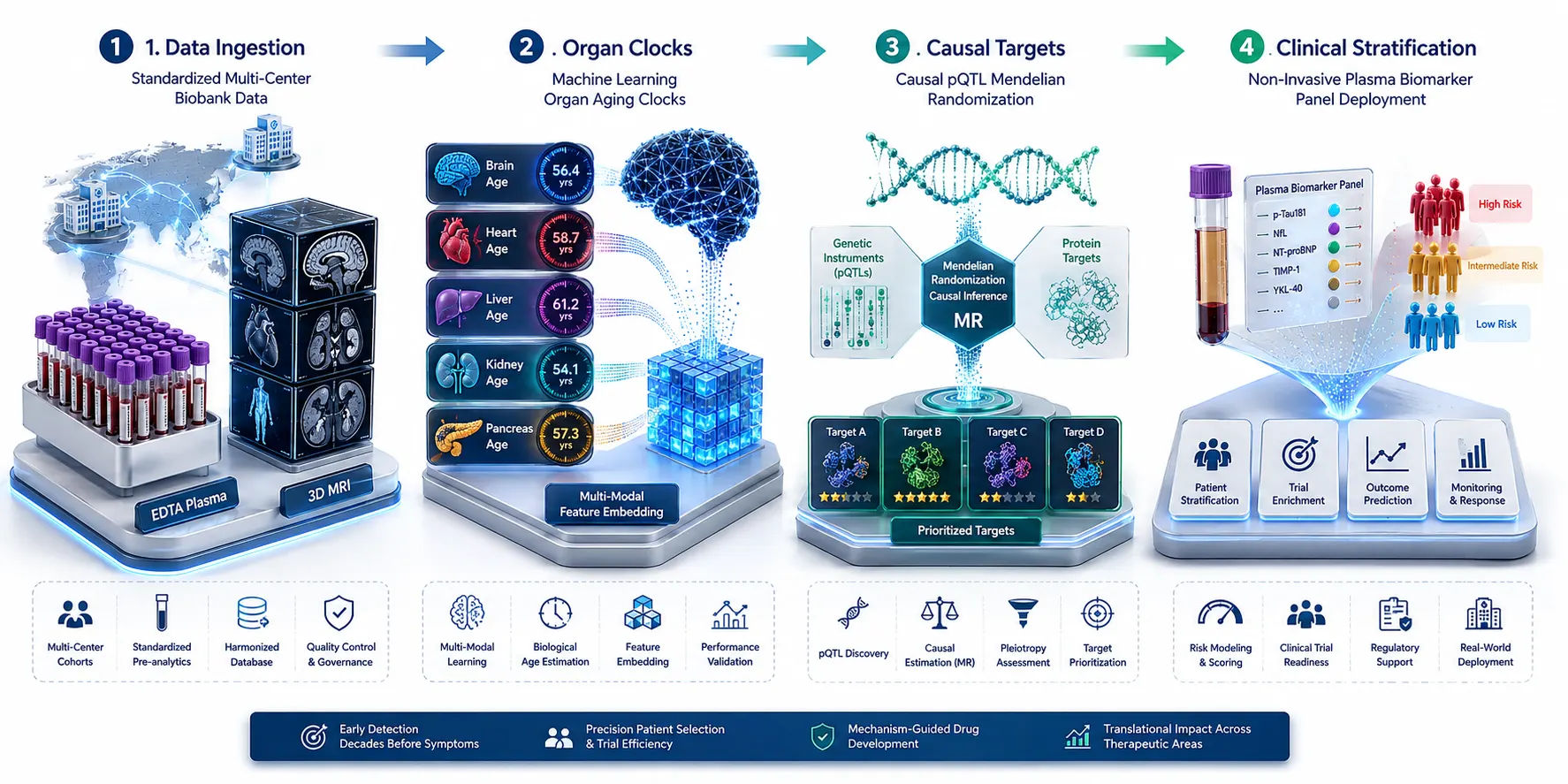 Figure 7: Translational Implementation Framework for Pharma R&D
8. Frequently Asked Questions (FAQ)
Q1: What imaging modalities are included in multi-organ imaging-derived phenotypes (IDPs)?
Multi-organ IDPs are extracted from standardized non-invasive imaging protocols, primarily multi-parametric Magnetic Resonance Imaging (MRI) covering the brain, heart, liver, kidneys, pancreas, and skeletal muscle, as well as Computed Tomography (CT) and Dual-Energy X-ray Absorptiometry (DEXA) for bone density and body composition.
Q2: How do researchers determine whether a plasma protein originates from a specific organ?
Researchers integrate plasma proteomic-imaging association networks with tissue-specific RNA expression biobanks (such as GTEx and Human Protein Atlas). If a plasma protein associated with brain MRI traits is overwhelmingly synthesized in neural tissue, it is classified as organ-derived. If it is primarily synthesized in the liver, it is classified as a remote hepatic mediator.
Q3: How do plasma proteomic organ aging clocks differ from epigenetic DNA methylation clocks?
Epigenetic DNA methylation clocks measure global nuclear DNA methylation changes across nucleated blood cells, reflecting systemic biological aging. In contrast, plasma proteomic organ aging clocks utilize secreted proteins specific to distinct organs (e.g., brain, heart, liver, kidney), providing granular resolution of asynchronous, organ-specific biological decay.
Q4: Can plasma proteomic biomarkers replace multi-organ MRI scans in clinical trials?
Multi-organ MRI scans remain the structural gold standard for organ imaging. However, once plasma proteomic signatures are rigorously validated against MRI IDPs and clinical endpoints, non-invasive plasma biomarker panels can serve as highly scalable, cost-effective surrogate endpoints for large-scale patient screening, monitoring, and stratification in biopharmaceutical clinical trials.
Q5: What sample volume and preparation protocols are required for high-throughput plasma proteomics?
High-throughput affinity platforms (such as SomaScan) typically require minimal EDTA plasma volumes (often 20 to 50 microliters), while mass spectrometry-based LC-MS/MS workflows require 100 to 200 microliters following standardized depletion of high-abundance blood proteins.
Q6: How does Mendelian Randomization help in drug target discovery?
Mendelian Randomization uses genetic variants (pQTLs) as unconfounded instrumental variables to determine whether a circulating plasma protein directly causes structural organ changes and disease risk, rather than merely being a consequence of disease. Causal proteins prioritized by MR have significantly higher success rates in biopharmaceutical clinical trials.
Q7: Are multi-organ proteomic aging clocks generalizable across different ethnic populations?
While foundational organ aging clocks trained on large biobanks (such as the UK Biobank) capture universal biological aging pathways, validating and recalibrating proteomic clocks across multi-ethnic cohorts ensures population-specific baseline adjustments and optimal diagnostic sensitivity.
Q8: Are these computational workflows intended for clinical diagnostic use?
Plasma proteomic aging clocks, multi-organ imaging models, and associated analytical workflows described here are developed for Research Use Only (RUO). They serve as powerful tools for target discovery, biomarker identification, mechanism-of-action evaluation, and translational research in biopharmaceutical and academic settings, and are not intended for direct clinical diagnostic procedures.
References:
- UK Biobank Imaging & Proteomics Study Group. (2026). The landscape of plasma proteomic links to human organ imaging. UK Biobank Research Publications, July 2026. https://www.ukbiobank.ac.uk/publications/the-landscape-of-plasma-proteomic-links-to-human-organ-imaging/ (Open Access).
- Pan-Organ Imaging Proteomics Group. (2025). The landscape of plasma proteomic links to human organ imaging. medRxiv preprint. https://www.medrxiv.org/content/10.1101/2025.01.14.25320532v1.full (CC BY 4.0 Open Access).
- Multi-Organ MRI & Proteomic Clocks Consortium. (2025). Multi-organ MRI digitizes biological aging clocks across proteomics. PMC Articles, PMC12265779. https://pmc.ncbi.nlm.nih.gov/articles/PMC12265779/ (CC BY 4.0 Open Access).
- Organ-Specific Aging Study Group. (2026). Accelerated multi-organ proteomic aging is detectable decades before disease onset. medRxiv preprint. https://www.medrxiv.org/content/10.64898/2026.01.16.26344299v1.full-text (CC BY 4.0 Open Access).
- Biobank-Scale Omics Initiative. (2025). Organ aging signatures in the plasma proteome track health and disease. PubMed Central, PMC10700136. https://pmc.ncbi.nlm.nih.gov/articles/PMC10700136/ (CC BY 4.0 Open Access).














