

1. Introduction: The Limitations of Classical Malaria Vaccines and the Need for T-Cell Omics
1.1 The Plasmodium Life Cycle Bottleneck: Pre-Erythrocytic, Blood-Stage, and Transmission-Blocking
Malaria, caused primarily by the obligate intracellular protozoan parasite Plasmodium falciparum and Plasmodium vivax, remains one of the most devastating infectious diseases worldwide, responsible for over 600,000 deaths annually. The complex, multi-stage life cycle of Plasmodium parasites presents severe challenges for conventional vaccine design. Inoculated by anopheline mosquitoes into human skin, sporozoites rapidly migrate through the bloodstream to the liver, where they infect hepatocytes—initiating the asymptomatic pre-erythrocytic (liver) stage. Within liver cells, a single sporozoite replicates into tens of thousands of merozoites that subsequently burst into the bloodstream to invade red blood cells, driving the symptomatic, asexual erythrocytic (blood) stage responsible for clinical illness, severe anemia, tissue sequestration, and mortality. Finally, a subset of blood-stage parasites differentiates into sexual gametocytes, which are ingested by female mosquitoes during a blood meal to undergo sexual reproduction and complete the transmission cycle.
Each developmental stage expresses distinct parasite protein subsets, displays dynamic antigenic variation, and occupies different host tissue microenvironments. Traditional single-stage vaccines frequently fail to provide durable population-wide protection, as parasites escaping pre-erythrocytic immune clearance rapidly proliferate during the blood stage. Achieving durable, broad-spectrum protective immunity against malaria necessitates targeting conserved antigen targets presented across multiple stages of the parasite life cycle.
1.2 Limitations of Antibody-Centric Subunit Vaccines (RTS,S and R21)
First-generation WHO-recommended malaria vaccines, such as RTS,S/AS01 and R21/Matrix-M, represent historic breakthroughs in global health. Both vaccines target the central repeat region and C-terminal domain of the Circumsporozoite Protein (CSP)—the predominant surface protein on pre-erythrocytic sporozoites. However, extensive clinical trial follow-up reveals three significant biological limitations of antibody-centric subunit vaccines:
- Antibody-Dominated Short-Lived Immunity: RTS,S and R21 primarily elicit high-titer neutralizing antibody responses against free sporozoites before liver entry. Circulating antibody titers decline rapidly within 6 to 12 months, leading to a progressive, steep drop in protective efficacy and requiring frequent booster doses.
- Strain Polymorphism & Immune Escape: Extensive field polymorphism in the Plasmodium falciparum CSP gene allows mutant parasite isolates to escape antibody neutralization, creating immune selection pressure in high-transmission endemic regions.
- Inadequate Induction of Cytotoxic T-Cell Immunity: Subunit antibody vaccines fail to clear intracellular liver-stage schizonts or infected host blood cells once invasion has occurred. Clearing intracellular parasites requires robust CD8+ cytotoxic T lymphocytes (CTLs) and CD4+ helper T cells that recognize parasite-derived peptides presented by host Human Leukocyte Antigen (HLA/MHC) Class I and Class II molecules.
1.3 The Dawn of Immunopeptidomics: Direct Mass Spectrometry Profiling of Naturally Presented HLA Peptides
To overcome the structural bottleneck of antibody-centric subunit vaccines, researchers are turning to immunopeptidomics—the direct, unbiased identification of the naturally processed and presented peptide repertoire (the immunopeptidome) displayed on host cell surfaces by HLA Class I and Class II molecules. Rather than relying on speculative in silico binding predictions or recombinant protein screening, immunopeptidomics utilizes high-sensitivity Liquid Chromatography-Tandem Mass Spectrometry (LC-MS/MS) to isolate, sequence, and quantify parasite-derived peptides directly from infected host cells. This approach reveals exact endogenously presented T-cell epitopes capable of driving protective cellular immunity.
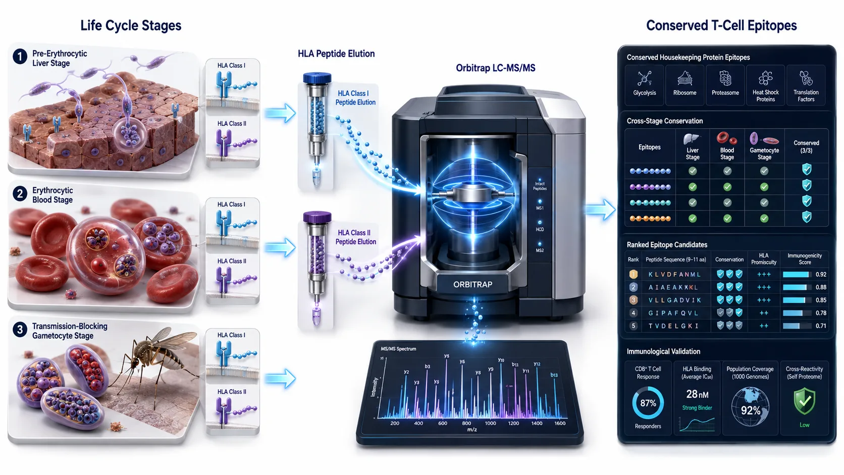 Figure 1: Plasmodium Life Cycle & Immunopeptidomics Antigen Discovery
2. Technical Workflow of High-Sensitivity Immunopeptidomics for Parasitic Infections
2.1 Immunoaffinity Purification (IP) vs. Mild Acid Elution (MAE)
Isolating minute quantities of parasite-derived HLA peptides from an overwhelming background of host self-peptides requires specialized sample preparation methodologies:
- Immunoaffinity Purification (IP): Host cells (e.g., infected hepatocytes, reticulocytes, or primary phagocytes) are lysed in mild non-ionic detergents. HLA Class I (HLA-A, B, C) or Class II (HLA-DR, DQ, DP) complexes are captured using pan-HLA monoclonal antibodies (such as W6/32 or L243) immobilized on Sepharose or magnetic beads. Bound peptide-MHC complexes are washed, and immunopeptides are acid-eluted for desalting and mass spectrometry analysis.
- Mild Acid Elution (MAE): Intact infected host cells are exposed to a gentle citric acid/phosphate buffer (pH 3.3) for 60 to 90 seconds. This selective acid shock disassociates cell-surface HLA-bound peptides without inducing cell lysis or releasing high-abundance intracellular host proteins. MAE provides an antibody-independent, highly scalable approach for recovering surface-presented immunopeptides across multi-ethnic HLA backgrounds without allele-specific antibody constraints.
2.2 High-Resolution LC-MS/MS Platforms (Orbitrap Astral & TIMS-TOF) and De Novo Sequencing
Mass spectrometry hardware breakthroughs have revolutionized immunopeptidomics sensitivity. Ultra-high-speed, high-sensitivity mass spectrometers—such as the Thermo Scientific Orbitrap Astral and Bruker timsTOF Pro platforms—achieve sub-femtomole detection limits and acquisition rates exceeding 200 Hz. Coupling Trapped Ion Mobility Spectrometry (TIMS) or high-resolution Orbitrap mass analyzers with Data-Dependent Acquisition (DDA) or Data-Independent Acquisition (DIA) enables identifying low-abundance parasite peptides present at fewer than 10 to 50 copies per cell.
Utilizing a specialized Quantitative Proteomics Service or high-resolution Mass Spectrometry Imaging Service workflow provides the high-sensitivity LC-MS/MS instrumentation, rigorous chromatographic separation, and spectral accuracy required to sequence rare parasite-derived immunopeptides.
2.3 Resolving Parasite vs. Host HLA Ligandomes: False-Discovery Rate (FDR) Control
Peptide identification from parasite-infected human cells presents complex computational challenges. Searching tandem mass spectra against combined human (Homo sapiens) and parasite (Plasmodium falciparum) proteome databases increases search space complexity, elevating false-positive identification risks. Advanced bioinformatics workflows utilize hybrid database search engines (e.g., Comet, PEAKS, or MaxQuant) combined with deep learning rescoring algorithms (such as Percolator, MS2Rescore, or Prosit) to enforce strict 1% False Discovery Rate (FDR) thresholds at both peptide and spectral match levels.
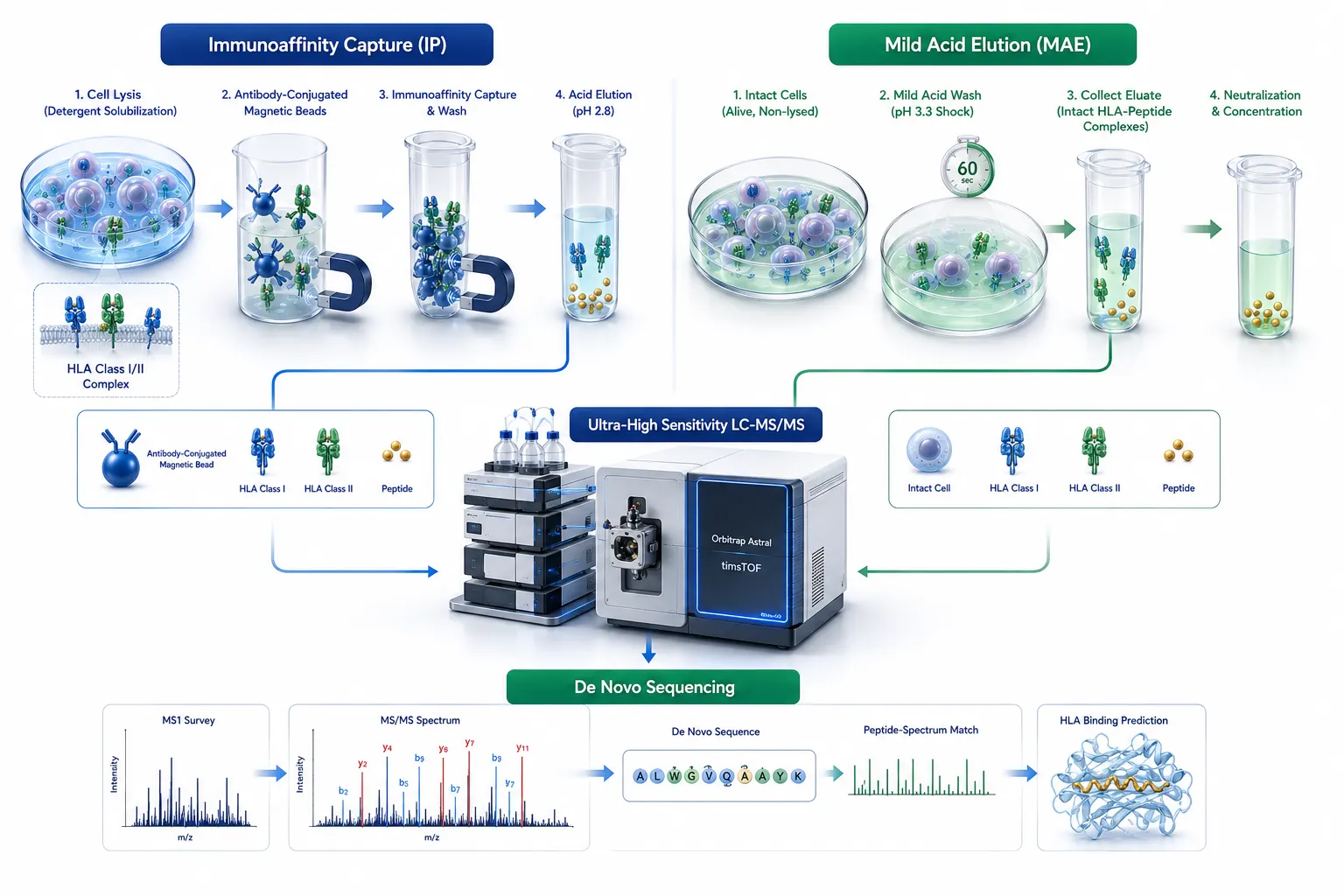 Figure 2: Immunoaffinity Purification (IP) vs. Mild Acid Elution (MAE)
3. Uncovering Cross-Stage and Cross-Species Conserved Plasmodium Antigens
3.1 Unveiling HLA Class I Presentation on Infected Reticulocytes and Phagocytes
For decades, a dogma in malariology held that mature human erythrocytes, lacking nuclei and protein synthesis machinery, could not present HLA Class I molecules or present blood-stage parasite antigens to CD8+ T cells. However, recent immunopeptidomics breakthroughs have dismantled this assumption:
- Infected Reticulocyte HLA-I Presentation: Unmature red blood cells (reticulocytes), which constitute the primary host niche for Plasmodium vivax and a subset of Plasmodium falciparum infections, retain functional HLA Class I molecules on their cell surface.
- Direct Parasite Presentation: Immunopeptidomics profiling of P. vivax-infected reticulocytes directly isolated from human clinical cases revealed over 450 unique parasite-derived HLA Class I peptides presented directly on the infected cell membrane, proving that blood-stage parasites are actively visible to cytotoxic CD8+ T lymphocytes.
3.2 Housekeeping Proteins as Universal Vaccine Targets
Crucially, immunopeptidomics reveals that a significant proportion (over 40% to 50%) of naturally presented parasite HLA peptides do not derive from variable, stage-specific surface antigens (like CSP, MSP1, or PfEMP1). Instead, they originate from highly conserved parasite housekeeping proteins—including ribosomal proteins, heat shock proteins (HSP70/HSP90), translation elongation factors, and metabolic enzymes (e.g., enolase, GAPDH). Because these housekeeping proteins are constitutively expressed across all developmental stages (liver stage, asexual blood stage, and sexual gametocytes) and are sequence-conserved across diverse Plasmodium strains and species (P. falciparum, P. vivax, P. knowlesi), they represent ideal targets for universal, cross-stage malaria vaccines.
3.3 Broad HLA Allele Coverage: Classical HLA-A/B/C and Non-Classical HLA-E Promiscuous Epitopes
To develop a globally effective malaria vaccine, candidate epitopes must bind promiscuously across diverse human HLA alleles prevalent in endemic regions (such as sub-Saharan Africa, Southeast Asia, and South America). Immunopeptidomics mapping has identified conserved parasite peptides that bind across multiple classical HLA supertypes (HLA-A*02, HLA-A*24, HLA-B*07, HLA-B*35, HLA-C*06). Furthermore, studies have identified parasite peptides presented by the non-classical HLA-E molecule. Because HLA-E exhibits minimal genetic polymorphism across human populations, HLA-E-restricted parasite epitopes offer an unprecedented mechanism for universal, population-wide T-cell vaccine coverage.
Applying advanced Bioinformatics Analysis Service pipelines allows researchers to construct comprehensive peptide-HLA binding matrices, assess allele population coverage, and evaluate cross-species sequence conservation across global parasite isolates.
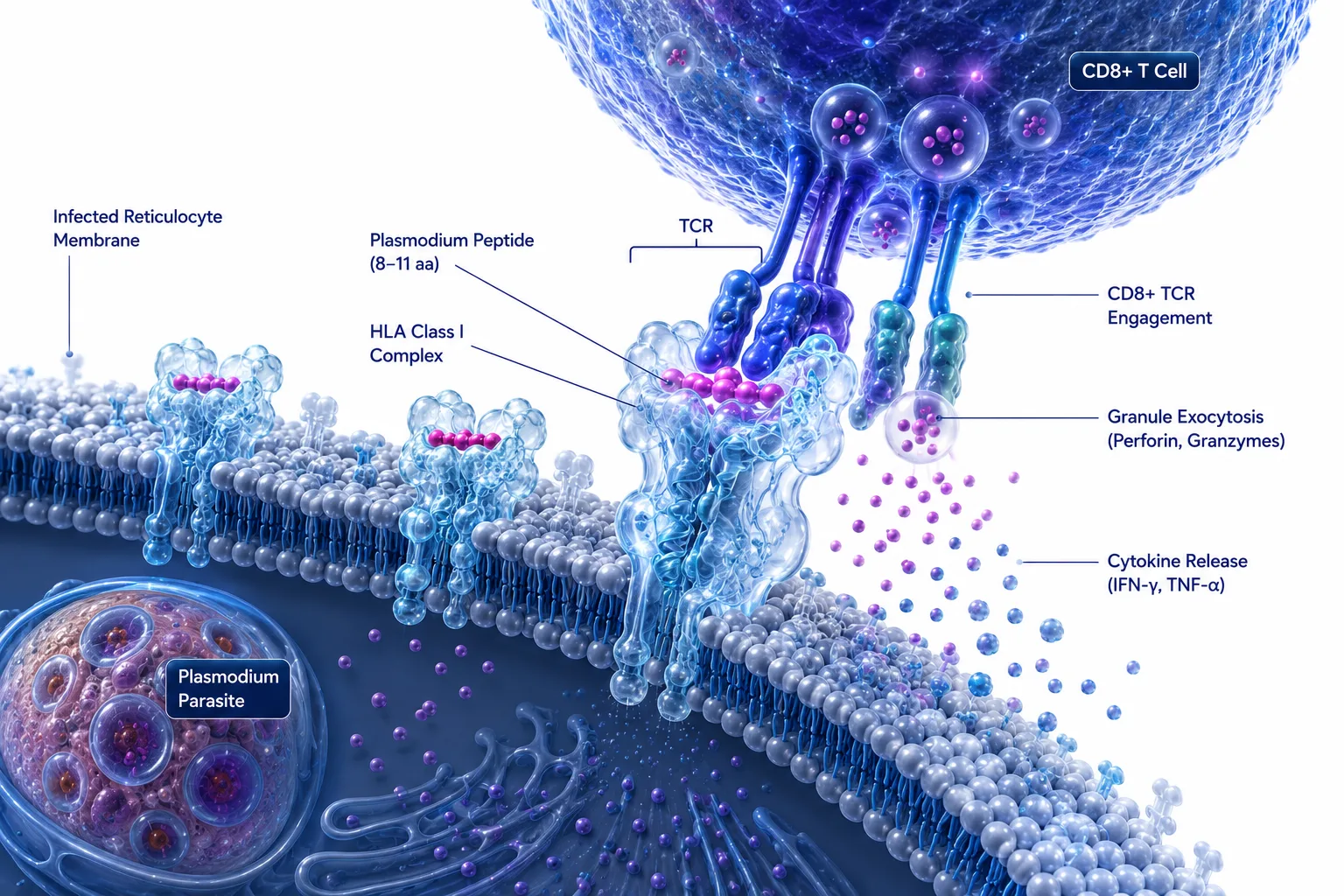 Figure 3: HLA Class I Presentation Mechanism on Infected Reticulocytes
4. From Mass Spectrometry Discovery to T-Cell Immunogenicity Validation
4.1 Structural Biophysics & HLA Binding Affinity Modeling
Mass spectrometry immunopeptidomics identifies peptides presented on host HLA molecules, but physical presentation must be coupled with structural stability and T-cell receptor (TCR) engagement. Computational biophysics algorithms (e.g., NetMHCpan-4.1, NetMHCIIpan, and NetPrio) evaluate peptide-HLA binding affinities (IC50 < 500 nM) and predicted structural stability. Structural modeling of peptide-HLA crystal complexes verifies that conserved parasite peptides fit snugly into the HLA peptide-binding groove, presenting solvent-exposed amino acid side chains for efficient TCR recognition.
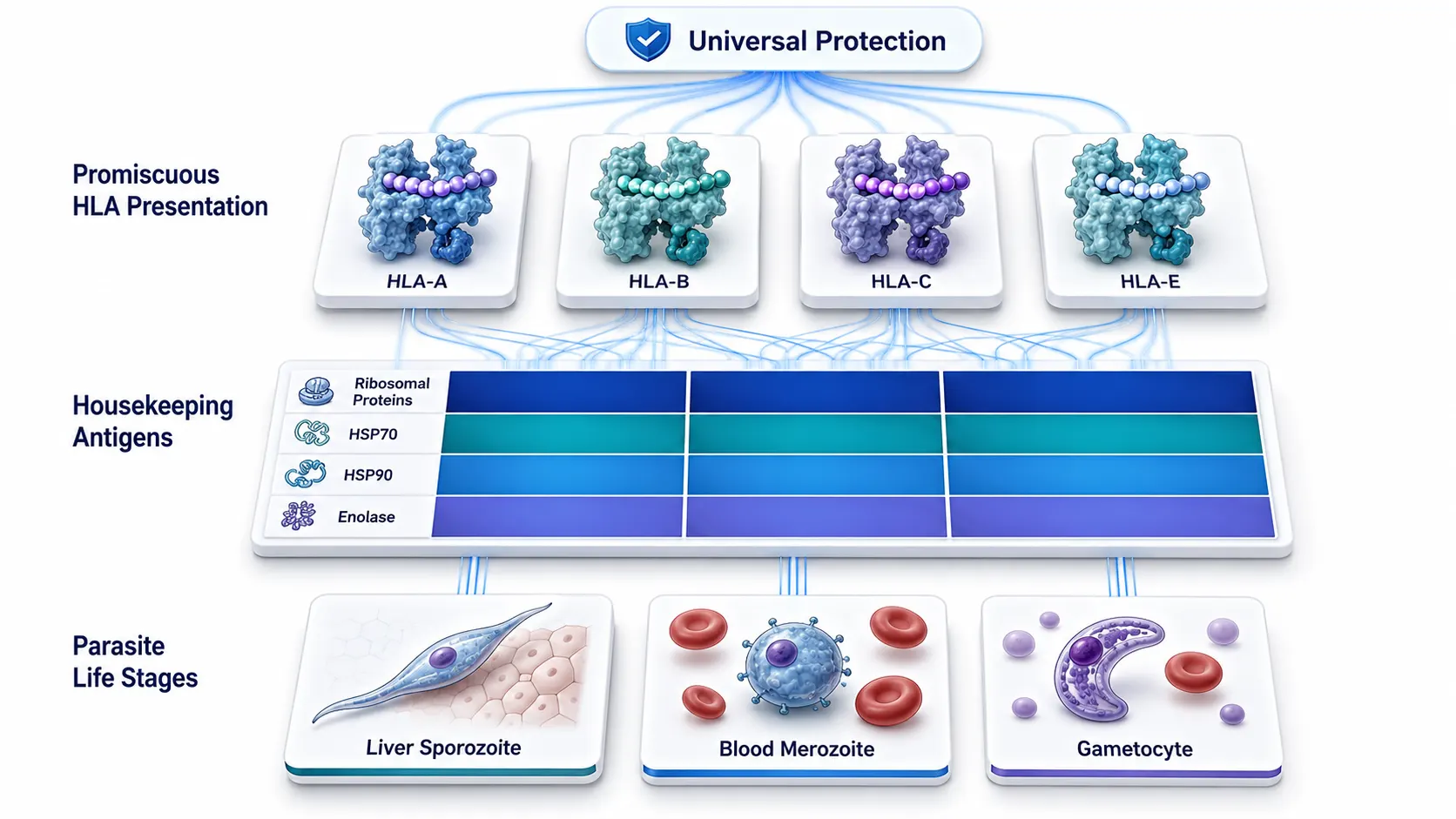 Figure 4: Cross-Stage & Cross-Species Conserved Antigen Landscape
4.2 High-Throughput T-Cell Assays: Ex Vivo ELISpot, ICS, and Cytotoxic Killing
Candidate immunopeptidomics-discovered epitopes undergo rigorous ex vivo immunological validation using peripheral blood mononuclear cells (PBMCs) isolated from malaria-exposed individuals, naturally immune adults in endemic regions, or irradiated sporozoite-immunized human volunteers:
- Ex Vivo IFN-gamma ELISpot: Quantifies the frequency of antigen-specific memory T cells responding to synthetic parasite peptide stimulation.
- Intracellular Cytokine Staining (ICS): Multi-color flow cytometry differentiates CD8+ vs. CD4+ T-cell activation, measuring multi-functional cytokine production (IFN-gamma, TNF-alpha, IL-2) and degranulation markers (CD107a).
- In Vitro Parasite Killing Assays: Demonstrates that CD8+ T cells stimulated with immunopeptidomics-identified peptides directly recognize and kill infected hepatocytes or reticulocytes, reducing intracellular parasite load.
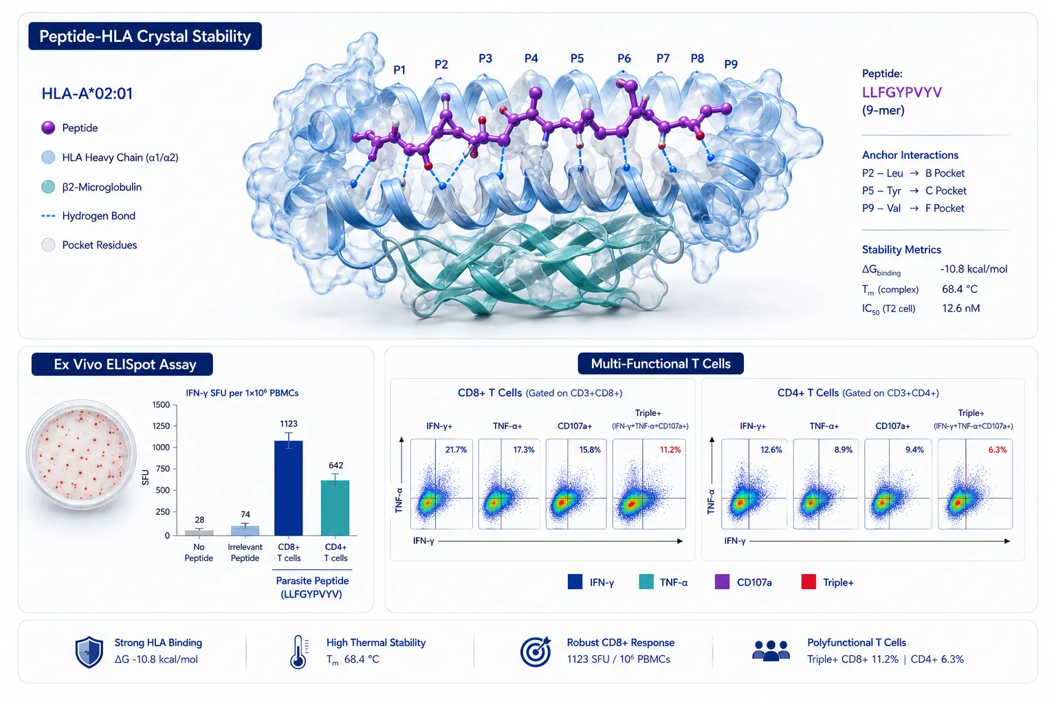 Figure 5: High-Throughput T-Cell Immunogenicity & HLA Stability Validation
5. Next-Generation Multivalent Vaccine Platforms (mRNA-LNPs & Viral Vectors)
5.1 Multi-Epitope mRNA-LNP Formulations Encoding Immunopeptidomics Antigens
The clinical success of lipid nanoparticle-encapsulated mRNA (mRNA-LNP) platforms in infectious disease oncology provides a powerful delivery vehicle for immunopeptidomics-discovered targets. Rather than encoding an entire full-length polymorphic parasite protein, multi-epitope mRNA constructs can be engineered to encode string-of-beads epitopes—linking multiple highly conserved, immunopeptidomics-validated CD8+ and CD4+ T-cell epitopes separated by optimized cathepsin or proteasomal cleavage linkers (e.g., AAY, AAK, or GPGPG linkers).
mRNA-LNP formulations encoding these multi-stage conserved epitopes drive robust intracellular antigen expression, endogenous proteasomal processing, and heavy HLA Class I surface presentation, eliciting potent, long-lasting T-cell memory responses.
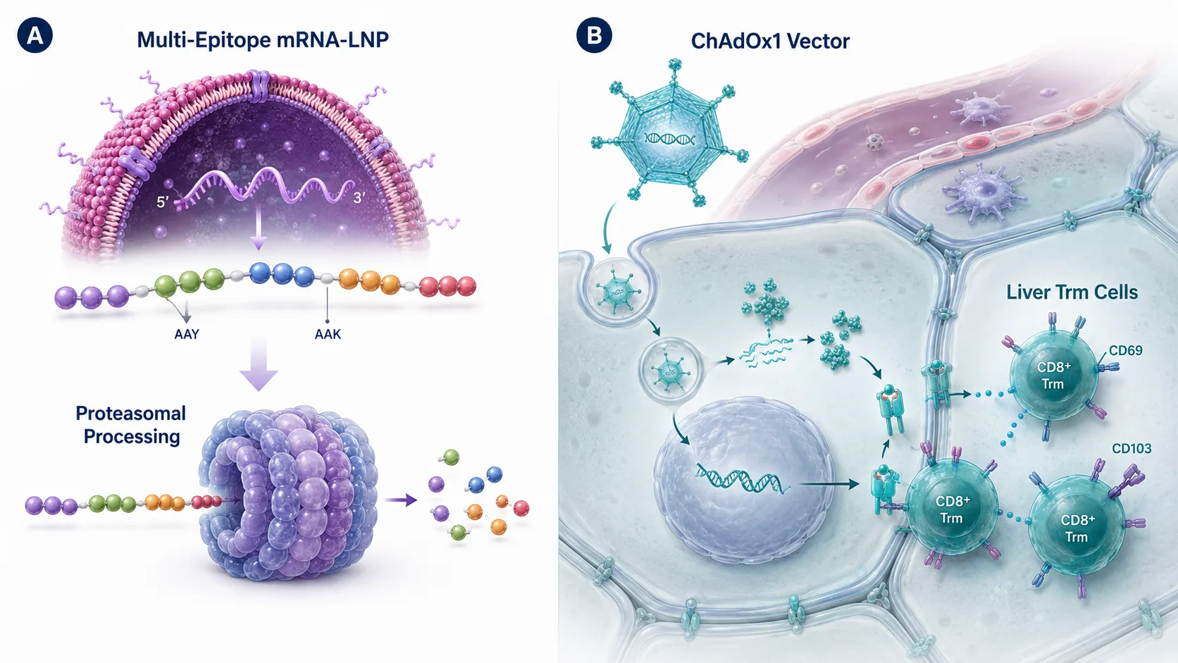 Figure 6: Multivalent mRNA-LNP & Viral Vector Vaccine Delivery Architecture
5.2 Viral Vectors (ChAdOx1 / MVA) and VLPs for Durable Memory CD8+ T-Cell Induction
Recombinant viral vector prime-boost regimens—such as replication-deficient chimpanzee adenovirus (ChAdOx1) combined with Modified Vaccinia Ankara (MVA) or bivalent Virus-Like Particles (VLPs)—represent another highly effective platform for T-cell induction. Heterologous viral prime-boost regimens encoding immunopeptidomics-derived parasite antigens generate high frequencies of liver-resident tissue memory T cells (Trm cells). These Trm cells provide an active frontline immune shield within liver sinusoids, instantly intercepting and destroying invading sporozoites before they can initiate systemic blood-stage malaria.
Partnering with an experienced Biomarker Discovery Service provider allows vaccine developers to validate candidate T-cell epitopes and characterize multi-epitope immune responses during preclinical and clinical evaluation.
6. Methodological Comparison: Recombinant Protein Screening vs. Mass Spectrometry Immunopeptidomics
| Analytical Feature | Recombinant Protein / Overlapping Peptide Screening | Mass Spectrometry Immunopeptidomics |
|---|---|---|
| Identification Mechanism | In silico binding predictions & synthetic peptide arrays | Direct physical identification of naturally presented HLA peptides |
| Biological Authenticity | High false-positive rate (Many predicted peptides are never presented) | 100% Endogenous Presentation (Confirmed cell-surface HLA bound) |
| Coverage of Post-Translational Modifications | Minimal (Fails to capture native parasite PTMs) | High (Directly identifies phosphorylated, acetylated & cleaved peptides) |
| Stage & Species Conservation | Often restricted to major surface antigens (CSP, MSP1) | Uncovers conserved housekeeping antigens across all life stages |
| HLA Allele Diversity | Limited by available synthetic peptide libraries | Unbiased (Captures classical HLA-A/B/C & non-classical HLA-E) |
| Developmental Throughput | High cost & slow synthesis for multi-protein libraries | Ultra-high throughput (Identifies 10,000+ peptides per MS run) |
7. Translational Implementation Framework for Vaccine R&D
For vaccine research teams, biopharmaceutical developers, and global health organizations integrating immunopeptidomics into malaria antigen discovery, we recommend a four-stage implementation framework:
- Infected Cell Ingestion & Sample Isolation: Culture Plasmodium-infected human liver cells, primary phagocytes, or infected reticulocytes. Perform specialized immunoaffinity purification (IP) or mild acid elution (MAE) to recover cell-surface HLA Class I and Class II peptide complexes.
- High-Sensitivity LC-MS/MS & Spectral Sequencing: Analyze immunopeptide eluates using ultra-high-speed mass spectrometry (e.g., Orbitrap Astral or timsTOF Pro). Apply multi-genome hybrid database searches and deep learning rescoring algorithms at 1% FDR.
- Cross-Stage Conservation & HLA Binding Profiling: Cross-reference identified parasite peptides against stage-specific transcriptomic/proteomic datasets to prioritize conserved housekeeping antigens. Evaluate HLA allele promiscuity and structural peptide-HLA stability.
- T-Cell Functional Validation & Multivalent Formulation: Validate peptide immunogenicity using ex vivo ELISpot, ICS, and parasite-killing assays with clinical donor PBMCs. Formulate prioritized multi-epitope constructs into mRNA-LNP or viral vector delivery platforms.
For comprehensive experimental execution, leveraging an integrated Immunoproteomics Service and evaluating specific post-translational modifications via PTM Analysis Service workflows ensures optimal HLA-peptide isolation, high-sensitivity mass spectrometry identification, and rigorous antigen validation for next-generation malaria vaccines.
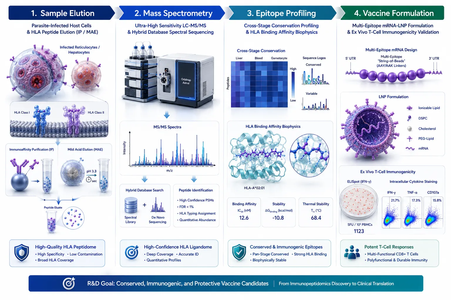 Figure 7: Translational Implementation Framework for Malaria Vaccine R&D
8. Frequently Asked Questions (FAQ)
Q1: What is immunopeptidomics, and how does it differ from conventional proteomics?
Conventional proteomics quantifies total cellular or plasma protein abundance following enzymatic digestion. Immunopeptidomics specifically isolates and identifies the subset of non-cleaved, naturally processed peptide fragments bound to cell-surface Major Histocompatibility Complex (MHC/HLA) Class I and Class II molecules, directly revealing what the host immune system "sees."
Q2: Why have traditional antibody-focused malaria vaccines shown limited long-term efficacy?
Classical vaccines (like RTS,S and R21) focus on inducing antibodies against the circumsporozoite protein (CSP) on sporozoites. Antibody titers wane quickly over time, and parasites exhibit high genetic polymorphism. Furthermore, antibodies cannot eliminate intracellular parasite stages inside liver hepatocytes or blood reticulocytes, which require cytotoxic T-cell immunity.
Q3: How do parasites inside red blood cells present antigens on HLA molecules?
While mature erythrocyte cells lack HLA machinery, immature reticulocytes (which are preferentially infected by P. vivax and P. falciparum) retain surface HLA Class I molecules. Furthermore, host antigen-presenting cells (dendritic cells and macrophages) ingest parasite debris and cross-present parasite antigens on HLA-I and HLA-II molecules.
Q4: What are "housekeeping proteins," and why are they promising vaccine targets?
Housekeeping proteins (e.g., ribosomal proteins, heat shock proteins, metabolic enzymes) are essential for basal parasite survival and are constitutively expressed across all life stages (liver stage, blood stage, sexual stage) and across different Plasmodium species. Because they are highly conserved and less prone to immune-driven mutation, immunopeptides derived from housekeeping proteins offer universal, cross-stage protection.
Q5: What sample quantity is required for mass spectrometry immunopeptidomics?
With next-generation mass spectrometers (such as Orbitrap Astral or timsTOF platforms), immunopeptidomics can be performed on 10^7 to 10^8 primary cells or tissue biopsies, yielding thousands of unique HLA peptides from sub-femtomole peptide input levels.
Q6: Can immunopeptidomics identify HLA Class II peptides presented to CD4+ T cells?
Yes. Using HLA Class II-specific antibodies (such as anti-HLA-DR, DQ, or DP) during immunoaffinity purification enables selective isolation of longer (12–25 amino acid) HLA Class II-bound peptides presented to helper CD4+ T cells, which are crucial for driving durable B-cell antibody responses and memory CD8+ T-cell expansion.
Q7: How does non-classical HLA-E presentation benefit global vaccine design?
Classical HLA genes (HLA-A, B, C) are highly polymorphic across human populations. In contrast, the non-classical HLA-E gene displays very low polymorphism globally. Parasite peptides presented by HLA-E can be recognized by T cells across virtually all individuals worldwide, overcoming population-level HLA restriction.
Q8: Are these computational workflows intended for clinical diagnostic use?
Immunopeptidomics workflows, HLA peptide discovery pipelines, and vaccine target selection protocols described here are developed for Research Use Only (RUO). They serve as powerful tools for target discovery, biomarker identification, mechanism-of-action evaluation, and vaccine candidate development, and are not intended for direct clinical diagnostic procedures.
References:
- Globally Representative Immunopeptidomics Consortium. (2025). A globally representative immunopeptidomics approach to identify malaria antigens. bioRxiv preprint. https://www.biorxiv.org/content/10.1101/2025.11.25.690604v1.full (CC BY 4.0 Open Access).
- Cross-Stage Malaria T-Cell Antigen Study. (2026). Identification of cross-stage, cross-species malaria CD8+ T cell antigens presented by infected reticulocytes. Nature Publications, July 2026. https://ciencia.ucp.pt/en/publications/identification-of-cross-stage-cross-species-malaria-cd8-t-cell-an-2/ (Open Access).
- Infectious Disease Immunopeptidomics Group. (2021). Leveraging immunopeptidomics to study and combat infectious diseases. PMC Articles, PMC8407116. https://pmc.ncbi.nlm.nih.gov/articles/PMC8407116/ (CC BY 4.0 Open Access).
- Pre-Erythrocytic Malaria Vaccine Consortium. (2020). Current challenges in the identification of pre-erythrocytic malaria vaccine antigens. PMC Articles, PMC7046804. https://pmc.ncbi.nlm.nih.gov/articles/PMC7046804/ (CC BY 4.0 Open Access).
- PathMHC Antigen Discovery Group. (2025). Targeting infection-specific peptides in immunopeptidomics studies for vaccine target discovery. Journal of Experimental Medicine, PMC12478359. https://pmc.ncbi.nlm.nih.gov/articles/PMC12478359/ (CC BY 4.0 Open Access).














