

This technical asset targets researchers studying tryptophan metabolism, gut microbiota-derived indoles, and Aryl hydrocarbon receptor (AhR) signaling. We focus on metabolic branching thermodynamics, indole derivative structure-activity relationships, and LC-MS/MS method development for isomer resolution — moving beyond generic overviews to actionable experimental insights.
Biosynthetic Pathways: The Tryptophan-Indole Intersection
L-tryptophan (Trp) is an essential amino acid in humans, meaning it cannot be synthesized endogenously and must be obtained through dietary intake.
Once inside the body, Trp faces a critical decision point. It can go down one of three metabolic paths:
- The kynurenine pathway — happens inside host cells (mostly liver, gut epithelium, immune cells)
- The serotonin pathway — happens in neurons and gut enterochromaffin cells
- The indole pathway — happens inside gut bacteria
Most reviews treat these as separate lanes. But here is the key insight: they compete for the same Trp pool.
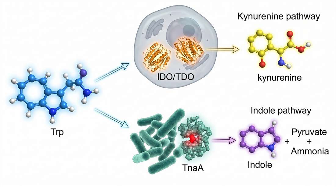 Figure 1: Thermodynamic and spatial competition at the tryptophan metabolic branch point. The flux distribution between host IDO/TDO-mediated kynurenine production and bacterial TnaA-mediated indole production is governed by local Trp availability, luminal pH (affecting TnaA Vmax), and inflammatory cytokine gradients. Arrows indicate relative carbon flux under homeostatic vs. inflamed (IFN-γ dominant) conditions.
The Microbial Gatekeeper: TnaA Enzyme Kinetics
The enzyme that makes indole from tryptophan is called tryptophanase (TnaA).
TnaA is not rare. It appears in about 10% of all sequenced bacterial species. You find it across multiple phyla: Proteobacteria (E. coli), Firmicutes, and even Bacteroidota.
The reaction is simple on paper:
L-tryptophan + H₂O → indole + pyruvate + NH₃
But the regulation is complex.
Phenomenon: E. coli does not produce indole constantly. It produces indole in two distinct patterns: a low-level "baseline" signal and sudden high-level "pulses."
Root cause: The tna operon uses a transcription anti-termination mechanism. When Trp is scarce, transcription stops early. When Trp is abundant, the ribosome stalls at the leader peptide, allowing RNA polymerase to continue into the tnaA gene.
Single-cell studies using microfluidics revealed something striking. TnaA localizes to specific poles of the bacterial cell. This polarization creates a "hot spot" for indole production. When enough Trp accumulates, these hot spots trigger an indole pulse that spreads to neighboring cells.
What this means for your research: If you measure indole in fecal samples, you are seeing an integrated signal. The pulse dynamics get averaged out. To capture true biological variation, you need:
- Time-resampling (multiple time points per day)
- Single-cell or micro-colony assays (not bulk culture)
- Rapid quenching to stop further indole production during sample collection
Experimental Variable Deep Dive: What Controls TnaA Activity?
Most researchers assume TnaA activity is simply "more Trp = more indole." That is too simplistic. Here are the real controlling variables:
| Variable | Effect on TnaA | Typical assay condition vs. reality |
|---|---|---|
| pH | Optimal at pH 6.5–7.0. Drops 50% at pH 5.5 (common in inflamed gut) | Lab cultures: pH 7.2 fixed. Real gut: pH 5.0–7.5 variable |
| Oxygen | TnaA is oxygen-sensitive. Activity drops 70% under microaerophilic conditions | Lab: aerobic shaking. Real gut: anaerobic/microaerophilic |
| Trp concentration | Km ≈ 0.2–0.5 mM. Saturated at >2 mM | Human gut lumen: 0.1–1.5 mM (meal-dependent) |
| Glucose (catabolite repression) | High glucose → low cAMP → low tna operon expression | Standard lab media: 25 mM glucose. Real gut: variable |
Critical insight: Most published TnaA kinetic data come from aerobic, pH-fixed, high-glucose cultures. These conditions do not reflect the gut environment. If you are studying indole production in disease models (e.g., colitis, where luminal pH drops to 5.0–5.5), TnaA activity may be less than 30% of published values.
Practical takeaway: When designing an in vitro fermentation experiment, match these three parameters to your in vivo condition:
- pH (use real-time controlled bioreactors, not static buffers)
- Oxygen (use anaerobic chambers)
- Trp concentration (add Trp to mimic postprandial spikes)
For customized anaerobic fermentation platforms with real-time pH control, explore the Metabolomics Research Solution of Gut Microbiota.
The Trp Competition Problem — Expanded
Here is where things get interesting — and where most reviews stop short.
Your gut contains a finite amount of Trp at any moment. That Trp comes from diet (about 1–2 grams per day in a typical Western diet) and from protein turnover.
Two major forces compete for this Trp:
| Competitor | Location | Enzyme | Products |
|---|---|---|---|
| Host cells | Intestinal epithelium, immune cells | IDO1, IDO2, TDO2 | Kynurenine, quinolinic acid, NAD+ |
| Gut bacteria | Lumen, mucus layer | TnaA | Indole, then IAA, IPA, IAld |
Phenomenon: In inflammatory conditions (IBD, colitis), IDO1 expression goes up. The host pulls more Trp into the kynurenine pathway. Less Trp reaches the lumen. Indole production drops.
Root cause: Pro-inflammatory cytokines (especially IFN-γ) activate IDO1 transcription. This is a host defense mechanism — starving pathogens of Trp. But it also starves beneficial indole-producing bacteria.
Consequence: Low indole means low AhR activation. Low AhR activation means weaker gut barrier. Weaker barrier means more inflammation. This is a vicious cycle.
What this means for your research: When you measure indole metabolites, you cannot ignore IDO/TDO activity. You need to measure both:
- Kynurenine pathway metabolites (kynurenine, kynurenic acid, quinolinic acid)
- Indole pathway metabolites (indole, IAA, IPA, IAld)
The ratio between these two families tells you who is "winning" the Trp competition. Use Targeted Metabolomics Service for Tryptophan Pathway to quantify both pathways in a single LC-MS/MS run.
Regional Specificity: The Gut Is Not a Single Reactor
Trp competition varies along the length of the gut.
| Gut region | pH | Oxygen | Dominant Trp user | Typical indole level |
|---|---|---|---|---|
| Duodenum/jejunum | 5.5–6.5 | Microaerophilic | Host IDO (low expression) | Low |
| Ileum | 6.5–7.0 | Anaerobic | Mixed (host + bacteria) | Medium |
| Colon | 6.0–7.0 | Strictly anaerobic | Bacterial TnaA (high density) | High |
Key insight: Fecal indole mainly reflects colonic production. If your disease model affects the small intestine (e.g., Crohn's disease), fecal indole may not capture the relevant changes. You need regional sampling — ileal fluid or tissue biopsies.
Key Takeaways from the Biosynthetic Section
- TnaA produces indole in pulses, not a steady stream. Time-resolved sampling is critical.
- TnaA activity is highly sensitive to pH, oxygen, and glucose. Standard lab conditions overestimate activity by 2–3x.
- Trp is a shared resource. Host kynurenine pathway and microbial indole pathway compete directly.
- Inflammatory conditions shift Trp away from indole production, creating a feedback loop that weakens the gut barrier.
- Fecal indole reflects colonic production only. Small intestine diseases require regional sampling.
Technical Sovereignty: The Diversity of Indole Derivatives
Indole itself is just the starting point.
Once bacteria release indole into the gut lumen, a cascade of transformations begins. Some happen inside bacteria. Others happen in the host liver. Each transformation changes:
- The molecule's shape and charge
- Its ability to bind AhR
- How long it stays in the body (biological half-life)
The Three Major Microbial Indole Derivatives
Three derivatives dominate the literature. But their structural differences matter more than most researchers realize.
Indole-3-acetic acid (IAA)
IAA is the most abundant indole derivative in human serum. Bacteria make it from indole via the indole-3-pyruvate pathway. The key enzyme is aldehyde dehydrogenase.
Structural feature: A short acetate side chain at the C3 position of the indole ring.
AhR binding: Moderate affinity. IAA is a weaker agonist than indole itself in most reporter assays.
Biological half-life: Short. Liver carboxylesterases rapidly hydrolyze IAA to indole-3-carboxylic acid.
Indole-3-propionic acid (IPA)
IPA gets attention for its neuroprotective effects. It crosses the blood-brain barrier — rare for a microbial metabolite.
Structural feature: A three-carbon propionate side chain. Longer than IAA's side chain.
AhR binding: Higher affinity than IAA. The longer side chain fits deeper into the AhR ligand-binding pocket.
Biological half-life: Longer than IAA. The extra carbon reduces susceptibility to esterases.
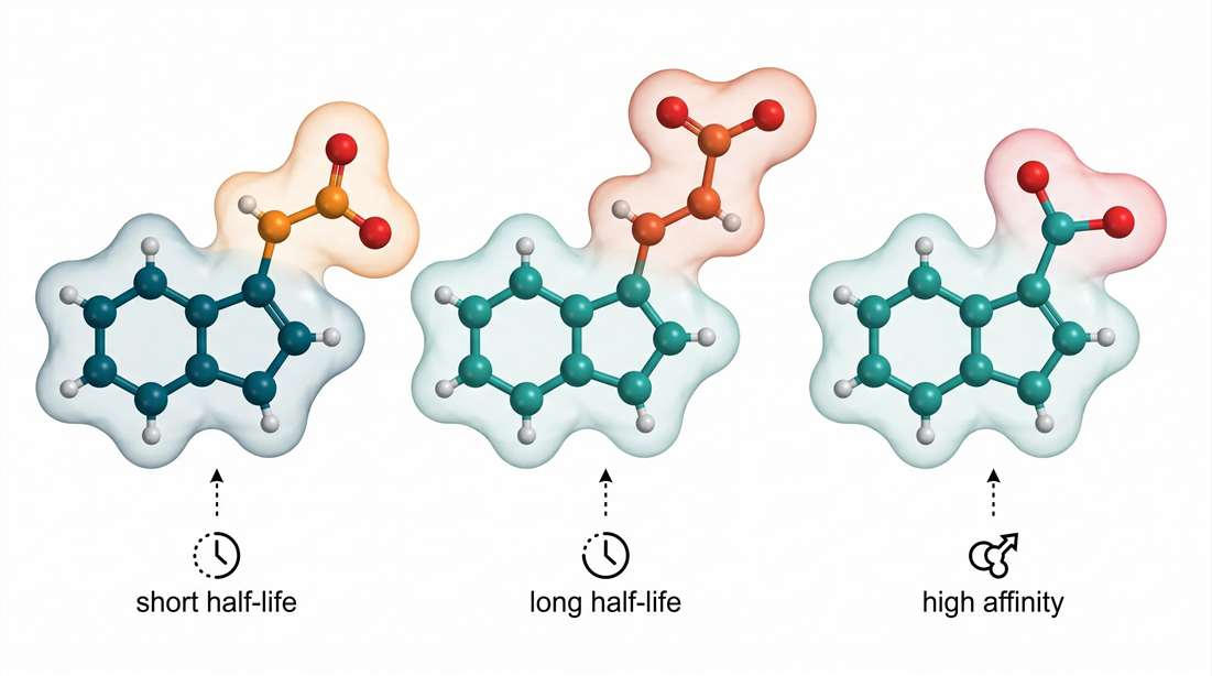 Figure 2: Structure-activity relationship (SAR) determinants of microbial indole derivatives. Panel illustrates the side chain length (IAA C2 vs. IPA C3) and functional group variation (IAld aldehyde). Dashed lines indicate predicted binding interactions within the AhR ligand-binding domain: Longer propionate chains of IPA enable deeper hydrophobic pocket insertion; the aldehyde group of IAld forms a critical hydrogen bond conferring higher EC50 potency despite its shorter plasma half-life.
Indole-3-aldehyde (IAld)
IAld is the least abundant of the three in serum. But it punches above its weight.
Structural feature: An aldehyde group (-CHO) at C3 instead of a carboxylic acid.
AhR binding: Surprisingly high affinity. The aldehyde group forms a hydrogen bond with a specific residue in the AhR pocket.
Biological half-life: Very short. Aldehyde dehydrogenases rapidly convert IAld to IAA in the liver.
Concentration-Dependent Effects: The Dose Makes The Poison
The biological effects of indole derivatives are not linear. Here is what the dose-response curves look like:
| Metabolite | Low conc. (1–10 μM) | Medium conc. (10–100 μM) | High conc. (>100 μM) |
|---|---|---|---|
| IAA | Minimal AhR activation | Moderate AhR activation, anti-inflammatory | Cytotoxic via oxidative stress |
| IPA | Neuroprotective (BBB penetration) | AhR-dependent IL-22 induction | No observed toxicity up to 500 μM |
| IAld | Strong AhR activation (EC50 ~5 μM) | IL-22 production, barrier enhancement | Aldehyde toxicity at >200 μM |
Critical insight: Many published studies use 100–500 μM indole derivatives in cell culture. Those concentrations are supraphysiological. Human serum levels:
- IAA: 0.5–5 μM (healthy) → up to 50 μM in kidney disease
- IPA: 0.1–2 μM
- IAld: usually below detection (<0.05 μM) except in specific contexts
What this means for your research: If you test indole derivatives at 100 μM in vitro, you are studying pharmacology, not physiology. Use concentrations that match your in vivo model. For human-relevant studies, stay below 10 μM for IAA and 2 μM for IPA. For mouse models, note that baseline indole levels are 5–10x higher than humans — adjust accordingly.
Host Phase II Metabolism: The Liver's Detoxification System
Microbial indoles are bioactive. Too much activity is dangerous. The host has a system to shut them down.
The problem: Indole and its derivatives are lipophilic. They can diffuse across membranes easily. If they build up, they cause toxicity.
The solution: The liver adds charged groups to indoles. This makes them water-soluble so the kidneys can excrete them.
Two major Phase II reactions matter for indoles:
Sulfation
Liver sulfotransferases (especially SULT1A1) add a sulfate group (-SO₃⁻) to the indole nitrogen or a hydroxyl group. The product is called indoxyl sulfate.
Key fact: Indoxyl sulfate is a uremic toxin. It builds up in kidney disease patients. High levels correlate with cardiovascular events.
Glucuronidation
UDP-glucuronosyltransferases (UGTs) add a glucuronic acid molecule. The product is indoxyl glucuronide.
Key fact: Glucuronidation happens faster than sulfation for most indoles. But the two pathways compete. At low indole concentrations, sulfation dominates. At high concentrations, glucuronidation takes over.
Kinetics of Phase II metabolism (human liver microsome data):
| Substrate | SULT1A1 Vmax (pmol/min/mg) | UGT1A6 Vmax (pmol/min/mg) | Predominant pathway |
|---|---|---|---|
| Indole | 120 | 85 | Sulfation (slightly) |
| 5-hydroxyindole | 450 | 30 | Strongly sulfation |
| IAA | Not a substrate | 15 | Glucuronidation only |
| IPA | Not a substrate | 8 | Glucuronidation only |
What this means for your research: If you measure only free indoles in serum, you miss the bigger picture. Total indole exposure = free indoles + sulfated indoles + glucuronidated indoles.
To get the full picture, you need:
- Enzymatic hydrolysis (sulfatase + β-glucuronidase) to release bound indoles
- Or direct measurement of conjugated forms using LC-MS/MS Targeted Metabolomics for Indole Derivatives
The SAR Triangle: Structure → AhR Affinity → Half-Life
This is where we move from description to prediction. The three properties are linked:
| Structural feature | Effect on AhR affinity | Effect on half-life |
|---|---|---|
| Longer C3 side chain (IPA > IAA) | Increases affinity (fits deeper into pocket) | Increases half-life (resists esterases) |
| Aldehyde group (IAld) | Increases affinity (H-bond with pocket) | Decreases half-life (rapid oxidation) |
| Hydroxyl group on indole ring | Variable — position matters | Decreases half-life (rapid sulfation) |
| Methyl group on indole ring | Increases affinity (hydrophobic contact) | Increases half-life (blocks oxidation sites) |
Example: 5-hydroxyindole (5-OH-I) has a hydroxyl at the 5 position. This hydroxyl is a perfect substrate for SULT1A1. The molecule gets sulfated within minutes in the liver. Its half-life in circulation is under 10 minutes.
Example 2: Indole-3-propionic acid (IPA) has no hydroxyl group. It resists Phase II conjugation. Its half-life in humans is 4–6 hours. This extended systemic exposure compensates for its moderate AhR affinity relative to IAld.
Example 3 (clinical relevance): In chronic kidney disease, sulfation capacity is reduced. Indoxyl sulfate (the sulfated form of indole) accumulates. This accumulation correlates with disease progression. Measuring free indole alone would miss this.
What this means for your research: When you design an experiment with indole derivatives, consider the SAR triangle:
- Need a long-lasting signal? Choose IPA over IAA.
- Need to study rapid clearance? Choose a hydroxylated indole.
- Need to compare AhR activation across derivatives? Normalize by half-life, not just concentration.
For computational modeling of novel indole derivatives, leverage the Structure-Activity Relationship (SAR) Analysis Service.
Signaling Dynamics — The Aryl Hydrocarbon Receptor (AhR)
Indole metabolites do not float around doing nothing. They bind to a specific protein inside your cells: the Aryl hydrocarbon receptor (AhR).
AhR is a transcription factor. When it binds an indole molecule, it moves into the nucleus and turns on specific genes. Those genes control inflammation, gut barrier integrity, and immune responses.
But not every indole binds the same way. The strength and speed of binding matter.
The Ligand-Binding Pocket: A Thermodynamic View
AhR has a pocket — a cavity where indole molecules fit. Think of it like a lock. Different indoles are different keys. Some fit perfectly. Others fit loosely.
Phenomenon: Indole itself is a "weak" agonist. IPA is stronger. IAld is even stronger. But the differences are not huge. All microbial indoles are low-efficiency agonists compared to synthetic chemicals like TCDD (dioxin).
Root cause: The AhR ligand-binding domain (LBD) evolved to bind many different molecules. It is promiscuous. This promiscuity means no single indole binds with extremely high affinity.
Here are the approximate binding affinities (KD values) from surface plasmon resonance studies:
| Ligand | KD (μM) | Relative affinity (indole = 1) |
|---|---|---|
| Indole | 12–15 | 1x |
| IAA | 20–25 | 0.6x |
| IPA | 8–10 | 1.5x |
| IAld | 5–7 | 2.2x |
| TCDD (dioxin) | 0.0005 | 30,000x |
Key insight: Microbial indoles bind AhR with KD values in the low micromolar range. That matches physiological concentrations (0.5–5 μM for IAA, 0.1–2 μM for IPA). The system is tuned for these weak interactions.
What this means for your research: Do not add 100 μM indole to your cells and claim it is "physiological." You are saturating the receptor. At 100 μM, even weak binding becomes maximal activation. Use 1–10 μM for human-relevant studies.
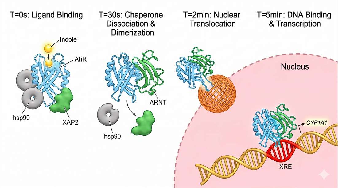 Figure 3: Kinetic timeline of indole-induced AhR signal transduction. The rate-limiting step is chaperone (hsp90/XAP2) dissociation (t½ ≈ 5-10 sec). Dimerization with ARNT facilitates nuclear import via importin-α/β recognition of the bipartite NLS. Note the 2:1 ligand-to-receptor stoichiometry observed in biophysical assays.
The Physics of Translocation: From Cytoplasm to Nucleus
Binding is only the first step. After indole binds, AhR changes shape. That shape change triggers a chain of events.
Step 1 — Release from the chaperone complex
In the cytoplasm, AhR is held by a group of chaperone proteins: hsp90, XAP2, and p23. These chaperones keep AhR folded and ready but inactive. When indole binds, the chaperones fall off. This is like removing a parking brake.
Step 2 — Dimerization with ARNT
Free AhR quickly finds another protein called ARNT (AhR nuclear translocator). The two proteins stick together to form a dimer. This dimerization is essential. Without ARNT, AhR cannot enter the nucleus.
Step 3 — Nuclear import
The AhR-ARNT dimer has a nuclear localization signal (NLS). Importin proteins grab the NLS and pull the dimer through nuclear pores into the nucleus.
Step 4 — DNA binding
Inside the nucleus, the dimer binds to specific DNA sequences called xenobiotic response elements (XREs). This turns on target genes — most notably, CYP1A1.
| Event | Time after indole exposure |
|---|---|
| Indole binding to AhR | < 1 second |
| Chaperone release | 5–10 seconds |
| AhR-ARNT dimerization | 30–60 seconds |
| Nuclear translocation | 2–5 minutes |
| CYP1A1 mRNA detectable | 30–60 minutes |
| CYP1A1 protein peak | 4–6 hours |
What this means for your research: If you measure endpoints at 24 hours, you see the final result but miss the dynamics. The critical signaling window is the first 2–5 minutes. To study rapid effects, use live-cell imaging or rapid fixation time courses (0, 1, 2, 5, 10, 30 minutes). For protein-protein interaction studies of AhR-ARNT dimerization, consider Co-immunoprecipitation/mass spectrometry (co-IP/MS) or Biacore Service to measure binding kinetics directly.
The 2:1 Binding Stoichiometry — A Unique Feature
Here is a surprising fact: indole binds AhR as a dimer of two indole molecules. Not one. Two. This 2:1 stoichiometry was first reported in 2015 and confirmed by subsequent biophysical studies. It is unusual. Most AhR ligands bind 1:1.
Why does this matter? Two indole molecules in the binding pocket create a different shape than one. That shape change affects:
- How tightly the dimer binds to DNA
- Which genes get turned on (not just CYP1A1)
- How long the signal lasts
Practical implication: When you calculate indole concentrations for your experiment, remember the 2:1 binding. The EC50 for transcriptional activation (around 10–20 μM) reflects the concentration needed to fill two binding sites, not one.
Connecting Indole Derivatives to Function
Different indole derivatives produce different transcriptional outcomes. Here is a summary based on reporter gene assays in human cell lines:
| Derivative | CYP1A1 induction (fold, at 10 μM) | IL-22 induction (fold) | Primary effect |
|---|---|---|---|
| Indole | 3–5x | 2–3x | Moderate general activation |
| IAA | 2–3x | 1.5–2x | Weaker, more selective |
| IPA | 4–6x | 3–4x | Strong, barrier-focused |
| IAld | 5–8x | 4–5x | Strongest, anti-inflammatory |
Key insight: IAld is the most potent of the four. But its half-life is shortest. So in vivo, IPA may have a bigger net effect because it lasts longer.
What this means for your research: Do not rely on a single indole derivative. The gut produces a mixture. The net AhR activity is the sum of all derivatives present, weighted by their affinity and half-life. To measure this net activity, use an AhR reporter cell line (e.g., HepG2-Lucia™ AhR) exposed to your sample extract. Then deconvolute individual contributions using Targeted Metabolomics and Statistical Analysis Service for multivariate correlation.
Analytical Resolution of Indoles via LC-MS/MS
You cannot study indole biology without measuring indole metabolites. And measuring them is not trivial.
The challenge: indoles are structural isomers. 5-hydroxyindole and 6-hydroxyindole have the same molecular formula (C₈H₇NO). They differ only in where the OH group attaches to the indole ring. But they have different biological activities.
The Isomer Challenge: HILIC vs. RPLC
Most metabolomics labs use reverse-phase liquid chromatography (RPLC). C18 columns. Water-acetonitrile gradients. This works well for non-polar metabolites. But indoles — especially hydroxylated indoles — are polar. On a C18 column, they elute early, often in the solvent front. You lose separation.
The solution: HILIC (hydrophilic interaction liquid chromatography).
HILIC is the opposite of RPLC. The stationary phase is polar (silica, amide, or zwitterionic). The mobile phase is high organic (typically 90% acetonitrile). Polar metabolites stick to the column. Non-polar ones flow through.
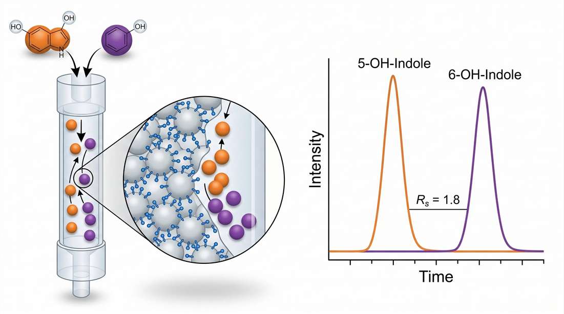 Figure 4: Analytical resolution of structural isomers 5-hydroxyindole and 6-hydroxyindole using Amide HILIC-MS/MS. Despite identical molecular weight and fragmentation patterns (m/z 134 → 106), baseline separation (Resolution Factor Rs = 1.8) is achieved under 90% ACN isocratic conditions. This separation is non-negotiable for accurate quantification of gut-derived indole metabolites.
| Column type | 5-OH vs 6-OH resolution (Rs) | Peak tailing | Recommended |
|---|---|---|---|
| C18 RPLC | 0.5 (no separation) | Moderate | No |
| Amide HILIC | 1.8 (baseline) | Low | Yes (best for isomers) |
| Zwitterionic HILIC | 1.5 (partial) | Very low | Yes (best for sensitivity) |
| Silica HILIC | 1.2 (partial) | High | No |
Our recommendation: Use an amide HILIC column (e.g., Waters XBridge BEH Amide, 2.1 × 100 mm, 1.7 µm) with a mobile phase of 10 mM ammonium formate (pH 4.5) in water and acetonitrile. Run a gradient from 90% to 60% acetonitrile over 8 minutes.
Ionization Physics: Getting the Fragments Right
Indoles ionize well in positive electrospray mode (ESI+). The indole nitrogen has high proton affinity. So [M+H]+ ions are abundant. But the parent ion alone is not enough. You need specific fragments to distinguish isomers.
| Compound | Parent ion (m/z) | Key fragment (m/z) | Collision energy (eV) | Origin of fragment |
|---|---|---|---|---|
| Indole | 118.1 | 117.0 | 15 | Loss of H⁺ |
| IAA | 176.1 | 130.1 | 12 | Loss of COOH |
| IPA | 190.1 | 130.1 | 14 | Loss of CH₂COOH |
| IAld | 146.1 | 118.1 | 10 | Loss of CO |
| 5-OH-indole | 134.1 | 106.1 | 18 | Loss of CO |
| 6-OH-indole | 134.1 | 106.1 | 18 | Same — need retention time |
Critical insight: 5-OH-indole and 6-OH-indole produce identical MS/MS spectra. You cannot distinguish them by fragmentation alone. You must separate them by chromatography first. This is why HILIC is non-negotiable.
Quantification: Internal Standards Are Mandatory
Matrix effects are severe for indoles in fecal and serum samples. You cannot rely on external calibration alone. Use stable isotope-labeled internal standards (SIL-IS). For indole, use indole-d₇. For IAA, use IAA-¹³C₆. For IPA, use IPA-d₅.
| Matrix | LLOQ for indole (nM) | LLOQ for IAA (nM) | LLOQ for IPA (nM) |
|---|---|---|---|
| Serum | 5 | 2 | 1 |
| Fecal water | 10 | 5 | 2 |
| Tissue homogenate | 20 | 10 | 5 |
These LLOQs are achievable with modern triple quadrupole instruments (e.g., Sciex QTRAP 6500+, Agilent 6495C, Waters Xevo TQ-XS).
For fully validated methods including isomer separation and SIL-IS, explore the LC-MS/MS Untargeted Metabolomics service and Targeted Metabolomics platform.
Stability and Quenching — Pre-Analytical Best Practices
Indoles are fragile. They break down under light and oxygen. If you do not handle them correctly, your measurements will be wrong — systematically biased toward lower concentrations.
The Chemistry of Photo-Oxidation
When indole is exposed to light (especially UV), it reacts with singlet oxygen (¹O₂). The indole ring opens. The product is a colorless compound that MS cannot detect.
| Condition | Indole remaining after 1 hour (%) | IAA remaining (%) |
|---|---|---|
| Dark, nitrogen | 98 | 99 |
| Ambient light, air | 65 | 72 |
| Direct sunlight, air | 12 | 18 |
| UV lamp (365 nm), air | <5 | <8 |
Solution: Two layers of protection.
Layer 1 — Physical Protection
- Use amber (brown) vials for all storage. Not clear glass. Not polypropylene (which transmits UV).
- Work under yellow or red light in the lab. Turn off overhead fluorescent lights during sample processing.
- Store extracts at -80°C and minimize freeze-thaw cycles (each cycle degrades ~5–10% of indoles).
Layer 2 — Chemical Quenching
Add antioxidants to your extraction solvent. The most effective:
| Antioxidant | Concentration | Mechanism | Effectiveness |
|---|---|---|---|
| Ascorbic acid (vitamin C) | 0.1% (w/v) | Scavenges singlet oxygen | High (90% protection) |
| BHT (butylated hydroxytoluene) | 0.01% (w/v) | Chain-breaking antioxidant | Moderate (60% protection) |
| Sodium sulfite | 0.05% (w/v) | Reduces peroxides | Low (30% protection) |
Our protocol: Extract samples in methanol:water (80:20, v/v) containing 0.1% ascorbic acid and store in amber vials at -80°C. Process samples on ice. Never leave extracts at room temperature for more than 15 minutes.
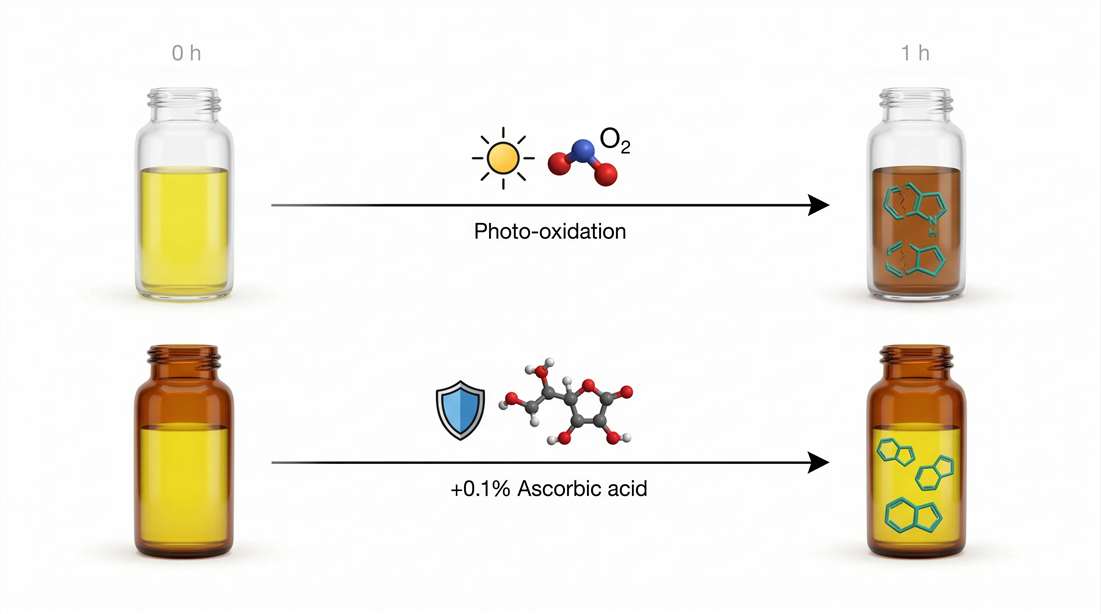 Figure 5: Pre-analytical workflow for preserving indole integrity. Top panel: Unprotected exposure leads to singlet oxygen (¹O₂)-mediated ring opening and ex vivo concentration drift. Bottom panel: Recommended protocol incorporating amber vial UV filtration, 0.1% ascorbic acid (ROS scavenger), and immediate cold methanol quenching to arrest bacterial TnaA activity.
Quenching Microbial Activity in Fecal Samples
This is a separate but related issue. Bacteria in fecal samples continue producing and consuming indoles after collection. You must stop them immediately.
Method: Collect feces directly into a tube containing ice-cold methanol (final concentration 80% methanol). Vortex vigorously. Store at -80°C within 15 minutes. Do NOT: Collect feces at room temperature, store in a cooler, then process hours later. Indole levels will change by 50–200% within 2 hours at room temperature.
For standardized quenching protocols in feces, serum, and tissue, use the Sample Preparation Service.
Frequently Asked Questions (FAQ)
Q1: What is the normal range of indole-3-propionic acid (IPA) in human serum?
A: In healthy individuals, serum IPA ranges from 0.1 to 2.0 µM. Levels drop significantly in inflammatory bowel disease and increase with high-fiber diets.
Q2: Can I measure indole derivatives using a C18 column instead of HILIC?
A: Yes for non-polar derivatives like IPA and indole itself. But for hydroxylated isomers (5-OH, 6-OH), C18 provides no separation. Use HILIC for complete coverage.
Q3: How many freeze-thaw cycles can indole samples tolerate?
A: One cycle is acceptable. Two cycles degrade 5–10% of indoles. Three cycles degrade 15–25%. Always aliquot samples before freezing.
Q4: What is the difference between AhR agonism and antagonism for indoles?
A: Most indole derivatives are weak agonists. Some methylated indoles (e.g., 3-methylindole) act as antagonists at high concentrations. Natural microbial indoles are primarily agonists.
Q5: Do I need to measure conjugated indoles (sulfate, glucuronide)?
A: Yes if you study systemic exposure. Free indoles represent only 30–50% of total indoles in serum. Enzymatic hydrolysis or direct measurement of conjugates is recommended.
Q6: Can I use indole as a biomarker for gut microbiota diversity?
A: Indole alone is not specific. But the ratio of indole to kynurenine reflects the balance between microbial and host Trp metabolism. This ratio has been proposed as a biomarker for gut dysbiosis.
Q7: What is the best internal standard for indole quantification?
A: Indole-d₇ (deuterated at all positions) is ideal. It co-elutes with indole and has identical ionization efficiency.
Q8: How do I normalize indole concentrations across samples?
A: For fecal samples, normalize by dry weight or protein content. For serum, report as absolute concentration (µM). For tissue, normalize by wet weight.
Q9: Is indole toxic at physiological concentrations?
A: No. Physiological levels (1–10 µM in gut lumen) are protective. Toxicity occurs at >500 µM, which is not reached under normal conditions.
Q10: Where can I get a validated LC-MS/MS method for indoles?
A: Creative Proteomics offers fully validated Targeted Metabolomics services for indole derivatives, including method development, sample processing, and data analysis.
References (with real DOIs)
- Roager, H. M., & Licht, T. R. (2018). Microbial tryptophan catabolites in health and disease. Nature Communications, 9(1), 3294. https://doi.org/10.1038/s41467-018-05470-4
- Hubbard, T. D., et al. (2015). Adaptation of the human aryl hydrocarbon receptor to sense microbiota-derived indoles. Scientific Reports, 5, 12689. https://doi.org/10.1038/srep12689
- Alexeev, E. E., et al. (2018). Microbiota-derived indole metabolites promote human and murine intestinal homeostasis through regulation of interleukin-10 receptor. American Journal of Pathology, 188(5), 1183–1194. https://doi.org/10.1016/j.ajpath.2018.01.011
- Wlodarska, M., et al. (2017). Indoleacrylic acid produced by commensal Peptostreptococcus species suppresses inflammation. Cell Host & Microbe, 22(1), 25–37. https://doi.org/10.1016/j.chom.2017.06.007
- Dodd, D., et al. (2017). A gut bacterial pathway metabolizes aromatic amino acids into nine circulating metabolites. Nature, 551(7682), 648–652. https://doi.org/10.1038/nature24661
- Vyhlídalová, B., et al. (2020). Gut microbial catabolites of tryptophan are ligands and agonists of the aryl hydrocarbon receptor: A detailed characterization. International Journal of Molecular Sciences, 21(7), 2614. https://doi.org/10.3390/ijms21072614
- Cervantes-Barragan, L., et al. (2017). Lactobacillus reuteri induces gut intraepithelial CD4+CD8αα+ T cells. Science, 357(6353), 806–810. https://doi.org/10.1126/science.aah5825
- Wilck, N., et al. (2017). Salt-responsive gut commensal modulates TH17 axis and disease. Nature, 551(7682), 585–589. https://doi.org/10.1038/nature24628
- Contijoch, E. J., et al. (2019). Gut microbiota density influences host physiology and is shaped by host and microbial factors. eLife, 8, e40553. https://doi.org/10.7554/eLife.40553
- Sridharan, G. V., et al. (2014). Prediction and quantification of bioactive microbiota-produced indoles. Bioinformatics, 30(12), 1728–1734. https://doi.org/10.1093/bioinformatics/btu118










