- Service Details
- Demo
- Case Study
- FAQ
- Publications
What are Prostaglandins?
The prostaglandins (PG) are a set of physiologically active lipid compounds with diverse hormone-like effects in animals. Prostaglandins are derived enzymatically from fatty acids. A prostaglandin contains 20 carbon atoms, including a 5-carbon ring. They are a sub-family of eicosanoids and of the prostanoid class of fatty acid derivatives. Prostaglandins have two derivatives: prostacyclins and thromboxanes. Prostacyclins are powerful locally acting vasodilators and can inhibit the aggregation of blood platelets. Through their role in vasodilation, prostacyclins also play important roles in inflammation. They are often synthesized in the walls of blood vessels and serve the physiological function of preventing needless clot formation, as well as regulating the contraction of smooth muscle tissue. On the contrary, thromboxanes are vasoconstrictors and facilitate platelet aggregation. Their names come from their role in clot formation (thrombosis).
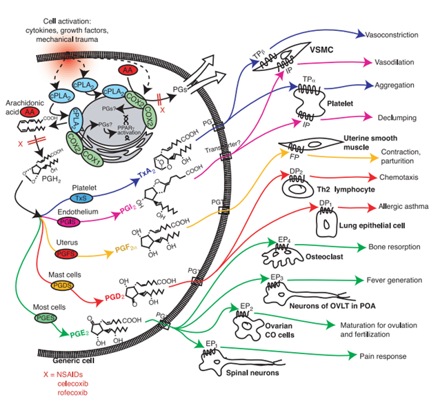
Prostaglandins have been found in almost every tissue and organin humans and other animals. They are produced by almost all nucleated cells. They are autocrine and paracrine lipid mediators that act upon endothelium, platelets, uterine and mast cells. They are usually synthesized in the cell from the essential fatty acids (EFAs). An intermediate arachidonic acid is produced from diacylglycerol via phospholipase-A2, then brought to either the lipoxygenase pathway orthe cyclooxygenase pathway. The lipoxygenase enzyme pathway is active in leukocytes and in macrophages and synthesizes leukotrienes. Alternatively, the cyclooxygenase pathway produces prostacyclin, thromboxane and prostaglandin D, E and F.
There are typically ten known prostaglandin receptors on diverse cell types. Prostaglandins ligate a subclass of cell surface seven-transmembrane receptors, G-protein-coupled receptors (GPCRs). These receptors are termed as DP1-2, EP1-4, FP, IP1-2, and TP, corresponding to the receptors that ligate the corresponding prostaglandin.
These various receptors means that prostaglandins act on a large group of cells and have a wide variety of effects such ascausing constriction or dilation in vascular smooth muscle cells, causing aggregation or disaggregation of platelets, sensitizing spinal neurons to pain, inducing labor, decreasing intraocular pressure, regulating inflammation, regulating calcium movement, regulating hormones, and controlling cell growth, etc.
Prostaglandins Analysis in Creative Proteomics
At Creative Proteomics, we offer metabolomics services, including prostaglandin analysis, designed to meet the diverse needs of our clients in both research and clinical settings. Our services are characterized by their precision, reliability, and customization to ensure that we deliver the highest quality results.
Quantitative Analysis of Prostaglandins
Quantitative analysis is essential for measuring the exact concentration of prostaglandins in various biological samples. Our quantitative analysis service includes:
- Absolute Quantification: Using internal standards, we accurately measure the levels of specific prostaglandins.
- Relative Quantification: Comparing the levels of prostaglandins across different samples to identify changes or trends.
Qualitative Analysis of Prostaglandins
Qualitative analysis focuses on identifying the presence and types of prostaglandins in samples. Our services include:
- Identification of Prostaglandin Species: Detecting different types of prostaglandins present in the sample.
- Structural Characterization: Detailed analysis of the molecular structure of prostaglandins to understand their specific roles and functions.
Metabolite Profiling
Metabolite profiling involves the comprehensive analysis of prostaglandin metabolites, providing insights into metabolic pathways and biological processes. Our metabolite profiling service includes:
- Pathway Analysis: Mapping the metabolic pathways involving prostaglandins to understand their biological roles.
- Biomarker Discovery: Identifying potential biomarkers for disease diagnosis and therapeutic targets.
Custom Analytical Solutions
We understand that every research project has unique requirements. Our custom analytical solutions are tailored to meet specific client needs, including:
- Custom Method Development: Developing and validating custom analytical methods for specific prostaglandins or sample types.
- Specialized Sample Preparation: Tailoring sample preparation techniques to ensure optimal analysis of prostaglandins in various matrices.
Data Analysis and Interpretation
Interpreting the data obtained from prostaglandins analysis is crucial for deriving meaningful insights. Our data analysis and interpretation service includes:
- Statistical Analysis: Applying advanced statistical methods to analyze the data and identify significant findings.
- Bioinformatics Support: Providing bioinformatics tools and expertise to interpret complex data sets and generate actionable insights.
Analytical Techniques for Prostaglandins Analysis
High-Performance Liquid Chromatography (HPLC)
HPLC is a fundamental technique in prostaglandins analysis due to its high resolution, sensitivity, and versatility. At Creative Proteomics, we utilize the Agilent 1260 Infinity II HPLC System for:
- Separation of Prostaglandins: Efficiently separating different prostaglandin species within complex biological samples.
- Quantification: Accurate measurement of prostaglandin concentrations using calibrated standards.
- Detection Methods: Employing various detectors such as UV, fluorescence, and mass spectrometry (MS) to enhance sensitivity and specificity.
Liquid Chromatography-Mass Spectrometry (LC-MS)
LC-MS combines the separation power of liquid chromatography with the detection capabilities of mass spectrometry. Our Thermo Scientific Q Exactive Orbitrap LC-MS/MS system is highly effective for:
- High Sensitivity and Specificity: Detecting and quantifying prostaglandins at very low concentrations.
- Identification of Metabolites: Detailed profiling of prostaglandin metabolites and their biological pathways.
- Structural Elucidation: Providing molecular weight and structural information of prostaglandins and their derivatives.
Gas Chromatography-Mass Spectrometry (GC-MS)
GC-MS is ideal for analyzing volatile and thermally stable prostaglandins. At Creative Proteomics, we use the Agilent 7890B GC System coupled with 5977B MSD for:
- High Resolution: Excellent separation of prostaglandin compounds in complex mixtures.
- Quantitative Accuracy: Precise quantification of prostaglandins in various biological matrices.
- Volatile Compound Analysis: Effective for analyzing prostaglandins and their metabolites that can be vaporized without decomposition.
Tandem Mass Spectrometry (MS/MS)
MS/MS involves multiple stages of mass spectrometry, providing detailed structural information about prostaglandins. Our Thermo Scientific TSQ Altis Triple Quadrupole Mass Spectrometer is beneficial for:
- Structural Characterization: Offering comprehensive insights into the molecular structure of prostaglandins.
- Metabolite Identification: Identifying and characterizing prostaglandin metabolites with high accuracy.
- Enhanced Selectivity: Improving the selectivity and sensitivity of prostaglandin analysis through multiple fragmentation stages.
Ultra-Performance Liquid Chromatography (UPLC)
UPLC is an advanced form of liquid chromatography that provides faster and higher resolution separations. At Creative Proteomics, we use the Waters ACQUITY UPLC System for:
- Rapid Analysis: Significantly reducing analysis time while maintaining high resolution.
- High Throughput: Suitable for large-scale studies and high-throughput screening.
- Enhanced Sensitivity: Improving the detection limits of prostaglandins in various samples.
Sample Requirements for Prostaglandins Analysis
| Sample Type | Recommended Volume/Amount |
|---|---|
| Plasma/Serum | 200-500 µL |
| Urine | 10-20 mL |
| Tissue Homogenates | 50-100 mg |
| Cell Culture Media | 1-2 mL |
| Synovial Fluid | 200-500 µL |
| Amniotic Fluid | 500 µL |
| Cerebrospinal Fluid | 200-500 µL |
| Bronchoalveolar Lavage Fluid | 1-2 mL |
| Saliva | 1-2 mL |
| Milk | 1-2 mL |
Important Considerations:
- Sample Collection: Ensure proper collection and handling to avoid contamination or degradation of prostaglandins.
- Storage Conditions: Store samples at -80°C if they are not analyzed immediately to preserve the integrity of prostaglandins.
- Avoid Repeated Freeze-Thaw Cycles: Minimize freeze-thaw cycles to prevent degradation of prostaglandins.
- Labeling and Documentation: Clearly label samples with relevant information, including sample type, collection date, and any pre-treatment details.
Report
- A full report including all raw data, MS/MS instrument parameters and step-by-step calculations will be provided (Excel and PDF formats).
- Analytes are reported as uM or ug/mg (tissue), and CV's are generally<10%.

PCA chart

PLS-DA point cloud diagram

Plot of multiplicative change volcanoes

Metabolite variation box plot

Pearson correlation heat map
Metabolomic profiling implicates mitochondrial and immune dysfunction in disease syndromes of the critically endangered black rhinoceros (Diceros bicornis).
Journal: Scientific reports
Published: 2023
Background
The study aimed to characterize the metabolome of black rhinoceroses (Diceros spp.) to provide foundational data on serum metabolites across different subspecies, sexes, and health phenotypes. This research is crucial for understanding metabolic variations in ex-situ managed rhinos and could aid in developing diagnostic biomarkers and health management strategies.
Materials & Methods
Study Population and Sample Collection
Fourteen Eastern black rhinos (Diceros b. michaeli) and sixteen Southern black rhinos (Diceros b. minor) from multiple institutions in the United States were included in this study. A total of 97 serum samples were collected longitudinally, approximately every three months, aiming for four samples per rhino within a year. Veterinary staff collected the samples, which were shipped to the Smithsonian's National Zoo & Conservation Biology Institute and stored at −80 °C until analysis.
Untargeted Metabolomic Analysis
Serum samples (0.1 mL aliquots) were sent to Creative Proteomics for untargeted metabolomic analysis using liquid chromatography coupled with mass spectrometry (LC–MS). Standardized protocols were followed for sample preparation and LC–MS analysis.
- Instrumentation: Analysis was performed using an Ultimate 3000LC coupled with the ThermoQ Exactive MS platform.
- Sample Processing: Samples underwent extraction and derivatization according to established protocols before analysis.
- Metabolite Identification: Compound Discover 3.0 software (Thermo) was utilized for metabolite identification based on retention time and ion signals in both positive and negative ionization modes.
- Statistical Analysis: Data were processed using SIMCA-P 14.1 for visualization and outlier detection. Partial least squares discriminant analysis (PLS-DA) and orthogonal partial least squares discriminant analysis (OPLS-DA) were employed to identify significant metabolites distinguishing between subspecies, sexes, and health phenotypes.
- Biomarker Discovery: Significant metabolites were identified based on VIP scores (VIP > 1.5) and t-tests (p < 0.05). Hierarchical clustering, principal component analysis (PCA), and volcano plots were used to visualize and interpret metabolic variations.
Quality Control
Quality control measures included the use of standard reference materials and replicate analyses to ensure data accuracy and reproducibility.
Results
Metabolic Profiles of Eastern and Southern Black Rhinos
Metabolomic analysis revealed distinct serum metabolic profiles between Eastern (Diceros b. michaeli) and Southern (Diceros b. minor) black rhinos. A total of 636 metabolites were consistently detected across all samples, with 352 identified in negative ion mode and 284 in positive ion mode.
 Serum metabolome profile for eastern versus southern subspecies comparison relative to the eastern subspecies.
 Serum metabolome profile for sex comparison relative to females.
 Serum metabolome profile for health phenotype comparison (healthy vs. inflammatory) relative to inflammatory phenotype.
Subspecies Comparison
Differential Metabolite Composition: Principal Component Analysis (PCA) and hierarchical clustering highlighted significant differences in metabolite composition between Eastern and Southern black rhinos. Notably, metabolites involved in lipid metabolism, amino acid derivatives, and oxidative stress pathways showed distinct patterns between the two subspecies.
Specific Metabolites: Metabolites such as arachidonic acid derivatives and iron-related compounds were found to be significantly different between subspecies, suggesting potential metabolic adaptations or differences in physiological responses.
Sex-Based Differences
Sexual Dimorphism in Metabolites: Analysis revealed sex-specific metabolic differences within each subspecies. Male and female black rhinos exhibited distinct metabolic profiles, particularly in metabolites related to hormonal regulation, lipid metabolism, and antioxidant defense mechanisms.
Health Phenotype Analysis
Metabolic Variations in Health Phenotypes: Comparison between healthy and inflammatory phenotype cohorts showed significant alterations in metabolite profiles associated with immune function, oxidative stress, and inflammatory markers. Principal metabolites contributing to phenotype discrimination included cytokines, oxidative stress markers, and metabolic intermediates indicative of inflammatory responses.
Biomarker Potential
Candidate Biomarkers: Several metabolites emerged as potential biomarkers for health status differentiation among black rhinos, highlighting their utility in monitoring health and disease progression in managed populations.
Statistical Analysis
Statistical Significance: Statistical tests, including partial least squares discriminant analysis (PLS-DA) and t-tests, confirmed the robustness of identified biomarkers and their significance in differentiating between subspecies, sexes, and health phenotypes.
 Receiver operating characteristic (ROC) curve-based approach for identifying potential biomarkers and evaluating their performance using area under the curve (AOC).
Reference
- Corder, Molly L., et al. "Metabolomic profiling implicates mitochondrial and immune dysfunction in disease syndromes of the critically endangered black rhinoceros (Diceros bicornis)." Scientific Reports 13.1 (2023): 15464.
What methods are used for Prostaglandins analysis, and what are their advantages and disadvantages?
Prostaglandins are typically analyzed using advanced analytical techniques such as Gas Chromatography-Mass Spectrometry (GC-MS), High-Performance Liquid Chromatography (HPLC), and Enzyme-Linked Immunosorbent Assay (ELISA).
- GC-MS: This method offers high specificity and sensitivity, capable of quantifying various prostaglandin types accurately. However, it requires complex sample preparation involving derivatization.
- HPLC: Known for its good sensitivity and specificity, HPLC requires specialized equipment and longer analysis time compared to other methods.
- ELISA: Provides high sensitivity and is suitable for clinical samples. However, it may have limited specificity and potential cross-reactivity with structurally similar compounds.
How do you extract and measure Prostaglandins from biological samples like serum or urine?
The extraction and measurement of prostaglandins from biological samples involve several steps:
Sample Collection: Biological samples such as serum or urine are collected under controlled conditions to prevent degradation or contamination of prostaglandins.
Extraction:
- Organic Solvent Extraction: Prostaglandins are often extracted using organic solvents like ethyl acetate or methanol. This process involves mixing the sample with the solvent, followed by centrifugation to separate the organic phase containing the prostaglandins.
- Solid-Phase Extraction (SPE): SPE is a more refined technique where the sample is passed through a column containing a solid adsorbent material that selectively binds prostaglandins. The bound prostaglandins are then eluted using an appropriate solvent.
Measurement:
- Post-extraction, the prostaglandins are quantified using techniques such as GC-MS, HPLC, or ELISA. The choice of method depends on the required sensitivity, specificity, and available instrumentation. GC-MS and HPLC offer high specificity and can separate and quantify multiple prostaglandins, while ELISA is more straightforward and suitable for high-throughput screening.
What are the applications of Prostaglandins analysis in inflammation and immune research?
Prostaglandins play a pivotal role in inflammation and immune responses, making their analysis critical in these research areas:
Inflammation: Prostaglandins, particularly PGE2, are key mediators of inflammation. By analyzing their levels, researchers can gauge the extent and progression of inflammatory responses. This is crucial for understanding diseases like rheumatoid arthritis, where prostaglandins contribute to pain and swelling.
Immune Response: Prostaglandins modulate immune cell functions, such as cytokine production, leukocyte migration, and T-cell differentiation. Their analysis helps in deciphering the complex interactions between prostaglandins and immune cells, which is vital for developing targeted therapies for autoimmune diseases and chronic inflammatory conditions.
What is the biosynthesis pathway of Prostaglandins, and what factors influence their synthesis and release?
Prostaglandins are synthesized from arachidonic acid through the cyclooxygenase (COX) pathway:
Biosynthesis Pathway: The pathway begins with the release of arachidonic acid from cell membrane phospholipids by phospholipase A2. Arachidonic acid is then converted to prostaglandin H2 (PGH2) by the action of COX-1 and COX-2 enzymes. PGH2 serves as a precursor for various prostaglandins (e.g., PGE2, PGD2, PGF2α) and thromboxanes, depending on the specific synthases present in the cells.
Regulation Factors:
- Cytokines and Growth Factors: Pro-inflammatory cytokines (e.g., IL-1β, TNF-α) and growth factors can upregulate COX-2 expression, enhancing prostaglandin synthesis.
- Hormonal Influence: Hormones such as glucocorticoids can suppress COX-2 expression, thereby reducing prostaglandin production.
- Inflammatory Stimuli: Infection, tissue injury, and other inflammatory stimuli can activate phospholipase A2, leading to increased release of arachidonic acid and subsequent prostaglandin synthesis.
In which diseases and pathophysiological processes do Prostaglandins play a critical role?
Inflammatory Diseases: In conditions like rheumatoid arthritis and inflammatory bowel disease, prostaglandins contribute to inflammation, pain, and tissue damage.
Cancer: Prostaglandins, especially PGE2, play roles in tumor growth, angiogenesis, and metastasis. They can promote the proliferation of cancer cells and modulate the tumor microenvironment.
Cardiovascular Diseases: Prostaglandins regulate vascular tone and platelet aggregation, influencing blood pressure and cardiovascular homeostasis. Dysregulation of prostaglandin synthesis is associated with hypertension, atherosclerosis, and thrombosis.
Reproductive Health: Prostaglandins are crucial in reproductive processes, including ovulation, menstruation, and labor. Abnormal prostaglandin levels can lead to disorders like dysmenorrhea and preterm labor.
How do you interpret Prostaglandins measurement results, and how are they correlated with physiological states or diseases?
Interpreting prostaglandin measurements involves correlating their levels with specific physiological or pathological conditions:
Elevated Levels: High prostaglandin levels typically indicate active inflammation or disease exacerbation. For instance, elevated PGE2 levels in synovial fluid are indicative of active rheumatoid arthritis.
Clinical Correlation: By comparing prostaglandin levels with clinical symptoms and other biomarkers, clinicians can assess disease severity, monitor treatment response, and predict disease progression. For example, decreasing prostaglandin levels during treatment may suggest effective control of inflammation.
Baseline and Comparative Analysis: Establishing baseline prostaglandin levels in healthy individuals allows for comparative analysis in diseased states. This helps in identifying deviations that signify pathological changes.
How are changes in Prostaglandins associated with drug treatments or nutritional interventions?
Drug Treatments:
- Nonsteroidal Anti-Inflammatory Drugs (NSAIDs): NSAIDs inhibit COX enzymes, reducing prostaglandin synthesis and alleviating inflammation and pain. Monitoring prostaglandin levels can help evaluate the efficacy of NSAID therapy and optimize dosage.
- Corticosteroids: These drugs suppress COX-2 expression and phospholipase A2 activity, leading to decreased prostaglandin production. Changes in prostaglandin levels during corticosteroid therapy can reflect the anti-inflammatory effects of the treatment.
Nutritional Interventions:
- Omega-3 Fatty Acids: Diets rich in omega-3 fatty acids can modulate arachidonic acid metabolism, reducing pro-inflammatory prostaglandin synthesis while increasing the production of anti-inflammatory eicosanoids. This dietary approach can be beneficial in managing chronic inflammatory conditions.
- Antioxidants and Polyphenols: Nutrients with antioxidant properties can influence prostaglandin synthesis by modulating inflammatory pathways and reducing oxidative stress.
Thermotolerance capabilities, blood metabolomics, and mammary gland hemodynamics and transcriptomic profiles of slick-haired Holstein cattle during mid lactation in Puerto Rico
Contreras-Correa, Z. E., Sánchez-Rodríguez, H. L., Arick II, M. A., Muñiz-Colón, G., & Lemley, C. O.
Journal: Journal of Dairy Science
Year: 2024
https://doi.org/10.3168/jds.2023-23878
Metabolomic profiling implicates mitochondrial and immune dysfunction in disease syndromes of the critically endangered black rhinoceros (Diceros bicornis)
Corder, M. L., Petricoin, E. F., Li, Y., Cleland, T. P., DeCandia, A. L., Alonso Aguirre, A., & Pukazhenthi, B. S.
Journal: Scientific Reports
Journal: 2023
https://doi.org/10.1038/s41598-023-41508-4
Transcriptomics, metabolomics and lipidomics of chronically injured alveolar epithelial cells reveals similar features of IPF lung epithelium
Willy Roque, Karina Cuevas-Mora, Dominic Sales, Wei Vivian Li, Ivan O. Rosas, Freddy Romero
Journal: bioRxiv
Year: 2020
https://doi.org/10.1101/2020.05.08.084459
The Computational Approach to Plant Oxylipins Profiling: Databases and Tools
Hamadani, A., Ganai, N. A., & Mansoor, S.
Journal: In Phyto-Oxylipins
Year: 2023