Exosome Lipidomics Service
Creative Proteomics provides comprehensive exosome lipidomics services using advanced platforms like LC-MS, UHPLC, Shotgun Lipidomics, and GC-MS to analyze lipid compositions, identify biomarkers, and support biological and clinical research.
Submit Your Request Now
×
- Define
- What We Provide
- List of Analytes Detectable
- Technology Platforms
- Demo
- FAQs
- Case Study
- Publications
What Is Exosome Lipidomics?
Exosome lipidomics refers to the study of lipids present in exosomes, which are small extracellular vesicles (typically 50-150 nm in size) released from cells. These vesicles originate from intracellular compartments called Multivesicular Bodies (MVBs, or Late Endosomes) and are involved in cell-to-cell communication by transferring various biomolecules, including lipids, proteins, DNA, RNA, mRNA, and miRNA.
Exosomes have a unique lipid composition that is similar to lipid rafts, which are specialized microdomains in cell membranes. They contain higher lipid content and exhibit greater resistance to detergents compared to other types of extracellular vesicles. The lipid molecules in the exosome membrane play a critical role in mediating interactions with receptors on target cells, such as G-protein coupled receptors, and can influence intracellular signaling pathways and biological processes in recipient cells. In addition to their structural role in exosome membranes, lipids are also crucial for exosome formation and their release into the extracellular space.
Understanding the lipid composition of exosomes is essential for deciphering their biology, as well as for exploring their potential in medical applications. Exosome lipidomics enables researchers to study vesicle biogenesis, identify lipid-based biomarkers, and investigate their role in disease monitoring and progression.
What Our Exosome Lipidomics Service Offers
Our exosome lipidomics analysis provides comprehensive analysis of lipid compositions within exosomes, offering insights into their structural and functional roles. We utilize advanced lipidomics technologies to deliver high-resolution profiling of exosomal lipids, allowing for in-depth examination of exosome biogenesis, lipid-based biomarkers, and signaling pathways.
Key Features of Our Service
- Comprehensive Lipid Profiling: We analyze a broad spectrum of lipid classes within exosomes, ensuring a complete lipidomic profile that includes phospholipids, sphingolipids, sterols, and more.
- High Sensitivity and Specificity: Using cutting-edge mass spectrometry, our service offers precise and sensitive detection of lipid species, enabling accurate identification even in low-abundance samples.
- Biological Insights: Our analysis reveals lipid interactions that are essential for exosome formation and release, shedding light on exosome-related signaling mechanisms in target cells.
- Biomarker Discovery: We help identify potential lipid-based biomarkers, facilitating research in diagnostics and therapeutic monitoring.
- Data Interpretation and Support: Our experts provide in-depth data interpretation and consultative support to help you fully understand and apply the lipidomic data to your research or clinical goals.
List of Analytes Detectable in Exosome Lipidomics
| Lipid Class | Lipid Subtypes |
|---|---|
| Phospholipids | - Phosphatidylcholine (PC) - Phosphatidylethanolamine (PE) - Phosphatidylserine (PS) - Phosphatidylinositol (PI) - Phosphatidylglycerol (PG) - Phosphatidic Acid (PA) |
| Sphingolipids | - Sphingomyelin (SM) - Ceramides (Cer) - Glycosphingolipids (e.g., Glucosylceramide, Lactosylceramide) |
| Glycerolipids | - Monoacylglycerol (MAG) - Diacylglycerol (DAG) - Triacylglycerol (TAG) |
| Sterol Lipids | - Cholesterol - Cholesteryl Esters (CE) |
| Fatty Acids | - Free Fatty Acids (FFA) - Fatty Acyl-CoA |
| Lysophospholipids | - Lysophosphatidylcholine (LPC) - Lysophosphatidylethanolamine (LPE) - Lysophosphatidylserine (LPS) |
| Other Lipid Types | - Gangliosides - Prenol Lipids - Ether-linked Lipids (e.g., Plasmalogens) |
Exosome Lipidomics Analysis Technology Platforms
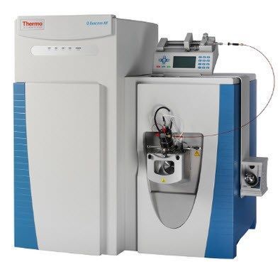
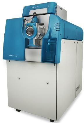
Liquid Chromatography-Mass Spectrometry (LC-MS): Provides high-resolution, accurate mass measurements, ideal for detecting and quantifying a wide range of lipid species in exosomes.
Model: Thermo Scientific™ Q Exactive™ HF-X Hybrid Quadrupole-Orbitrap™ Mass Spectrometer
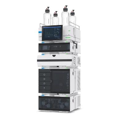
Ultra-High-Performance Liquid Chromatography (UHPLC): Ensures excellent separation of lipid molecules, enhancing the detection sensitivity and improving analytical precision in complex samples.
Model: Agilent 1290 Infinity II UHPLC
Shotgun Lipidomics with Direct Infusion-MS: Offers rapid, high-throughput analysis, ideal for broad lipid coverage and detailed profiling in large sample sets.
Model: AB Sciex TripleTOF™ 5600+ System
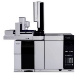
Gas Chromatography-Mass Spectrometry (GC-MS): Specially used for fatty acid analysis, providing robust data for lipid characterization and quantification.
Model: Agilent 7890B GC with 5977B MSD
Workflow of Exosome Lipidomics Analysis

Demo

PCA chart

PLS-DA point cloud diagram

Plot of multiplicative change volcanoes

Metabolite variation box plot

Pearson correlation heat map
FAQs
What sample quality is required for exosome lipidomics analysis?
For optimal results, exosome samples should be free of contamination from cellular debris, proteins, or non-exosomal vesicles. Purification through ultracentrifugation or commercial exosome isolation kits is recommended, as this ensures that lipidomic profiles reflect genuine exosomal content. High-quality isolation prevents background noise in mass spectrometry, leading to more accurate lipid quantification and identification.
How does exosome lipidomics analysis control for sample variability?
Sample variability is addressed through rigorous quality control (QC) steps. Our protocols include the use of internal lipid standards to normalize data and ensure consistent measurements across samples. Additionally, replicates are run to account for any instrumental or extraction variances. For human-derived samples, biological replicates (from different individuals or batches) are encouraged to capture inherent variability in biological systems.
Are there specific lipids that can only be detected in fresh samples?
Yes, certain lipid species, such as polyunsaturated fatty acids (PUFAs), are highly sensitive to oxidation and can degrade if samples are not properly stored. Storing samples at -80°C immediately after collection and minimizing freeze-thaw cycles are critical to preserving these labile lipids. For highly sensitive studies, we recommend analyzing samples within a few months of collection and using antioxidants during sample preparation if possible.
How are low-abundance lipids in exosomes detected?
Our platform employs advanced mass spectrometry, which allows for the detection of lipids at very low concentrations. The instruments used (e.g., Q Exactive HF-X and TripleTOF 5600+) offer high sensitivity, making it possible to identify and quantify trace lipids. In addition, specialized techniques like targeted lipidomics can focus on specific low-abundance lipid classes, enhancing detection sensitivity and accuracy.
How do you differentiate between exosomal lipids and other vesicular lipids?
Exosomal lipids have unique compositions, resembling lipid rafts with higher cholesterol, sphingomyelin, and ceramide content compared to other vesicles. To ensure specificity, samples are carefully purified, and lipid profiles are cross-referenced with known exosomal lipid markers. Mass spectrometry data is analyzed against established lipidomic databases to accurately identify exosome-specific lipids and differentiate from other vesicle types.
Can exosome lipidomics be used to identify disease-specific lipid biomarkers?
Yes, exosome lipidomics is a powerful tool for discovering lipid-based biomarkers associated with diseases. By comparing lipid profiles of exosomes from diseased versus healthy samples, we can identify lipid species with differential abundance. These lipid changes can serve as potential biomarkers for disease states or progression. For robust biomarker discovery, it's important to use a large, well-characterized cohort to ensure statistical significance and reproducibility.
How is lipidomics data interpreted and validated?
Data interpretation involves complex statistical analyses, such as principal component analysis (PCA) or cluster analysis, to identify lipid patterns and correlations with biological conditions. Our team provides detailed interpretation, identifying key lipids and their potential functional roles. Validation can involve cross-referencing findings with published databases or conducting targeted lipidomics on separate samples to confirm the results.
Learn about other Q&A .
Case Study
Lipidomic Profiles of Plasma Exosomes Identify Candidate Biomarkers for Early Detection of Hepatocellular Carcinoma in Patients with Cirrhosis
Journal: Cancer Prevention Research (Phila)
Published: 2021
- Background
- Materials and Methods
- Results
Hepatocellular carcinoma (HCC), a leading cause of cancer-related deaths, has a poor prognosis with a 5-year survival rate below 20%. Early detection in high-risk patients, such as those with cirrhosis, is crucial to improving outcomes. Current surveillance methods like ultrasound and serum α-fetoprotein (AFP) are inadequate due to low sensitivity and specificity, resulting in a high proportion of late-stage diagnoses.
Exosomes, small membrane-bound vesicles found in body fluids, have emerged as promising noninvasive biomarkers and mediators of cancer progression. They contain proteins, lipids, and nucleic acids that reflect biological changes in disease states. This study leverages ultra–high-resolution mass spectrometry to analyze lipidomic differences in circulating exosomes between cirrhotic patients with and without HCC. It identifies potential biomarkers for early HCC detection and explores altered metabolic pathways contributing to cancer progression.
Participants
- 72 cirrhotic patients (31 with HCC, 41 without HCC), matched by age, gender, and etiology.
- Diagnoses confirmed using established criteria; all HCCs were treatment-naïve.
Exosome Isolation
- Plasma samples underwent ultracentrifugation to isolate exosomes, which were snap-frozen and stored.
- Lipids extracted and analyzed using ultra–high-resolution mass spectrometry.
- Chromatography and mass spectrometry optimized for lipid detection and quantification.
Statistical Analysis
- Lipid data normalized and analyzed for differences between HCC and non-HCC groups.
- Logistic regression used to identify lipid classes associated with HCC.
- Pathway enrichment analysis performed to explore biological significance.
In this study, plasma samples were collected from 72 cirrhosis patients, including 31 with hepatocellular carcinoma (HCC) and 41 without (non-HCC). Exosomes were isolated from the plasma samples, and lipidomic profiling was performed using ultra-high-resolution mass spectrometry. No significant differences in total lipids were observed between HCC and non-HCC plasma, but a significant decrease in total lipids was noted in exosomes from HCC patients compared to non-HCC patients. In total, 2,864 lipid species belonging to 52 classes were identified, with triglycerides (TG) and phosphatidylcholines (PC) being the most abundant lipid classes.
Principal component analysis (PCA) revealed distinct lipid profiles between exosomes and plasma, as well as between HCC and non-HCC groups. HCC exosomes exhibited significant enrichments in lipid classes such as dilysocardiolipins (DLCL), cardiolipins (CL), sphingosines (SPH), and lysophosphatidylserines (LysoPS). Specific lipid species like DLCL, CL, and SPH were major contributors to the enrichment, with their high abundance strongly associated with HCC. Logistic regression analysis confirmed that increased levels of SPH, DLCL, LysoPS, and OAHFA were strongly associated with HCC, even after adjusting for potential confounders such as age, gender, and BMI.
On the other hand, lipid classes such as gangliosides (GD1a), fatty acids (FA), sulfatides (ST), and acylglucosyl sitosterol esters (AcHexSiE) were depleted in HCC exosomes. These lipid species were either less abundant or absent in HCC exosomes compared to non-HCC exosomes, with a strong association between the lack of ST and AcHexSiE detection and HCC. Additionally, a pathway analysis revealed that changes in lipid profiles in HCC exosomes were linked to several biological pathways, including glycerophospholipid metabolism, retrograde endocannabinoid signaling, and ferroptosis.
These findings highlight the potential of lipidomic profiling of exosomes as a diagnostic tool for differentiating HCC from cirrhosis, providing insights into the lipid metabolism alterations associated with liver cancer.
 PCAs of plasma and exosomal lipid profiles from HCC and non-HCC patients show four distinct clusters.
Volcano plot of lipid classes in HCC exosomes versus non-HCC ex osomes.
Reference
- Verderio, Claudia, Martina Gabrielli, and Paola Giussani. "Role of sphingolipids in the biogenesis and biological activity of extracellular vesicles." Journal of lipid research 59.8 (2018): 1325-1340.
- Multi‐omics identify xanthine as a pro‐survival metabolite for nematodes with mitochondrial dysfunction. 2019. https://doi.org/10.15252/embj.201899558
- Microbial dysbiosis associated with impaired intestinal Na+/H+ exchange accelerates and exacerbates colitis in ex-germ free mice. 2018. https://doi.org/10.1038/s41385-018-0035-2
- Annexin A2 modulates phospholipid membrane composition upstream of Arp2 to control angiogenic sprout initiation. 2023. https://doi.org/10.1096/fj.202201088R
- B cell-intrinsic epigenetic modulation of antibody responses by dietary fiber-derived short-chain fatty acids. 2020. https://doi.org/10.1038/s41467-019-13603-6
- Laboratory evaluation of larvicidal and oviposition deterrent properties of edible plant oils for potential management of Aedes aegypti (Diptera: Culicidae) in drinking water containers. 2019. https://doi.org/10.1093/jme/tjz021
Publications
Here are some publications in Lipidomics research from our clients: