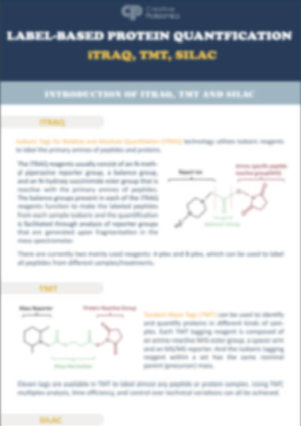
- Service Details
- Case Study
What is SILAC?
The Stable Isotope Labeling by Amino Acids in Cell Culture (SILAC) technique is a widely utilized metabolic labeling strategy in quantitative proteomics. The technology enables accurate and replicable determination and measurement of relative differential alterations in protein abundance. It involves the gradual replacement of "light" normal 12C or 14N amino acids (AA) with "heavy" 13C- or 15N-labeled AA during protein translation in vivo. The SILAC technique can be widely applied for comprehensive identification, characterization, and quantitation of proteins in complex organism mechanisms.
The principle of SILAC
The SILAC labeling technique relies on cell culture, where stable isotopes labeled AA substitute for natural amino acids in the proteome during the process of protein metabolism. The essential AA leucine (L), lysine (K), and methionine (M), as well as the non-essential AA arginine (R), are indispensable for various cultured cell lines. They have all been successfully utilized in SILAC labeling. The most utilized isotopically labeled AA in SILAC experiments are 13C and 15N-labeled K and R, as trypsin, a specific proteolytic enzyme, cleaves at the carboxyl side of K and R residues. Therefore, the combinations of SILAC labeling with K and R and tryptic digestion allows for overall proteomic quantification.
 Figure 1. The structure of frequently employed SILAC Label Reagents for K and R.
The fundamental workflow of SILAC
 Figure 2. The workflow of SILAC - based proteomics analysis [1].
For instance, two groups of identical cells are cultured in the same complete medium, except that one medium contains common K and R without any labeling, referred to as the 'light' medium, while the other complete medium contains labeled K and R such as K4R6, K8R10, etc., known as the 'heavy' medium. After 5-6 generations of cell culture, the heavy labeled amino acids are fully incorporated into the proteome (>99%), resulting in a discernible mass shift between the two groups of identical proteins. However, there is no observed disparity in other chemical properties. The two groups of cells were harvested in equal proportions, followed by protein extraction and subsequent equal mixing in a 1:1 mass ratio. The resulting mixture was then subjected to tryptic digestion and subsequent analysis using LC-MS/MS. After analyzing the MS data and the intensity ratio of introduced isotope-labeled peptides to unlabeled peptides, we could determine the quantification of identical proteins under two or three treatment conditions.
Applications
- Distinguish quantitative variations in protein expression across samples.
a. monitor quantitative differences at the protein level under different conditions.
b. identify differentially expressed proteins in organelles, such as nucleus, nucleolus. - Monitor protein turnover.
- Investigate the dynamic changes of protein post-translational modifications (PTMs).
- Distinguish protein-protein interactions (PPIs) networks to determine the true interacting partners and directly discriminate them from unlabeled environmental contaminants.
Advantages of SILAC-based Proteomics Analysis Service
- High labeling efficiency: The in vivo labeling efficiency can reach as high as 99% and remains unaffected by lysate or other factors.
- High sensitivity: The sample requirements are small, usually only tens of micrograms of protein per sample.
- High precision: The multiple samples are mixed, digested simultaneously, and subsequently identified. This sequential approach ensures consistent treatment of the samples, minimizing the influence of experimental procedures and equipment variations, thereby enhancing precision and reproducibility.
- Comprehensive services: Combined with other technologies, we can provide PPIs and PTMs analysis.
Our SILAC based Quantitative Proteomics Service
At Creative Proteomics, we offer a comprehensive SILAC service encompassing various cells culture under diverse conditions, as well as advanced proteomics analysis for SILAC labeled cells. Additionally, we can also offer the super-SILAC technique for tissue or exosome analysis. Moreover, we could provide PPIs and PTMs analysis utilizing the SILAC strategy to enhanced throughput and cost-effectiveness in quantitative proteomics experiments, as well as improved relative quantitation.
In Creative Proteomics, we are professional enough to provide a feasible experimental scheme tailored to your specific requirements to complete the SILAC quantitative proteomics research. Our experienced proteomics research team, strict quality control system, together with ultra-high resolution detection system and professional data pre-processing and analysis capability, ensuring high-quality and reliable results for quantitative proteomics analysis.
Technologies Platform
- Professional detection and analysis capability: Experienced research team, strict and matured techniques.
- High stability and reproducible: Obtain consistent and reproducible inter- and intra- assay results for data analysis.
- High labeling efficiency: The labeling efficiency of SILAC reagents is greater than 99%.
- High throughput: LC-MS/MS platform can identify and quantify thousands of proteins simultaneously.
- High resolution and sensitivity: Triple TOF 5600, Q-Exactive, Q-Exactive HF, Orbitrap Fusion™ Tribrid™.
Samples requirement
Diverse unlabeled cell samples and samples at any stage post SILAC labeling is within our capability.
- For SILAC labeled cells,
Cell: suspension cell > 1 x 10⁷;
adherent cell > 1 x 10⁷;
microorganism > 50 mg or 2 x 10⁷ cell;
Protein extract: Total protein >1 mg and concentration >1 μg/μL.
Note: If you provide protein lysate, please inform the buffer components whether it contains thiourea, SDS, or strong ion salts to ensure the test results. In addition, the sample should not contain components such as nucleic acids, lipids, and polysaccharides, which will affect the separation effect. - For unlabeled cells,
we will elaborately design the experimental protocols according to your needs. The quantity of cells required should be sufficient for resuscitation and cultivation. - For unlabeled protein samples, we could employ super-SILAC method,
Body fluid: serum/plasma > 500 μL;
Tissue: animal tissue > 50 mg;
fresh plant > 100 mg;
Results Delivery
- Detailed report, including experiment procedures, parameters, etc.;
- Raw data and data analysis results (e.g. proteins & peptides identified and quantified results, downstream bioinformatics analysis results, such as volcano plot, GO, PCA, KEGG, Protein interaction networks, etc.).
How to place an order
Please feel free to contact us via email for a comprehensive discussion regarding your specific requirements. Customer service representatives are available round the clock, seven days a week.

Reference
- Chen X, Wei S, Ji Y, et al. Quantitative proteomics using SILAC: principles, applications, and developments. Proteomics, 2015, 15(18): 3175-3192.
Improved SILAC quantification with data independent acquisition to investigate bortezomib-induced protein degradation
Journal: Journal of Proteome Research
Published: 2021
Main Technology: SILAC, data-dependent acquisition (DDA)
Background: We determine empirically that using DIA achieves similar peptide detection numbers as DDA and that DIA improves the quantitative accuracy and precision of SILAC by an order of magnitude. Finally, we apply SILAC-DIA-MS to determine protein turnover rates of cells treated with bortezomib, a 26S proteasome inhibitor FDA-approved for multiple myeloma and mantle cell lymphoma.
Pulse SILAC (pSILAC) is the predominant method to study protein turnover at the proteome scale, enabling the discrimination of preexisting and newly synthesized proteins. Using pSILAC, the cell's translational machinery is used to incorporate stable isotope labeled amino acids present in the cell media, thereby marking newly synthesized proteins. Over time, as proteins are synthesized and degraded (also called protein turnover), the levels of the newly synthesized proteins increase while the preexisting proteins decline allowing protein half-lives to be determined. This approach has been used to explore proteostasis in a number of biological systems, including differentiation, cancer homeostasis, drug treatment response, and the accumulation of "old" proteins in aging.
Methods: Comparison of PSM and peptide detections in SILAC-DDA vs SILAC-DIA.
Results: Here, we develop a SILAC-DIA-MS workflow using free, open-source software. We observe that SILAC-DIA produces more sensitive protein turnover models. Of the proteins determined differentially degraded by both acquisition methods, we find known ubiquitin-proteasome degrands such as HNRNPK, EIF3A, and IF4A1/EIF4A-1, and a slower turnover for CATD, a protein implicated in invasive breast cancer. With improved quantification from DIA, we anticipate this workflow making SILAC-based experiments like protein turnover more sensitive.
 Figure 1. An approach for quantifying pulse SILAC peptides using free, open-source software.