What Are Aldehydes
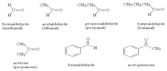
"Aldehydes" constitute a category of organic compounds primarily distinguished by the existence of a carbon atom engaged in a double bond with an oxygen atom, often referred to as the carbonyl group. Simultaneously, this carbon atom forms single bonds with both a hydrogen atom and an additional atom or molecular group, conventionally represented as 'R.' The carbonyl group, marked by the double bond connecting carbon and oxygen, is a unifying feature universally present in all aldehydes.
Many aldehydes are notable for their pleasant odors, and they are generally synthesized from alcohols through a process involving dehydrogenation, where hydrogen is removed. This synthesis process contributes to the nomenclature "aldehyde."
Aldehydes can be further classified based on factors such as their carbon chain length, structural characteristics, and the functional groups they contain. Common examples of aldehydes include formaldehyde, acetaldehyde, and butanal, categorized by their carbon chain length and specific functional groups. Furthermore, they can be subdivided into categories such as aliphatic aldehydes, aromatic aldehydes, and terpenoid aldehydes (which can be further divided into monoterpenoid aldehydes and sesquiterpenoid aldehydes) based on their structural features.
Why Analyze Aldehydes
The analysis of "Aldehydes" (aliphatic carbonyl compounds) holds significant importance in scientific and medical research for several compelling reasons and applications:
Environmental and Occupational Health Monitoring: Aldehydes, such as formaldehyde, can be present in the environment, including airborne formaldehyde and aldehydes generated during industrial and chemical processes. Analyzing these compounds aids in monitoring exposure levels in environmental and occupational settings, thereby assessing potential health risks.
Quality Control of Food and Drinking Water: Certain aldehydes may occur in food and drinking water, especially those introduced through food processing or water treatment processes. Detecting aldehydes ensures the safety and quality of food and drinking water, safeguarding public health.
Biomedical Research Significance: Aldehydes play a pivotal role in biomedical research, particularly concerning disease mechanisms and biomarkers. For instance, analyzing aldehyde modifications in DNA and proteins can elucidate the molecular basis of diseases, contributing to diagnostic and therapeutic research.
Investigating Environmental and Health Effects: Analyzing aldehyde exposure is instrumental in studying potential impacts on human health and ecosystems. This aids in determining the roles aldehydes may play in disease development, immune system function, and ecosystem health.
Quality Control and Product Development: In industrial and manufacturing processes, aldehyde analysis serves as a critical tool for quality control, ensuring that products meet specified standards. Furthermore, aldehydes serve as intermediates in synthesis and chemical reactions, making their detection and quantification crucial in the chemical and pharmaceutical industries.
Indoor Air Quality Assessment: Aldehydes like formaldehyde can be released into indoor air, particularly from building materials, furniture, and household items. Elevated concentrations of indoor aldehydes can lead to indoor air pollution. Detection aids in assessing indoor air quality and implementing necessary corrective measures.
Methods for Aldehyde Analysis
Gas Chromatography-Mass Spectrometry (GC-MS)
GC-MS has been widely employed for aldehyde analysis. For volatile aldehydes, direct GC analysis can be performed in conjunction with headspace (HS) extraction without the need for derivatization. However, many reported methods utilize derivatization to enhance the volatility, stability, and ionization efficiency of aldehydes. In contrast to the 2,4-dinitrophenylhydrazine (DNPH) derivatization reagents commonly used in LC or LC-MS, hydrazine/amine derivatization reagents with chlorine or fluorine on the benzene ring exhibit significant advantages in GC or GC-MS aldehyde analysis. Commonly used derivatization reagents include 2,4,6-trichlorophenylhydrazine (TCPH), pentafluorophenylhydrazine (PFPH), 2,3,4,5,6-pentafluorobenzylhydroxylamine (PFBHA), among others. These reagents improve the volatility and thermal stability of derivatives while generating more stable and specific detection ions.
Liquid Chromatography-Mass Spectrometry (LC-MS)
Conventional Derivatization LC-MS/MS Analysis LC-MS/MS with tandem mass spectrometry capability has become the mainstream approach for LC-MS analysis. This is primarily due to the significant improvement in detection specificity, achieved through multiple reaction monitoring (MRM) mode, which reduces interference and provides rapid, multi-channel analysis. Since the ionization efficiency of analytes greatly affects MS instrument sensitivity, derivatization techniques are often employed in LC-MS/MS to enhance the low ionization efficiency of aldehydes. Derivatization reagents for LC-MS typically possess high electron affinity or contain intramolecular positive charges. Derivatization reagents used in reported HPLC-UV and -FLD methods are mostly suitable for LC-MS due to their ionizable structural components. DNPH, based on the hydrazine reactive group, is a representative derivatization reagent widely used for aldehyde derivatization in LC-MS analysis. Other commonly used derivatization reagents include dansyl hydrazine (DnsHz), ortho-phenylenediamine, D-cysteine, 9,10-phenanthrenequinone (PQ), 3-nitrophenylhydrazine, and 3,4-diaminodiphenylketone, among others.
Chemical Isotope Labeling LC-MS/MS Targeted Analysis
External standard quantification is often affected by mass spectrometer signal fluctuations, leading to imprecise quantification. The addition of internal standards for quantitative purposes can provide effective calibration. However, the selection of suitable internal standards is often challenging, making it difficult to find compounds that meet the chromatographic separation, mass spectrometry response, and qualitative/quantitative requirements. Isotope internal standards can address the aforementioned issues. Nevertheless, it is challenging for routine laboratories to synthesize suitable isotope internal standards that meet requirements. Commercially available isotope internal standards are costly, and many analytes, including unknown metabolites and trace metabolic products, lack commercially available isotope internal standards. Synthesizing high-purity isotope-labeled derivatization reagents to derive isotope internal standards from standards or mixed samples can be used for targeted quantification, metabolic difference analysis, non-targeted scanning, and other applications in LC-MS, representing a frontier and key technology in the field of metabolomics analysis.
Chemical Isotope Labeling LC-MS Un-Targeted Scanning and Relative Quantification
Conventional derivatization or chemical isotope labeling techniques provide excellent sensitivity and specificity for LC-MS/MS quantitative analysis but lack the capability to screen unknown aldehydes. Metabolomics analysis faces a significant demand for non-targeted scanning and biomarker screening from a large number of biological samples. By combining chemical isotope labeling technology with various scanning modes (MRM, neutral loss scanning, precursor ion scanning, full scanning, etc.) in LC-MS, aliphatic, carboxylic, hydroxy, and amino groups of sample analytes can be divided into subgroups, significantly improving the sensitivity, coverage, and accuracy of non-targeted scanning. This represents a cutting-edge area of research in metabolomics analysis.
Derivatization Liquid Chromatography-High-Resolution Mass Spectrometry Analysis
Liquid chromatography-high-resolution mass spectrometry (LC-HRMS) has become a future direction in LC-MS analysis. Metabolomics analysis methods based on high-resolution mass spectrometry provide accurate mass information for parent ions and MS/MS fragment ions, enabling reliable identification of detected metabolites in complex biological matrices.

Aldehydes Quantitative Analysis At Creative Proteomics
Creative Proteomics provides tailored services for analysis and determination of aldehydes using chromatography techniques (GC-MS and HS-GC-MS)
| Aldehydes Quantified in Our Service | ||
|---|---|---|
| 10 Aldehyde | 4-HHE | 4-HNE |
| Acrolein | C2 Aldehyde | C3 Aldehyde |
| C4 Aldehyde | C5 Aldehyde | C6 Aldehyde |
| C7 Aldehyde | C8 Aldehyde | Crotonaldehyde |
| Malondialdehyde (MDA) | Formaldehyde | |
Sample requirement
| Sample Type | Minimum Amount | Typical Amount |
|---|---|---|
| Animal and Clinical Tissue Samples | 5-10mg | 50mg |
| Blood Samples (Serum, Plasma, Whole Blood) | 10μL | 50μL |
| Urine Samples | 10μL | 50μL |
| Fecal and Gastrointestinal Content | 10mg | 25mg |
| Fluid Samples (Cerebrospinal Fluid, Saliva, etc.) | 5-10μL | 25μL |
| Plant Tissue Samples (Roots, Stems, Leaves, Flowers, Fruits, etc.) | 10mg | 50mg |
| Cell and Microbial Cells | 1*10^5 cells | 1*10^6 cells |
| Culture Media and Fermentation Broth | 10μL | 50μL |
Report
A detailed technical report will be provided at the end of the whole project, including the experiment procedure, MS instrument parameters.
Analytes are reported as uM/ml, while CV's are generally 10%.
The name of the analytes, abbreviation, formula, molecular weight and CAS# would also be included in the report.