Tumor microenvironment usually refers to the internal and external environment in which tumor occurs, grows and metastasizes with tumor cells. The tumor microenvironment includes the structural, functional and metabolic components of the tumor tissue, and an in-depth understanding of the complex components and functions of the tumor microenvironment is of great clinical significance for tumor prevention and treatment.
The tumor microenvironment mainly consists of tumor cells, their surrounding immune cells, tumor-associated fibroblasts, microvasculature, nearby interstitium, and various cytokines and chemokines. Certain cell or protein functions interact with each other, promoting or inhibiting each other, and the functions performed in different environments may be different. Proteomics analysis can help the progress of research related to tumor microenvironment.
Here are some case studies.
Case 1 How exosome proteomic analysis can help tumor microenvironment research (1)
Tumor-Derived Exosomes play a decisive role in remodeling the pre-metastatic microenvironment and thus promoting the formation of metastatic cancer.
Researchers at Weill Cornell Medical College in New York believe that proteins in exosomes secreted by tumor cells (such as the receptor tyrosine kinase protein MET in melanoma exosomes, macrophage migration inhibitory factor MIF in pancreatic ductal adenoma exosomes, and integrin proteins in tumor cell exosomes) are key factors that mediate the tendency of tumor cells to metastasize to different organs.
To search for proteins that play a key role in exosomes of 231 BrT1 cell origin, this study used Label free LC-MS/MS technique to analyze and compare the protein composition of exosomes in the supernatants of different cell lines in culture. CEMIP (Cell Migration Inducing Hyaluronidase 1) was found to be significantly differentially expressed. High purity exosomes were isolated using density gradient centrifugation and were found to be significantly enriched for CEMIP protein. Subsequent in vivo experiments were performed to confirm the function of exosome-specific proteins. The results suggest that brain metastatic cancer cell-derived exosomal CEMIP can remodel the cerebrovascular network.
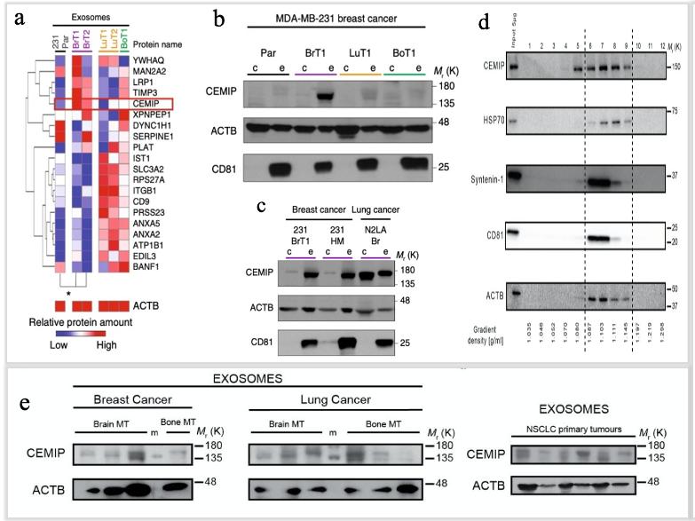
Exosomes from brain metastatic cells support brain metastatic colonization and are enriched in CEMIP protein (Rodrigues et al., 2019).
Case 2 Spatial proteomics fuels breast cancer tumor microenvironment research (2)
In this study, 15 formalin-fixed paraffin-embedded (FFPE) samples from eight patients with HER2+ breast cancer were analyzed. Two of the patients had three different sampling time points, three patients had two different sampling time points, and three patients had a single sampling time point. Proteomics assays were analyzed by Digital Spatial Profiler (DSP).
Differential expression analyses for DSP data were used to look for specific protein differences between three different groupings: (1) Primary vs metastatic tumor, (2) Breast vs brain tumors, and (3) timepoint (three different samples of a single patient).
The authors performed spatial analysis of ROIs of HER2+ breast cancer tumor areas (PanCK+) and stromal areas (PanCK-) with different levels of immune infiltration (i.e., COLD, WARM, HOT) to find the differences between primary and metastatic status. The DSP experimental design is shown in Figure A. It was found that the ROI immune infiltration degree of hot, warm, and cold were different for each patient. The highest number of immune cells was found in the primary foci and the lowest number in the metastatic tissue (Figure B). The expression of immune-related proteins was higher in the primary tumors, especially tumor-infiltrating lymphocytes: CD3, CD8 (Figure C). The differential protein analysis revealed potential immunotherapeutic biomarkers in HER2+ breast cancer patients.
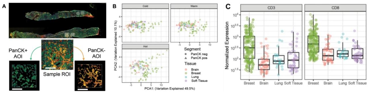
DSP protein expression analysis of primary VS metastases (Schlam et al., 2021)
This study highlights the importance of spatial resolution of the tumor and stromal regions within the TME to observe differences in the expression of immunotherapeutic biomarkers. This study suggests that proteomic immunomarker detection may provide guidance for patient management at different stages of HER2+ breast cancer patients' disease, and thus may provide individualized treatment plans to improve patient prognosis.
You may be interested in:


Resources
- Rodrigues, G., Hoshino, A., et al. (2019). Tumour exosomal CEMIP protein promotes cancer cell colonization in brain metastasis. Nature cell biology, 21(11), 1403-1412.
- Schlam, I., Church, S. E., e al. (2021). The tumor immune microenvironment of primary and metastatic HER2− positive breast cancers utilizing gene expression and spatial proteomic profiling. Journal of translational medicine, 19(1), 1-14.


