


Ferroptosis needs molecular executioner evidence, not only a generic oxidation signal
Build the project around a three-tier lipid evidence chain
Prevent ex vivo autoxidation before asking the mass spectrometer to solve it
Make LC-MS/MS identifications proportionate to the claim
Use an orthogonal control hierarchy that can falsify the interpretation
Choose targeted, discovery, and complementary assays by the decision they must support
Read the result as a pattern, then report the uncertainty that remains
Turn the design into a four-phase experimental specification
Frequently asked questions
Related Services
Meta Intent: A practical guide to designing oxidized lipidomics experiments that distinguish ferroptosis-associated phospholipid oxidation from generic oxidative stress and ex vivo analytical artifacts.
Ferroptosis is often introduced with a simple chain of events: iron-dependent lipid peroxidation accumulates, membrane integrity fails, and the cell dies. That description is useful, but it creates an analytical trap. A fluorescent lipid-oxidation probe, elevated reactive oxygen species, or a drop in viability can be entirely compatible with ferroptosis without establishing which oxidized lipids changed, whether those changes occurred in the sample rather than during extraction, or whether another death process produced the same signal. A defensible study therefore treats oxidized lipidomics as an evidence-building exercise, not as a decorative add-on to a viability assay.
The practical question is not simply, "Can I measure lipid oxidation?" It is, "Can I show that a chemically defined, biologically relevant oxidized phospholipid pattern tracks the proposed ferroptotic mechanism under a controlled analytical workflow?" The answer depends on experimental timing, sample handling, extraction chemistry, chromatographic separation, MS/MS evidence, and an orthogonal control set. This article translates that logic into a project design that can be adapted to cultured cells, tissue, and discovery-stage translational models.

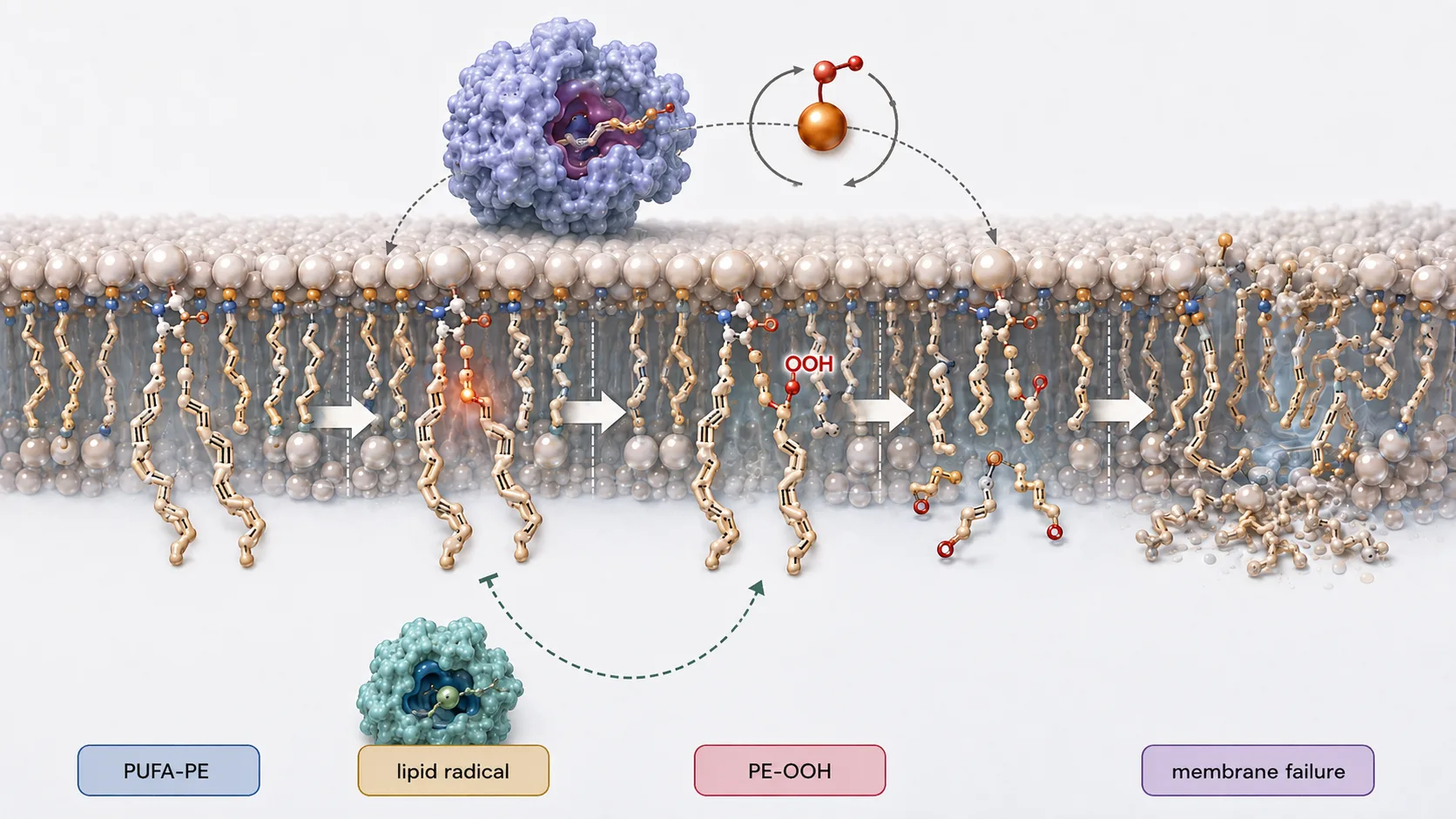
Figure 1. The ferroptotic lipid-peroxidation cascade: a susceptible PUFA-containing membrane phospholipid becomes a hydroperoxide and then a broader set of secondary oxidation products if repair capacity is exceeded.
Ferroptosis needs molecular executioner evidence, not only a generic oxidation signal
Membrane oxidation is a process, whereas ferroptosis is a mechanistic interpretation. Oxidation-sensitive dyes are valuable for following the kinetics of oxidative stress, but they report a composite signal shaped by probe localization, loading, efflux, oxidation chemistry, and instrument settings. They do not identify an acyl chain, phospholipid class, hydroperoxide position, or oxidation state. In particular, a positive C11-BODIPY response should be read as evidence of lipid-oxidation flux, not as a standalone molecular fingerprint of ferroptosis.1 LC-MS-based redox lipidomics supplies the missing molecular layer by measuring the lipid species that accumulate and the lipid-class context in which they accumulate.
For a ferroptosis-focused study, the most informative target is usually not every oxidized molecule detectable in the extract. It is a coherent group of PUFA-containing phospholipids and their oxygenated derivatives that changes with the phenotype. Hydroperoxy phosphatidylethanolamines are frequently of interest because PUFA-PE oxidation has a strong mechanistic connection to canonical ferroptotic pathways, but the relevant chemistry can differ by cell type, inducer, membrane composition, and repair state.5 The study should therefore be framed as a testable hypothesis: a defined family of oxidized phospholipids rises before or alongside loss of viability, and that pattern is weakened by a mechanistically appropriate rescue.
This approach prevents a common overclaim. A broad increase in oxidized phosphatidylcholines, lysophospholipids, or free fatty-acid oxidation products may be biologically meaningful, yet it may indicate generalized oxidative damage, membrane remodeling, inflammation, or extraction-induced autoxidation rather than the phospholipid oxidation pattern that supports a ferroptotic conclusion. Start with a narrow mechanistic question, then decide whether a Targeted Lipidomics assay, a discovery-oriented screen, or a staged combination is the best way to answer it.
Build the project around a three-tier lipid evidence chain
A strong project separates three related but non-interchangeable tiers of evidence. Tier one is the susceptible substrate pool: the abundance and composition of PUFA-containing phospholipids that can become oxidized. Tier two is the primary oxidation signature: oxygenated phospholipids, especially hydroxy and hydroperoxy forms, assigned with retention time, accurate mass, and informative fragmentation. Tier three is the phenotypic consequence: cell death kinetics, membrane damage, rescue response, and complementary redox readouts. The value comes from alignment across tiers, not from one exceptionally large fold change at one tier.
For example, a treatment may reduce the abundance of arachidonoyl- or adrenoyl-containing PE species because their substrate pool is being consumed. At the same time, the corresponding oxygenated PE signal may rise, then fall late in the time course as cells lose integrity or reactive intermediates decompose. A single late endpoint can therefore obscure the pathway. A compact kinetic series that captures an early, pre-terminal interval is often more useful than many replicates collected only after extensive cell lysis.3 The same logic applies to tissue: collect with a defined ischemia interval and document whether the observed oxidation state could have developed after perfusion or excision.
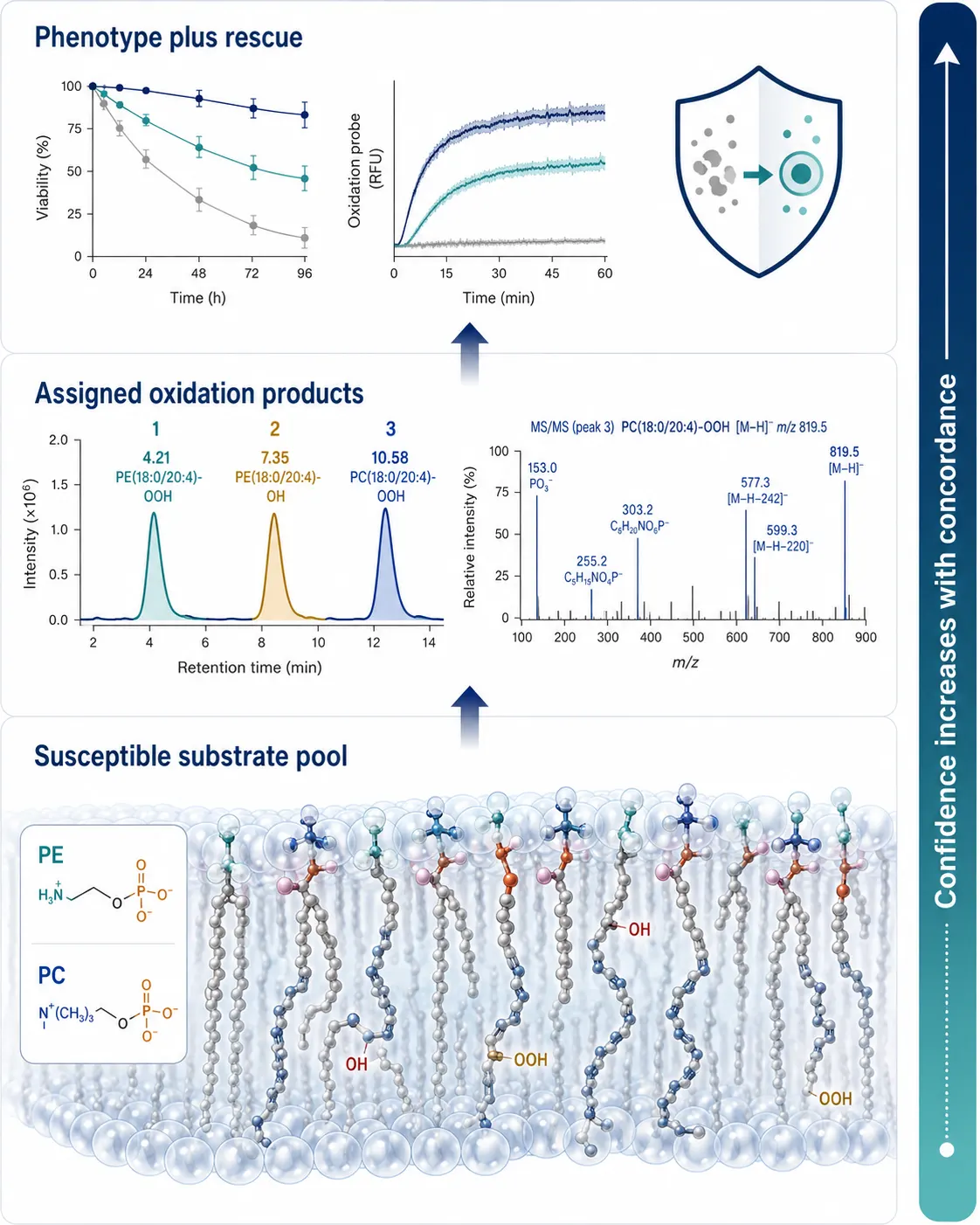
Figure 2. A three-tier evidence chain links the susceptible phospholipid pool, chemically assigned oxidation products, and the ferroptosis-associated phenotype.
Use the first pass to establish the lipid landscape rather than to force every feature into a candidate list. A broad Untargeted Lipidomics discovery run can reveal which phospholipid classes and PUFA-containing species are present in the model. A subsequent targeted panel should then quantify the subset that is most diagnostic for the biological question. This staged design is usually more defensible than declaring a complete oxidized lipidome from a small number of unvalidated features.
Prevent ex vivo autoxidation before asking the mass spectrometer to solve it
Oxidized lipids are unusually vulnerable to preanalytical distortion. Warm handling, delays before quenching, repeated freeze-thaw cycles, oxygen exposure, and metal contamination can all make a fragile biological signal look larger, smaller, or less reproducible than it was in vivo. The issue is not merely technical hygiene: an extraction artifact can mimic the exact increase in oxygenated lipids that the experiment was designed to find.
Write the chain of custody into the method before the first sample is collected. Define the biological stopping point, time from plate or tissue to quench, solvent temperature, order of addition, storage temperature, and maximum permitted handling delay. Keep conditions matched across treatment groups, randomize extraction order where practical, and intersperse pooled quality-control material throughout the queue. Antioxidants or metal chelators may be useful during extraction, but they are method controls rather than universal defaults. They should be shown not to suppress recovery, alter the targeted analyte distribution, or mask a treatment-specific difference.
Parallel artifact controls are especially valuable. One aliquot can follow the standard cold extraction; a matched aliquot can be exposed to a deliberately less protective handling condition to show the direction and scale of induced change. A reagent blank, process blank, and pooled QC address different questions and should not be treated as substitutes. When the study needs to support hydroperoxide assignments, a parallel reduction experiment with triphenylphosphine or another validated reducing condition can provide a powerful chemical check: hydroperoxide candidates should convert in the expected direction to their corresponding alcohol products without creating a misleading wholesale shift in the rest of the lipidome.
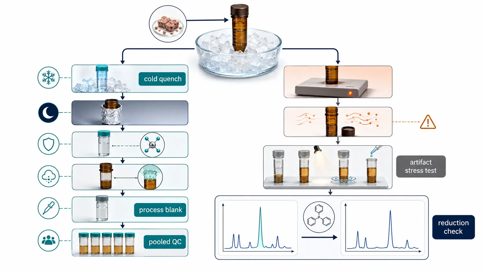
Figure 3. Artifact control begins at collection: matched cold handling, blanks, pooled QC, and a parallel chemical-reduction check help separate biological oxidation from ex vivo autoxidation.
Sample amount and matrix complexity determine how much cleanup is necessary. Cell pellets may support direct extraction with a short workflow, whereas lipid-rich tissue, plasma, or organoid-conditioned material may require more deliberate cleanup and recovery monitoring. A sample preparation strategy should be selected around the target lipid class and matrix rather than copied from an unrelated metabolomics protocol. If phospholipid substrate depletion is central to the hypothesis, pair the oxidation panel with phospholipid analysis so that an apparent absence of a hydroperoxide is not confused with absence of its precursor.
Make LC-MS/MS identifications proportionate to the claim
Accurate mass alone rarely warrants a structural claim for oxidized phospholipids. Many isobaric species share a plausible elemental composition, and in-source fragmentation can create signals that resemble authentic oxidation products. The more specific the biological interpretation, the more specific the orthogonal evidence should be. At a minimum, retain raw chromatographic behavior, adduct identity, mass accuracy, isotope pattern where applicable, and MS/MS fragments that support both the phospholipid backbone and the oxidized acyl chain.
Chromatographic resolution matters because oxidation can alter polarity and retention in ways that are informative but not universal. Reversed-phase separations can resolve broad acyl-chain and oxygenation differences, while C30 or other selectivity-enhancing phases may help where isomer separation is central to the project. The right choice depends on the chemical question, matrix, and anticipated isomer burden. For a method intended to distinguish hydroxy from hydroperoxy products, establish the retention and fragmentation behavior with authentic or closely matched standards whenever possible. Without that confirmation, describe features conservatively as putative oxygenated phospholipids rather than naming an exact positional isomer.
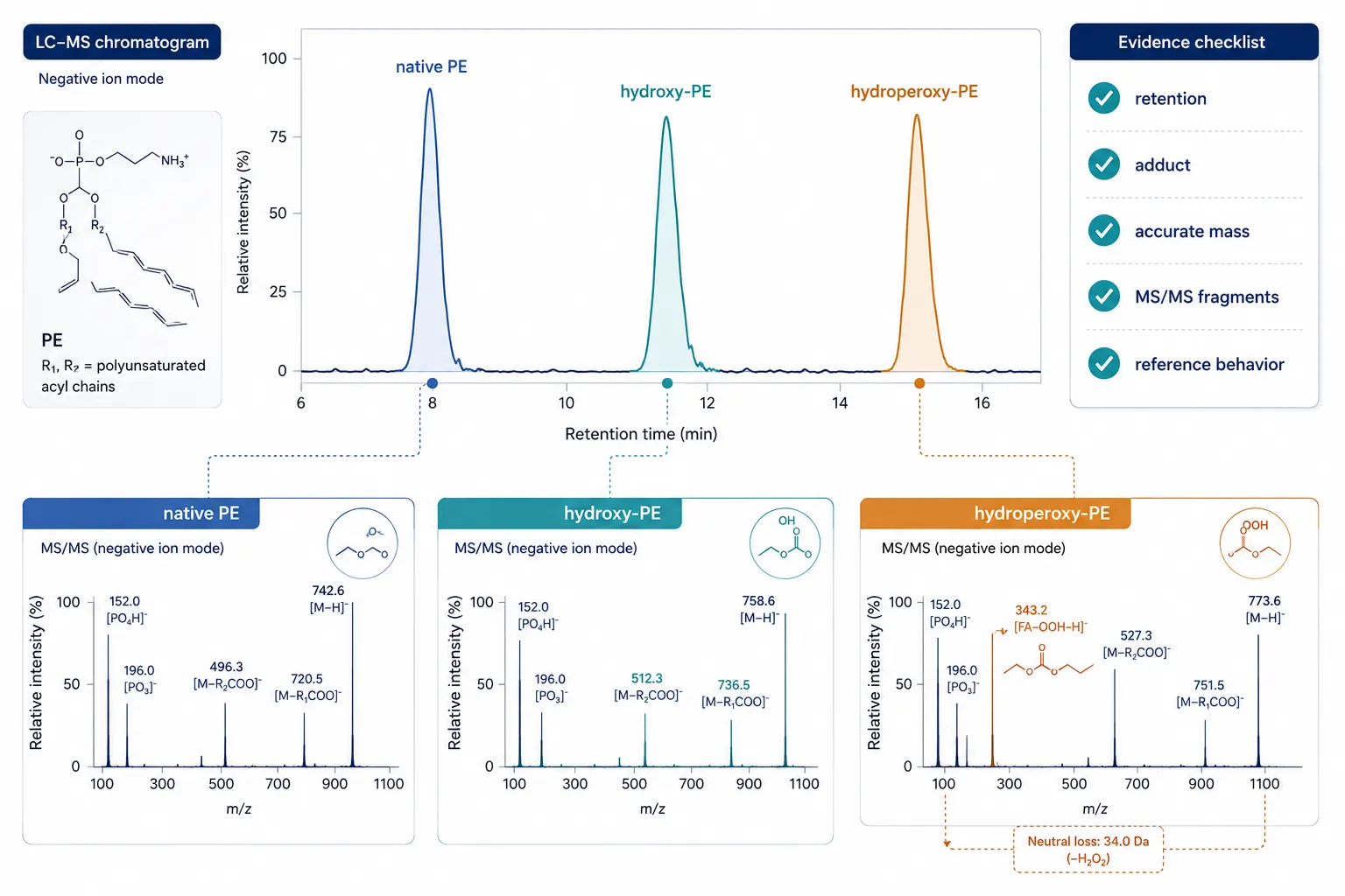
Figure 4. Defensible oxidized phospholipid assignment combines chromatographic separation with precursor, adduct, and MS/MS evidence that supports both lipid class and oxidized acyl chain.
Quantification should also reflect uncertainty. Stable-isotope internal standards are preferred when they are available for the relevant class and behavior. Class-matched surrogates can still improve process control, but they do not erase differences in ionization, retention, or recovery among individual oxidized species. Report whether values are absolute concentrations, relative abundances, or internal-standard-normalized responses. For exploratory datasets, an explicit confidence tier is more useful than false precision: confirmed with a standard, strongly supported by MS/MS and reduction behavior, or putative from accurate mass and fragmentation alone.
Use an orthogonal control hierarchy that can falsify the interpretation
Ferroptosis evidence becomes much stronger when the controls can disprove the preferred story. A useful hierarchy includes a vehicle control, a treatment condition, a time-resolved rescue condition, and a mechanism-diverting comparator chosen for the model. The rescue should reduce the phenotype and shift the lipid signature in a coherent way. Conversely, a comparator that preserves viability but leaves the putative oxidized phospholipid pattern unchanged may reveal that the lipid change is a stress marker rather than the driver of death.
Do not rely on one inhibitor or one dye. Combine a cell-death measurement with a lipid-oxidation readout and the molecular LC-MS panel. Where biologically justified, include perturbations that change iron availability, phospholipid PUFA remodeling, peroxide repair, or lipoxygenase-related pathways. The aim is not to assemble the longest inhibitor list; it is to generate a small set of contrasts that make competing explanations testable. In projects studying fatty-acyl remodeling, integrating fatty acid metabolism analysis can clarify whether the observed oxidized lipid profile reflects a changed PUFA substrate supply, altered oxidation, or both.
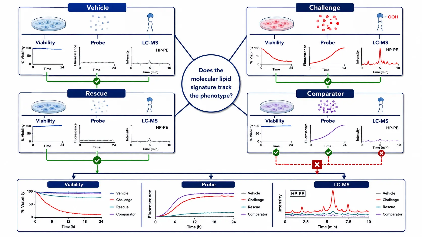
Figure 5. Orthogonal validation is a control lattice, not a single rescue experiment: each arm is evaluated against phenotype, oxidative flux, and the molecular lipid signature.
Choose targeted, discovery, and complementary assays by the decision they must support
There is no universally superior oxidized lipidomics workflow. Select the method based on the decision that follows from the data. Use targeted LC-MS/MS when the question is whether a defined set of hydroxy or hydroperoxy phospholipids changes with a perturbation and needs reproducible relative or absolute quantification. Use a discovery workflow when the lipid class, acyl composition, or unexpected oxidation product is unknown. Use both when discovery defines the candidate space and a focused panel is needed to validate the final comparison.
Whole-lipidome coverage is not equivalent to mechanistic depth. A broad Lipidomics Service can establish membrane composition and treatment-associated remodeling, yet a highly targeted assay may be the better choice for a repeatable hydroperoxide endpoint. Similarly, eicosanoids can capture inflammatory oxidative mediators that complement but do not replace membrane phospholipid evidence; an eicosanoids analysis layer is useful when the project asks how ferroptotic stress intersects with extracellular lipid mediator release.
Four planning questions usually make the choice clear. First, is the project discovering an unknown signature or verifying a pre-specified one? Second, is the claimed endpoint a lipid class, an acyl chain, a positional isomer, or a total oxidation burden? Third, can an authentic standard or matched surrogate be obtained for the critical analytes? Fourth, what action will follow from a positive, negative, or ambiguous result? If the fourth answer is unclear, the method may be accumulating data without reducing uncertainty.
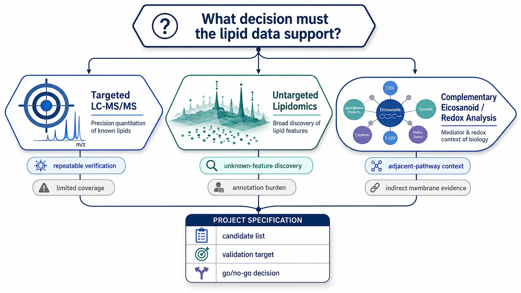
Figure 6. Method selection should begin with the decision to be made: targeted assays support repeatable verification, discovery workflows map unknown chemistry, and complementary assays answer adjacent biological questions.
Read the result as a pattern, then report the uncertainty that remains
A practical result review starts by asking whether the individual observations tell the same story. Suppose a treatment increases lipid-dye oxidation, lowers viability, depletes a defined PUFA-PE substrate, and increases a reduction-sensitive hydroperoxide feature with supportive MS/MS data. If a rescue condition reverses the phenotype and reduces that feature in the same time window, the combined evidence supports a ferroptosis-associated molecular interpretation. In contrast, if total oxidation rises but the candidate oxidized phospholipids are unstable across replicate extractions, present in blanks, or unaffected by a phenotype-rescuing condition, the more defensible conclusion is that the study detected oxidative stress without yet identifying the molecular executioner.
Use data visualization to make this reasoning inspectable. Show individual biological replicates rather than only group means. Plot time on the x-axis whenever the project asks a kinetic question. Put substrate and product measurements near each other so that apparent accumulation can be interpreted with precursor availability in mind. For a focused panel, report the direction, magnitude, confidence tier, missing-value handling, and the number of samples that passed each quality gate. A heat map may be appropriate for discovery, but it should not replace extracted-ion chromatograms and representative MS/MS spectra for the few features carrying the strongest mechanistic claim.
Normalization needs the same discipline. Protein, cell count, tissue mass, DNA, or total phospholipid can each be useful denominators, but they answer slightly different questions. Choose the primary normalization before seeing the group separation, then state why it matches the sampling unit. If terminal membrane failure changes protein recovery or cell number, a second sensitivity analysis may be necessary to show whether the conclusion depends on the chosen denominator. For studies that compare multiple experiments or matrices, retain the raw internal-standard-normalized response alongside the biological normalization so analytical performance and biological effect are not conflated.
Finally, distinguish an analytical negative from a biological negative. A missing candidate may reflect low abundance, poor extraction, in-source decomposition, coelution, a mismatched acquisition method, or a genuine lack of formation. A small feasibility run with pooled material, standard spikes where available, dilution linearity, and replicate injections can identify which explanation is most likely before scarce biological samples are consumed. This is often the point at which a targeted method can be refined, a discovery method can be broadened, or the original mechanism can be revised without overstating an inconclusive signal.
Turn the design into a four-phase experimental specification
Phase one is biological scoping. Define the cell state, perturbation, expected time window, and a primary competing explanation. Record the exact matrix and the minimum sample amount that can support both phenotype and lipid analysis. Phase two is preanalytical validation. Test cold handling, extraction recovery, carryover, process blanks, pooled QC behavior, and whether protective additives alter the analytes that matter. Phase three is analytical confirmation. Establish retention behavior, MS/MS evidence, confidence tiers, and the minimum set of internal standards or surrogates. Phase four is biological interpretation. Examine whether the lipid signature, rescue behavior, and phenotypic data converge over time.
This structure makes the final report easier to defend because it records what was controlled before the dataset became interesting. It also identifies when to stop. If blanks carry the candidate feature, if QC drift exceeds the pre-set tolerance, if hydroperoxide-reduction behavior is inconsistent, or if the lipid pattern does not track the phenotype across independent experiments, do not advance that feature as a mechanistic marker. Preserve it as an observation and revise the question.
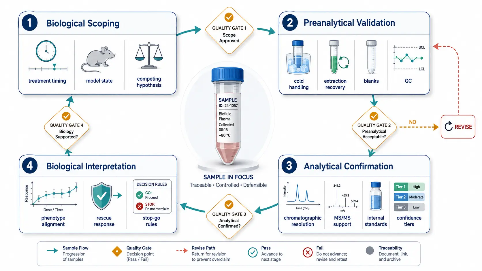
Figure 7. A four-phase specification makes the experimental chain auditable from biological question to final mechanistic interpretation.
The workflow also fits naturally into a broader oxidative-stress research program. If protein thiol status, redox enzyme activity, or oxidative protein damage are part of the same causal question, Redox Proteomics can provide a complementary layer rather than a redundant one. For sterol oxidation questions, see the matrix companion on oxysterol LC-MS workflows built around artifact control. These measurements should be integrated around one hypothesis, with shared sample timing and a clear statement of which analytical layer is expected to falsify the model.
Frequently asked questions
Is a lipid-oxidation dye sufficient to claim ferroptosis?
No. It is useful as a flux or screening readout, but it does not identify the oxidized molecular species or exclude other oxidative death processes. Pair it with phenotype controls and targeted molecular evidence when the mechanistic claim matters.
Should hydroperoxy phospholipids always be the primary endpoint?
Not always. They are mechanistically compelling but chemically labile. In some matrices, a validated set of hydroxy products, substrate depletion measures, and reduction-confirmation experiments provides a more reliable evidence package.
Can I use one extraction protocol for cells, plasma, and tissue?
Only after matrix-specific validation. Different matrices alter recovery, suppression, oxidation risk, and background composition, so a protocol that works for a cell pellet may not be defensible for plasma or lipid-rich tissue.
How should I report a feature without an authentic standard?
Use a confidence tier and state the evidence used for the annotation. Avoid assigning an exact positional isomer unless retention, fragmentation, and an appropriate reference support that level of specificity.
Why do treatment and rescue groups sometimes show the same total oxidation burden?
Total oxidation can hide a shift between lipid classes or oxidation states. Inspect species-level patterns and time-resolved behavior rather than relying on one aggregate intensity measure.
When should I add a second omics layer?
Add it when it tests a defined alternative explanation, such as changed PUFA substrate supply or altered protein redox regulation. Do not add another platform merely because the first dataset is complex.
References:
- Wiernicki B, et al. Excessive phospholipid peroxidation distinguishes ferroptosis from other cell death modes including pyroptosis. Cell Death & Disease. 2020. doi: 10.1038/s41419-020-03118-0
- Chen D, et al. iPLA2beta-mediated lipid detoxification controls p53-driven ferroptosis independent of GPX4. Nature Communications. 2021. doi: 10.1038/s41467-021-23902-6
- Mortensen MS, Ruiz J, Watts JL. Polyunsaturated Fatty Acids Drive Lipid Peroxidation during Ferroptosis. Cells. 2023;12(5):804. doi: 10.3390/cells12050804
- Perez MA, et al. Ether lipid deficiency disrupts lipid homeostasis leading to ferroptosis sensitivity. PLOS Genetics. 2022;18(9):e1010436. doi: 10.1371/journal.pgen.1010436
- Kagan VE, et al. Redox Epiphospholipidome in Programmed Cell Death Signaling: Catalytic Mechanisms and Regulation. Frontiers in Endocrinology. 2021;11:628079. doi: 10.3389/fendo.2020.628079
For research use only. This article describes analytical study design and does not provide clinical diagnostic or treatment guidance.