
In the field of cancer research, mass spectrometry imaging (MSI) technology is promising, but its application is limited by many problems, such as raw data preprocessing, image accuracy and image recognition ability.
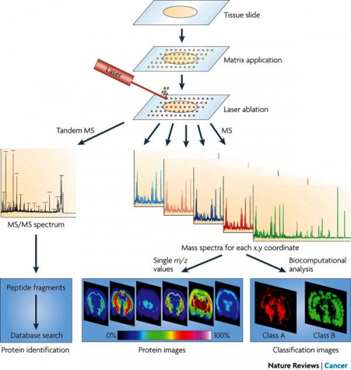
Mass spectrometry imaging is mainly used to directly scan biological samples by mass spectrometry to analyze the structural, spatial and temporal distribution of chemical components in cells or tissues. This imaging method is not limited to one or several specific protein molecules. Each protein molecule can be found in tissue samples and accurate information on their spatial distribution is provided. As early as a few years ago, some scientists put forward the idea of using this technology to determine the type of biological tissue, but there had been no practical and effective method.
This latest method uses desorption electrospray ionization technology to optimize data preprocessing, improve image accuracy, and enhance biochemical characteristics of different tissue types by extracting specific molecular imprints from biological tissues, so as to improve the ability of image recognition.
The researchers said that with the newly developed integrated biological information platform, a large amount of specific information data from human tissues obtained by mass spectrometry imaging technology was used for the database building of various types of tissues. By analyzing multiple samples and comparing with the results of traditional histological analysis, the computer could learn to identify different types of tissues. They designed their own workflow for colorectal cancer tissue testing, with good results.
Compared with standard histology which takes weeks to produce complete results, a single detection using mass spectrometry requires only a few hours to obtain more detailed information, which not only shows whether the tissue is cancerous, but also tells which type or subtype of cancer it is. This information is important for doctors to choose the most effective treatment.
The researchers pointed out that there had been little change in the analysis methods of histopathological samples since 19th century, when staining technique was used to show the structure of tissues. To this day, staining is still the dominant method of histological analysis in hospitals. And it's getting more and more complex and expensive. Mass spectrometry may change the basic paradigm of histology, and scientists will define the tissue types based on chemical composition instead of tissue structure. In the future, the detection will no longer rely on the eyes of experts, but on the basis of massive data. The information obtained by one detection assay is much more than that obtained by traditional histologic detection assay.
Mass spectrometry imaging technique is undoubtedly a new way of completely automated histological analysis, and the new technologies that scientists have been studying continuously, are also gradually helping resolve the new problems encountered in the practical application of mass spectrometry imaging technology.
Mass spectrometry imaging technology is a new journey of fully automated histologic analysis. The newly developed integrated biological information platform can be used to construct various kinds of tissue database with the large number of specific information data of human tissues obtained by mass spectrometry imaging technology, which makes the resolution of cancerous tissue relatively simple and efficient.