


Define the mechanism before choosing the experiment
Use comparative controls that diagnose the source of a signal
Constrain the database search around a defined adduct
Control error at the modified-peptide level
Verify the spectrum before claiming a covalent ligand
Separate molecular presentation from functional recognition
Four-phase implementation plan
Frequently asked questions
Meta Intent: A practical preclinical decision guide for testing whether a defined covalent drug adduct is presented on HLA-bound peptides, and for separating a credible candidate from an enlarged-search-space false positive.
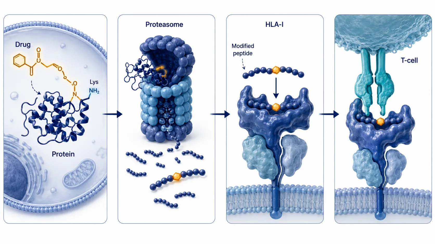
Covalent therapeutics can create a chemical event that conventional antigen-discovery workflows do not model: a small molecule or metabolite becomes attached to a protein residue, survives antigen processing, and is represented on a cell-surface peptide-HLA complex. Such a ligand may be a covalent neoepitope. It is an experimentally testable hypothesis, not an automatic consequence of target engagement. The peptide must be generated, loaded by a compatible HLA molecule, detected by MS, and distinguished from non-covalent repertoire changes or preparation artifacts.
This distinction matters when comparing related covalent scaffolds in discovery. An immunoproteomics workflow can measure the naturally presented ligandome, but the search must be designed around the drug's known chemistry, the HLA background, and the desired decision. An unbounded mass-shift search can create more candidate explanations than evidence.

Figure 1. A covalent neoepitope is supported only when a defined drug-peptide adduct survives processing and is detected as an HLA-presented ligand.
Define the mechanism before choosing the experiment
Drug-induced immune recognition can follow more than one mechanism. In haptenation, a reactive compound forms a covalent adduct on a protein or peptide. In altered-repertoire mechanisms, a drug changes peptide loading or HLA-pocket behavior without becoming a covalent peptide modification. These alternatives need different database models and controls. A mass shift on a peptide is relevant to the first mechanism; a changed motif distribution without a drug adduct may be relevant to the second.
Begin with the electrophile, anticipated leaving group, plausible nucleophilic residues, and whether intact drug or a metabolite is expected to react. Calculate a defined monoisotopic delta for the actual retained adduct, rather than using a nominal drug mass. The chemical proposal should state the allowed residues and whether more than one adduct form is credible. This is the basis for an interpretable high-resolution PTM profiling strategy.
Use comparative controls that diagnose the source of a signal
At minimum, compare untreated material, vehicle, and drug-treated material with matched biological replicates. Add an inactive or non-reactive structural analogue when it is chemically meaningful. That control is especially valuable because it helps distinguish covalent adduct formation from a general transcriptional, stress, or viability effect. Record viability, exposure duration, dose, and recovery, then choose a treatment range that supports the intended mechanistic comparison rather than merely maximizing stress.
Choose the cellular model for the claim, not merely for convenience. A monoallelic HLA cell line can make allele attribution and motif review much clearer. A disease-relevant line may better represent target abundance, metabolism, and antigen-processing state, but it can contain multiple HLA alleles and a more variable ligandome. Primary cells add biological relevance but may constrain input material and treatment design. A practical program can begin in a controlled discovery model, then test the most credible candidate in a second model selected for the intended biological question.
Replicates should be biologically independent cultures rather than repeated injections of one HLA eluate. Injection replicates help establish instrument consistency, whereas biological replicates determine whether drug treatment repeatedly produces a candidate ligand. Keep culture, lysis, immunoprecipitation, desalting, and LC-MS batches balanced across conditions. If treated samples are processed on a separate day or column batch, a batch-specific difference can be mistaken for a treatment-specific peptide repertoire.
High-resolution HLA typing is not administrative metadata. HLA alleles constrain motif expectations, anchor-residue plausibility, and the peptide population that can be compared across conditions. Use a monoallelic or well-characterized line when the question is allele-specific; use a more complex biological system only when its added relevance outweighs the attribution challenge. An orthogonal protein identification service may help evaluate source-protein expression, but expression alone does not prove presentation.
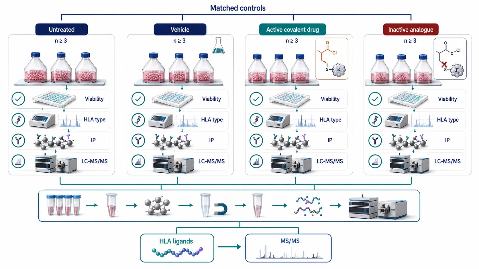
Figure 2. Matched untreated, vehicle, active-drug, and inactive-analogue controls help identify whether a candidate depends on covalent chemistry.
Constrain the database search around a defined adduct
Immunopeptidomics is normally a non-enzymatic search: HLA ligands have variable termini and broad precursor space. Adding a large variable modification multiplies that space. Do not assign one drug mass shift to all amino acids merely because a search engine allows it. Restrict the modification to residues supported by the reaction chemistry, define a realistic number of modifications per peptide, use HLA-appropriate peptide lengths, and keep ordinary variable modifications limited.
A closed custom-modification search is the primary test when the retained adduct and residue specificity are known. An open modification search is a discovery supplement, useful for finding unexpected drug fragments, hydrolysis products, or alternative adducts. It should not replace the closed search or be used as a license to claim every nearby delta mass. Candidate open-search shifts need chemical plausibility, localization evidence, and follow-up in a constrained search.
Pair database searching with peptide-level and modification-localization review. A bioinformatics-for-proteomics analysis should preserve the custom delta, allowed residues, decoy strategy, score thresholds, and all candidate spectra, so the modification search remains reproducible.
Database content requires the same discipline. Start with an appropriate reviewed proteome and contaminant set, then document any added non-canonical proteins, engineered constructs, or drug-target sequences. Do not make an adduct search more permissive to compensate for an incomplete protein database. If the source sequence is uncertain, resolve that uncertainty separately through proteogenomics or targeted sequence confirmation. Otherwise, a modification assignment can become an accidental explanation for a sequence mismatch.
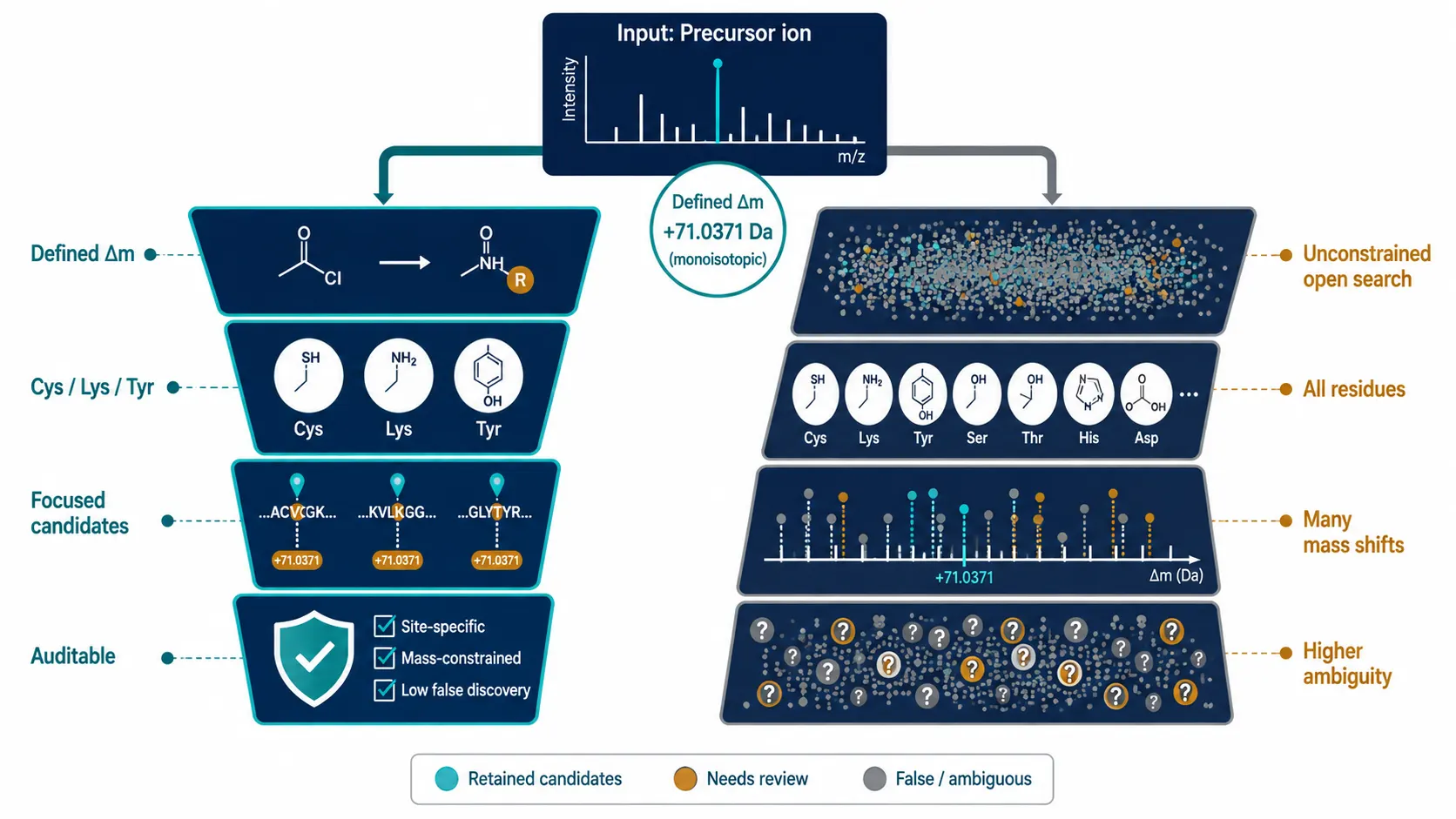
Figure 3. Restricting a defined drug mass shift to plausible residue chemistry keeps modified-immunopeptide searching interpretable.
Control error at the modified-peptide level
A global one-percent FDR can be inadequate evidence for a rare modified subgroup. High-confidence unmodified ligands may dominate global score distributions, while the small modified subset has different fragmentation, localization, and decoy behavior. Calculate and report modified-subgroup or delta-mass-specific error control where supported by the workflow, and review the result at PSM, peptide, and site-localization levels. A low global error estimate is not a substitute for inspecting the exact drug-adduct candidate.
Basic ligandome QC remains essential: length distribution, HLA motif coherence, replicate occurrence, contaminant review, and condition specificity. A candidate found only in one treated run, at the edge of a broad mass tolerance, with weak site localization, is a hypothesis for targeted follow-up, not a demonstrated neoepitope. The article on open modification search versus targeted PTM analysis provides a useful framework for that escalation.
Use decoys that reflect the search being evaluated. A target-decoy estimate calculated largely from ordinary unmodified peptides may not represent the behavior of rare, large, residue-restricted adducts. Where the software permits, stratify modified and unmodified candidates before applying score thresholds, then separately inspect the mass-shift and residue-localization subset. Report the number of modified PSMs, unique sequences, unique sites, and biological replicates that remain after filtering. These counts make it easier to distinguish repeated evidence for one plausible ligand from a long list of single-spectrum possibilities.
Candidate abundance should be interpreted with care. A low MS1 signal may reflect genuine low presentation, but it may also arise from poor ionization, stochastic precursor selection, sample loss, or an unstable adduct. Conversely, a strong signal can be a co-purified contaminant or a treatment-dependent stress peptide rather than an HLA-presented drug adduct. Retention-time alignment, HLA motif plausibility, and controlled immunoprecipitation blanks are therefore part of error control, not optional cosmetic QC.
Verify the spectrum before claiming a covalent ligand
Verification should test multiple independent features. Synthesize the modified peptide where feasible, ideally with an isotope-labeled analogue for coelution work. Compare retention time, precursor mass, isotope pattern, and fragment-ion coverage under the same LC-MS method. Look specifically for fragment ions that localize the adduct to the proposed residue and for chemically plausible diagnostic drug-related ions or neutral losses. Absence of one diagnostic ion does not automatically refute a candidate, but the observed spectrum must still support the proposed structure.
Targeted reacquisition can improve signal quality, but it cannot repair an incorrectly specified structure. When the adduct is labile, compare fragmentation modes and document which evidence survives. A de novo peptide sequencing service may help characterize unexpected backbone or fragment features, while a defined synthetic standard remains the strongest practical orthogonal comparator for a named drug-peptide adduct.
Manual review should ask a sequence of falsification questions. Is the precursor isotope envelope consistent with the proposed charge state and mass delta? Are the informative fragments sufficient to distinguish the proposed modification site from a nearby alternative? Does the modified spectrum explain more of the observed signal than the unmodified counterpart? Is the same peak present in vehicle or inactive-analogue controls? Could an in-source fragment, co-isolated precursor, carryover event, or non-specific lysate reaction create the apparent adduct? A candidate that fails one of these questions may still guide method development, but it should not move directly to biological interpretation.
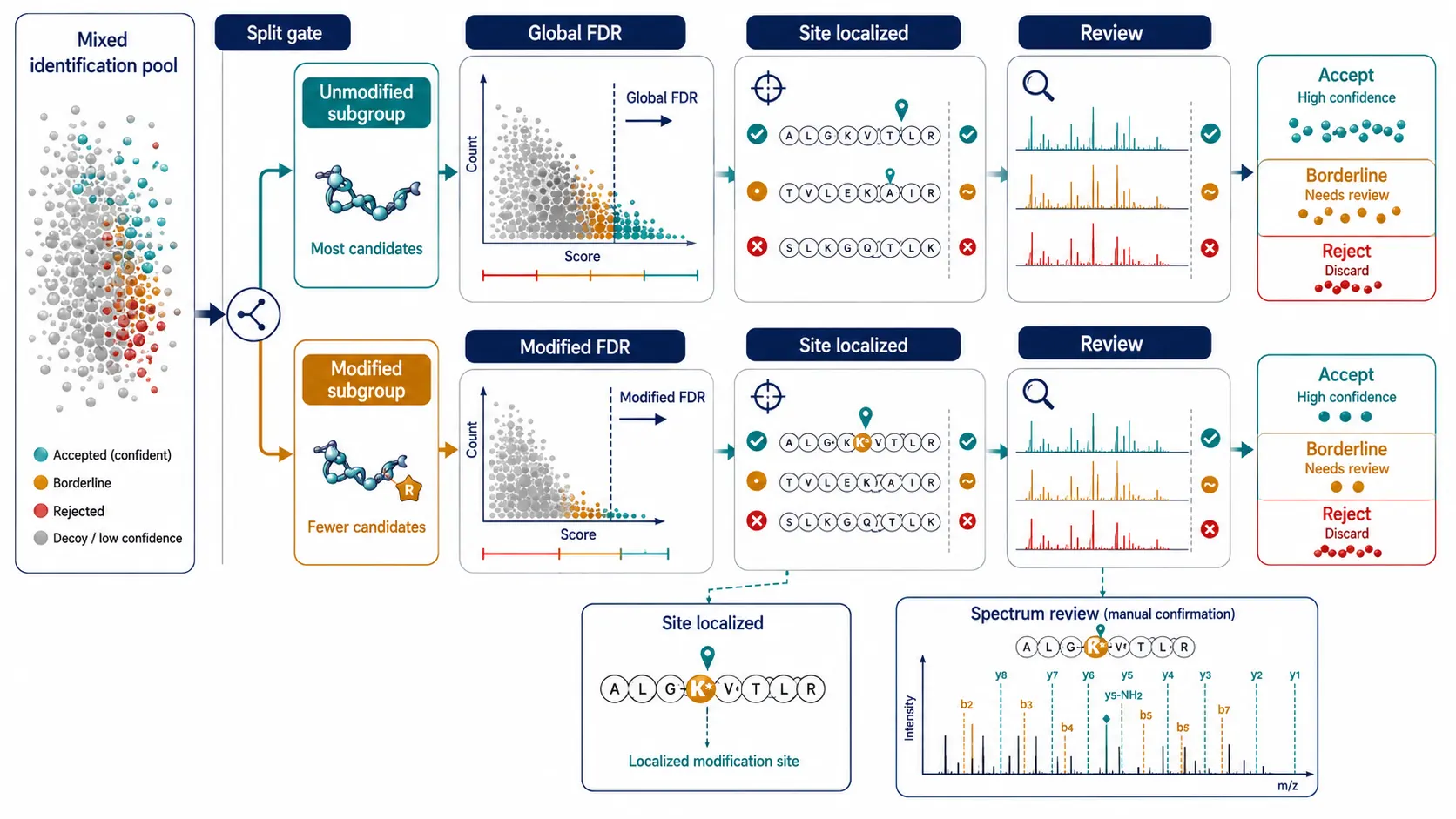
Figure 4. Synthetic-standard comparison turns a database-search candidate into a testable structural assignment.
Separate molecular presentation from functional recognition
Detection of an adducted HLA ligand does not establish T-cell recognition, and a drug-dependent T-cell response does not itself identify the presented molecular structure. Functional follow-up can use peptide-pulsed antigen-presenting cells, T-cell activation assays, or pMHC multimer reagents, selected to fit the scientific question. Controls should include the unmodified peptide, the drug-free condition, and where useful the inactive analogue. Interpret functional results as evidence of recognition in that assay, not as a universal prediction of immunogenicity.
For a compact discovery program, use four gates: chemistry and exposure plausibility; HLA-ligand capture with controlled treatment; closed adduct search plus subgroup error control; then synthetic-standard and functional confirmation. This prevents a broad immunopeptidomics experiment from being overread as a complete risk assessment.
Functional follow-up is best framed as a focused hypothesis test. Peptide-pulsing experiments can test whether an adducted peptide is recognized in a chosen cellular assay, but they may bypass aspects of endogenous processing. Tetramer or multimer staining can identify cells that bind a defined peptide-HLA complex, yet reagent quality and HLA restriction remain critical. Cytokine assays provide a response readout but do not reveal whether the same ligand is abundant on the original treated cell. Use each assay to answer its own question, then integrate the result with the mass-spectrometric evidence rather than treating any assay as a stand-alone prediction of human response.
| Route | Primary output | Best use | Main limitation |
| Standard ligandome | Unmodified HLA peptides | Repertoire baseline | Does not identify defined adducts |
| Closed defined-adduct search | Specified modified candidates | Known covalent chemistry | Misses unknown metabolites |
| Open modification search | Unexpected delta masses | Hypothesis generation | Large search space and localization risk |
| Synthetic-standard verification | Orthogonal structural support | Named candidate confirmation | Requires material and method development |
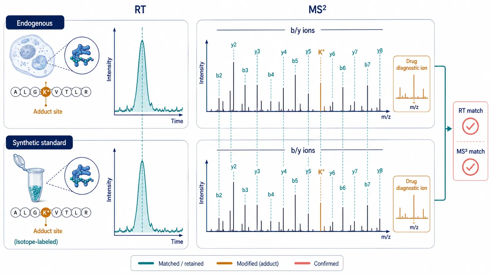
Figure 5. Defined-adduct discovery is strongest when broad profiling, constrained searching, and orthogonal verification are used as separate evidence layers.
Four-phase implementation plan
Plan: define the drug-derived adduct, reactive residue, HLA model, and matched controls. Generate: treat cells under interpretable exposure conditions, immunoprecipitate HLA complexes, and capture naturally presented ligands. Search: run a constrained custom-PTM search first, use open searching only for controlled discovery, and apply modified-subgroup error review. Verify: compare candidate spectra with standards and decide whether functional work is justified.
At the capture stage, keep lysis and HLA immunoprecipitation conditions consistent across all arms. Include process blanks and, when material permits, an antibody or bead control that helps identify non-specific peptide carryover. Elute HLA ligands under conditions compatible with the experiment, separate small peptides from heavy-chain and antibody material, and avoid exposing the eluate to an avoidable period of high temperature or reactive solvent. A drug adduct that is chemically labile can be lost or transformed after capture, producing a false negative or a mass shift that belongs to preparation rather than presentation.
Plan targeted follow-up before the discovery acquisition is complete. Save sufficient material, raw files, and a defined list of candidate-adduct hypotheses for reinjection or parallel reaction monitoring. Predefine what would justify synthesis of a modified standard: for example, a treated-condition candidate observed in more than one biological replicate with chemically plausible localization and acceptable subgroup-level error evidence. This guardrail keeps an expensive verification step focused on the candidates most likely to change the next experimental decision.
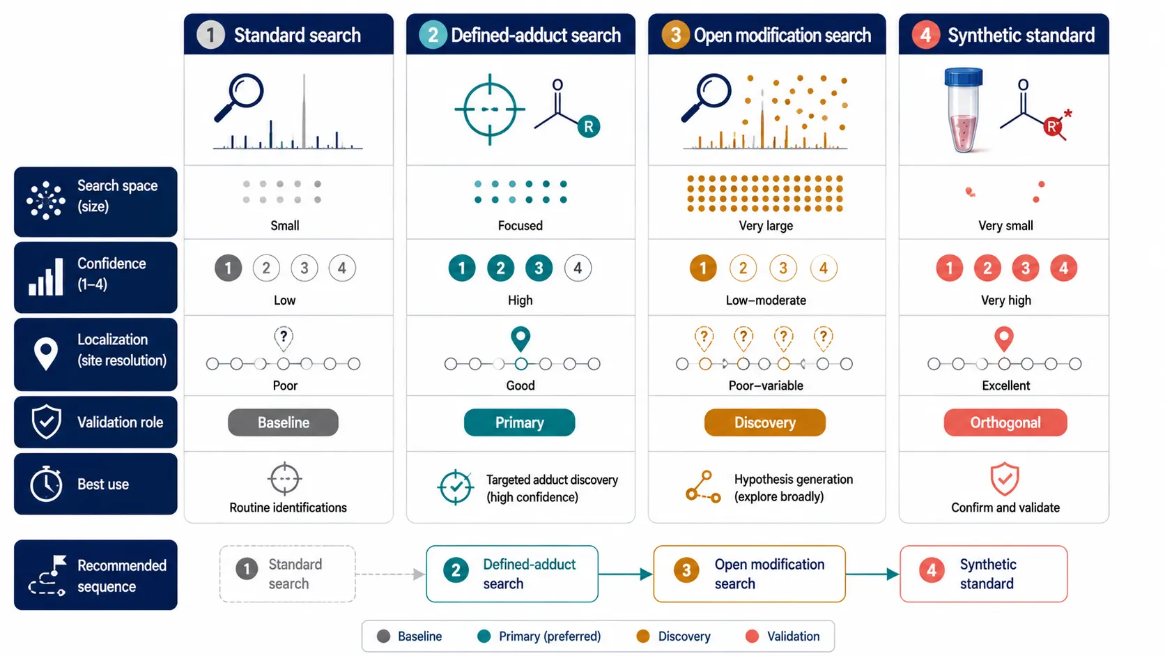
Figure 6. A staged SOP keeps the drug chemistry, mass-spectrometry evidence, and functional follow-up aligned with one defined claim.
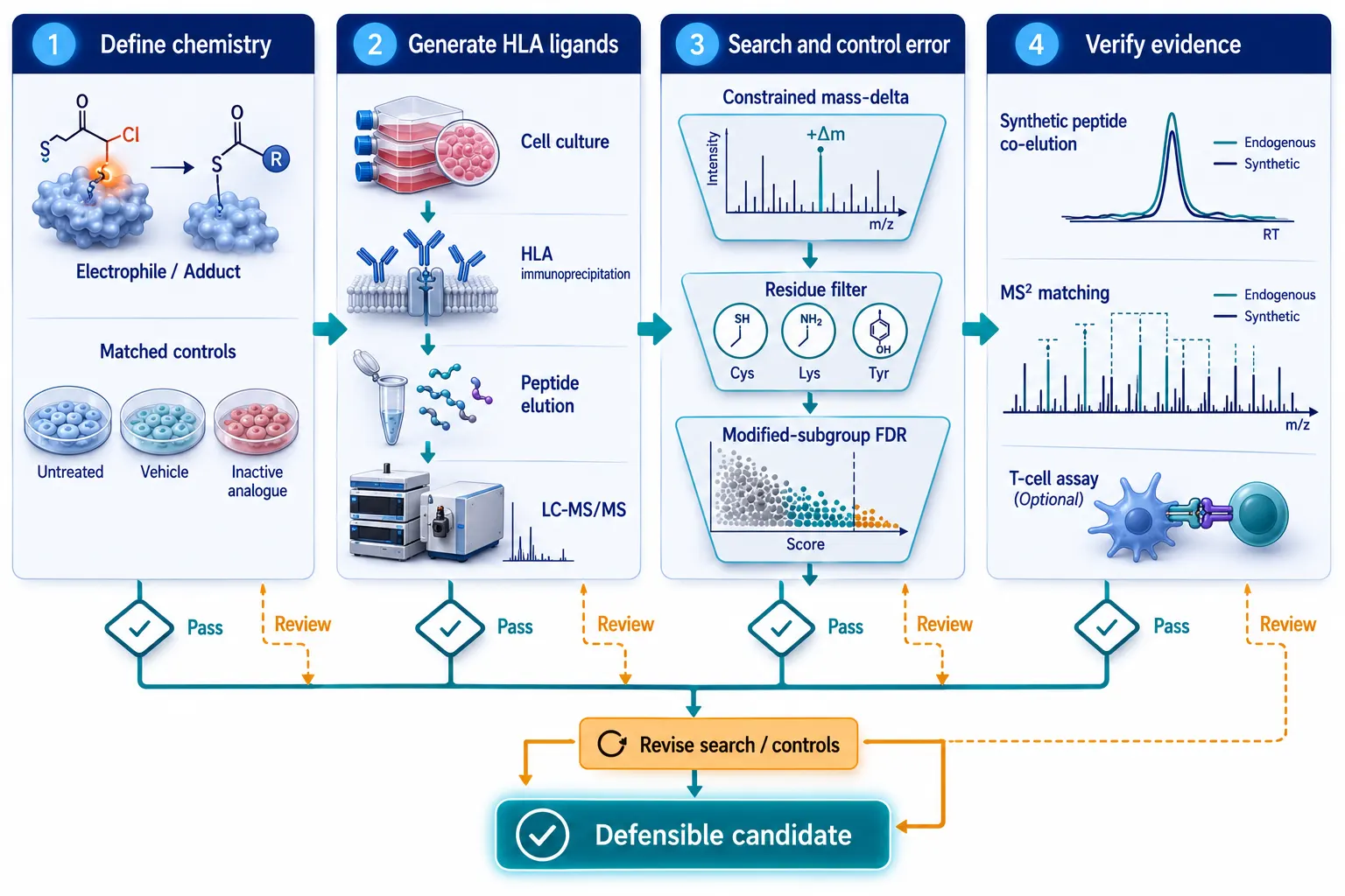
Figure 7. Defensible covalent neoepitope identification relies on convergent chemical, computational, spectral, and functional evidence.
Frequently asked questions
Can an open search replace a defined-adduct search?
No. Use a defined search as the primary test when the chemistry is known; open search is complementary discovery evidence.
Does a one-percent global FDR validate a modified ligand?
Not by itself. Review error behavior and localization within the modified subgroup, then inspect the individual spectrum.
Can a drug alter the ligandome without covalently modifying a peptide?
Yes. Altered loading or repertoire mechanisms require a different interpretation from a covalent hapten model.
Is an in-lysate drug incubation an adequate control?
It can help detect non-specific chemistry, but it does not reproduce cellular uptake, metabolism, processing, or HLA loading.
When is a synthetic modified peptide necessary?
It is the preferred orthogonal comparator when a specific covalent neoepitope is being assigned or advanced.
Can a detected ligand be called immunogenic?
No. Molecular presentation and functional T-cell recognition require separate evidence.
References:
- Alterations in the HLA-B*57:01 Immunopeptidome by Flucloxacillin and Immunogenicity of Drug-Haptenated Peptides. 2021.
- HLA-A*02:01 Presents Penicillin-Modified Cysteinylated Peptides for T Cell Recognition. 2025.
- Current Perspectives on MS-Based Immunopeptidomics: The Computational Angle to Tumor Antigen Discovery. 2023.
- caAtlas: An Immunopeptidome Atlas of Human Cancer. 2021.
- Creating MHC-Restricted Neoantigens with Covalent Inhibitors. 2023.
For research use only. This article describes preclinical research design and analytical strategy; it does not provide clinical risk prediction or treatment guidance.