In Situ Imaging of Endogenous Peptides in Intact Tissue Sections
Spatial peptidomics uses matrix-assisted laser desorption/ionization mass spectrometry imaging (MALDI-MSI) to visualize the spatial distribution of endogenous peptide-range ions directly in tissue sections without homogenization. Mass spectra are acquired across defined tissue coordinates, and selected m/z features are rendered as ion images while preserving anatomical structure. Sequence assignment is treated as a separate layer of evidence and can be supported by on-tissue MS/MS and/or orthogonal LC-MS/MS from matched regions.
Compared with homogenate peptidomics, MALDI-MSI preserves spatial information that would otherwise be lost during extraction and pooling. This enables comparison of peptide distributions across histological compartments, anatomical regions, or experimental groups while retaining the relationship between molecular signals and tissue morphology.
MALDI-based spatial proteomics commonly uses on-tissue enzymatic digestion to generate tryptic peptides as proxies for proteins. Spatial peptidomics instead focuses on endogenous peptides already present in the tissue and does not rely on routine proteolytic digestion. For studies centered on neuropeptides in brain or neuroendocrine tissue, see our dedicated spatial neuropeptidomics service.
Spatial Peptidomics vs. Related Analytical Approaches
The choice between MALDI imaging and other spatially resolved strategies depends on the biological question, the molecular class of interest, and the level of sequence confirmation required.
| Approach | Principle | Typical Output |
|---|---|---|
| MALDI-MSI spatial peptidomics | In situ imaging of endogenous peptide-range ions directly from tissue sections | Ion images of m/z features with ROI-level relative-intensity analysis; sequence assignment requires MS/MS and/or orthogonal LC-MS/MS evidence |
| Homogenate peptidomics | Peptide extraction from homogenized tissue followed by LC-MS/MS | Deep sequence identification and abundance profiling, but spatial localization is lost during homogenization |
| LCM + LC-MS/MS | Laser capture microdissection of defined regions followed by solution-phase LC-MS/MS | Region-specific peptide identification from selected areas, with lower spatial throughput than imaging |
| Immunohistochemistry / immunofluorescence | Antibody-based localization of predefined targets | Targeted localization of known molecules only, limited by antibody availability |
For deeper solution-phase identification from homogenized tissue or matched extracts, our endogenous peptidomics platform provides complementary LC-MS/MS peptidome profiling across a wide range of sample types.
Workflow for Spatial Peptidomics and MALDI-MSI
A typical MALDI-MSI workflow combines tissue preparation, matrix application, image acquisition, histology co-registration, and orthogonal identification of selected m/z features.
Study Design & Tissue QC
Define tissue regions, replicate structure, and analytical goals before sectioning
Cryosectioning & Slide Preparation
Prepare tissue sections while minimizing ex vivo proteolysis and analyte delocalization
Matrix Deposition & MALDI-MSI
Acquire peptide-range mass spectra across defined tissue coordinates
Histology Co-registration & Peptide Identification
Register ion images with histology and identify selected features by MS/MS or LC-MS/MS
ROI Analysis & Reporting
Perform segmentation, ROI comparisons, and peptide/precursor-protein annotation
1
Study Design and Tissue QC
Tissue type, regions of interest (ROIs), comparison groups, replicate design, preservation history, and the need for sequence identification are defined before sectioning. These factors determine the imaging and orthogonal identification strategy.
2
Cryosectioning and Slide Mounting
Fresh-frozen tissue is sectioned under conditions selected for the tissue type and imaging platform, then mounted on MALDI-compatible conductive slides. Sample handling is designed to minimize ex vivo proteolysis and analyte delocalization.
3
Matrix Deposition and MALDI-MSI Acquisition
Matrix chemistry, deposition conditions, acquisition platform, and pixel size are selected according to peptide mass range, tissue type, spatial resolution, and sensitivity requirements. MALDI-MSI then acquires spatially resolved spectra across the tissue section.
4
Histology Co-registration and Peptide Identification
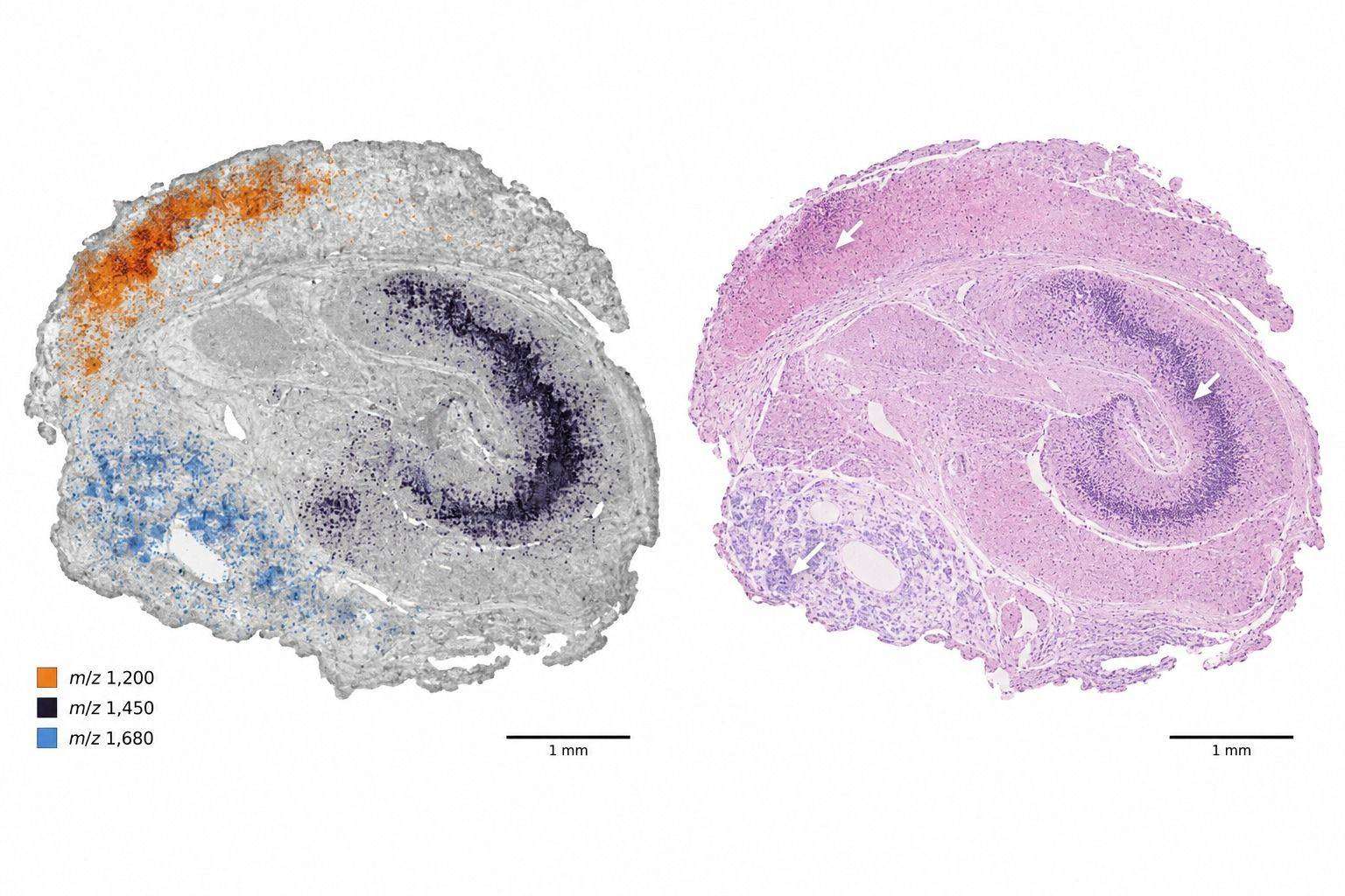
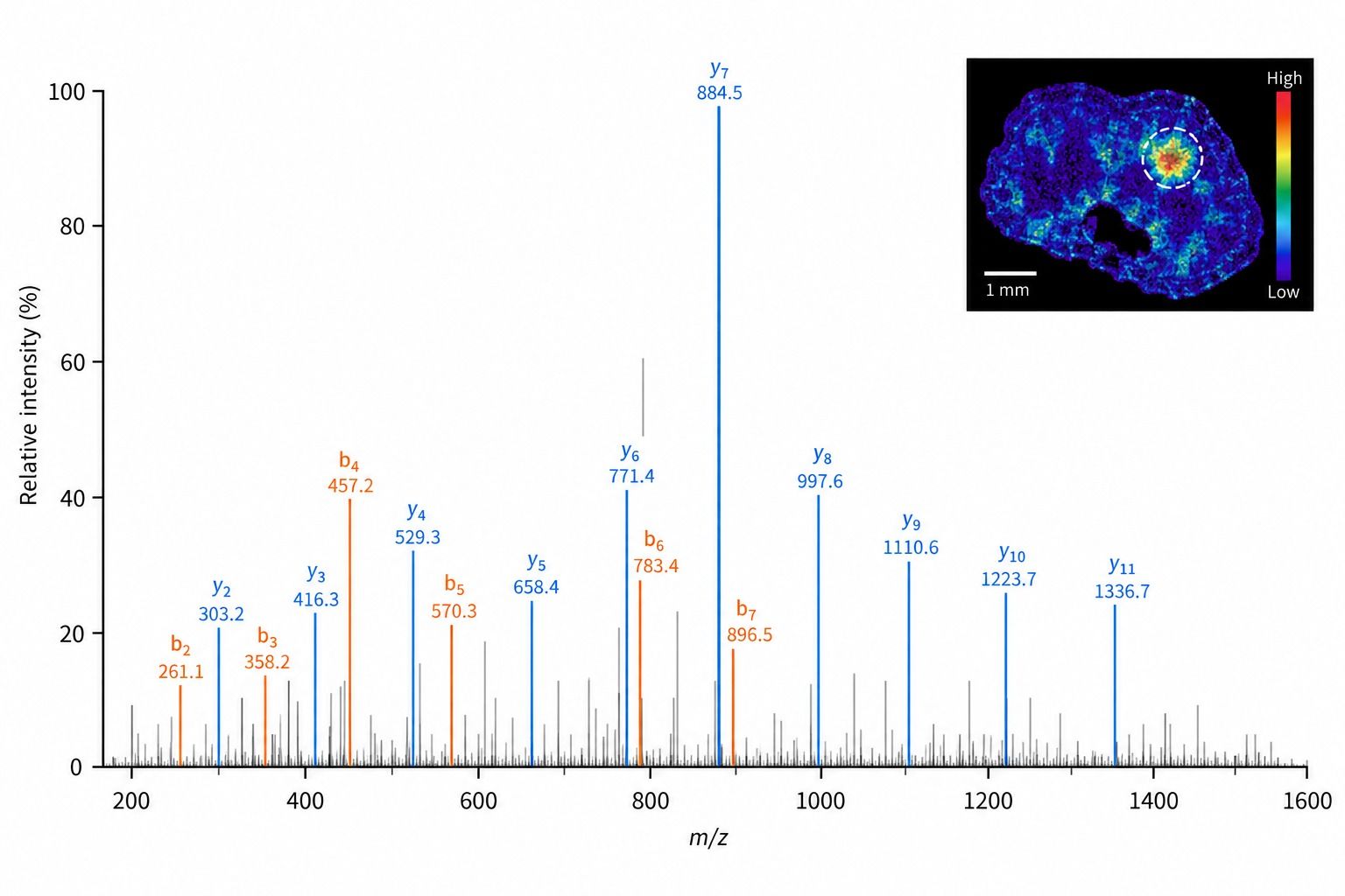
Ion images are co-registered with post-MSI or serial-section histology. Selected m/z features can be investigated by on-tissue MS/MS where feasible or supported by LC-MS/MS of laser capture microdissected (LCM) or region-specific extracts for sequence assignment.
5
ROI Analysis and Reporting
Data processing may include normalization, ion-image generation, spatial segmentation, ROI-level intensity analysis, colocalization, and precursor-protein annotation for identified peptides. Results are summarized with the level of identification evidence clearly separated from the imaging signal itself.
Spatial Peptide Imaging and Identification Strategies
The analytical strategy is selected according to whether the project prioritizes untargeted imaging, region-specific identification, histological correlation, or comparative ROI analysis.
For identified peptides, analysis can include precursor-protein annotation, PTM-aware interpretation, and integration with relevant endogenous-peptide or neuropeptide knowledge bases.
Tissue Applications for Spatial Peptidomics
Spatial peptidomics is most informative when peptide localization across anatomical or histological regions is part of the biological question. Common research applications include:
- Tumor heterogeneity and tumor-stroma interfaces, including spatial differences in endogenous peptide and proteolytic-fragment signals
- Region-specific proteolytic processing in experimental disease models or lesion-adjacent tissue
- Anatomical zonation in endocrine, reproductive, renal, hepatic, gastrointestinal, and other structurally compartmentalized tissues
- Spatial localization of endogenous peptide biomarkers, peptide hormones, and proteolytic products
- Integration with spatial transcriptomic, proteomic, or metabolomic data from serial or matched sections
Fresh-frozen tissue is generally preferred for endogenous-peptide MALDI-MSI because fixation and aqueous processing can alter peptide recovery and spatial distribution. Selected FFPE tissues may be evaluated using specialized digestion-free methods, but feasibility and identification confidence depend strongly on fixation and storage history. For hormone- and neuropeptide-focused spatial analysis of the central nervous system, our dedicated spatial neuropeptidomics workflow remains the preferred entry point.
Sample Submission and Study Design
Endogenous-peptide MALDI-MSI is highly sensitive to pre-analytical handling. Sample collection, preservation, sectioning, slide substrate, and any planned histology or LCM follow-up should therefore be defined before submission.
| Sample Type | What We Need to Know | Handling Principles |
|---|---|---|
| Fresh frozen tissue | Tissue origin, collection method, freezing history, and study question | Rapid freezing and minimal thawing are preferred to limit ex vivo proteolysis. Embedding medium, tissue orientation, storage, and transport conditions should be agreed before preparation. |
| Mounted tissue sections | Section thickness, slide type, and whether the section is intended for imaging, LCM, or both | Use MALDI-compatible conductive slides and avoid unplanned washing or fixation before the imaging workflow is defined. Section handling and transport are confirmed case by case. |
| FFPE blocks or sections | Fixation and storage history, block age, and expected tissue region | Feasibility is assessed case by case. Digestion-free endogenous-peptide imaging may be possible, but fixation history can alter peptide recovery, chemical state, and identification confidence. |
| Region of interest (LCM) | Region definition, collection buffer, and storage conditions | Collection format, buffer compatibility, storage, and transport are defined according to the downstream LC-MS/MS workflow and the amount of material available. |
Biological replication, section order, batch structure, and ROI definition should reflect the intended comparison. The acquisition and normalization strategy is then matched to the study design.
Data Analysis and Interpretation
MALDI-MSI produces a mass spectrum at each sampled tissue coordinate, so interpretation requires both spectral processing and image-level analysis.
- Mass calibration, spectral preprocessing, peak detection/alignment, and ion-intensity normalization
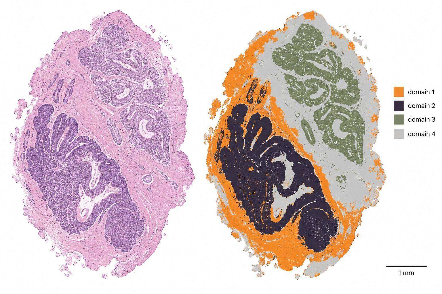
- Ion-image generation, spatial segmentation, and clustering of molecularly distinct regions
- Histology co-registration and annotation of regions of interest
- ROI-based relative-intensity comparisons and colocalization analysis of selected m/z features
- Peptide assignment using on-tissue MS/MS and/or orthogonal LC-MS/MS, with confidence reporting appropriate to the identification workflow
- Precursor-protein annotation and peptide-to-protein mapping for identified sequences
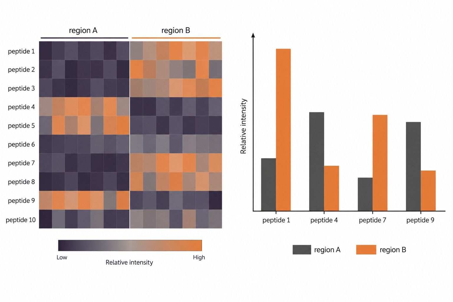
MALDI-MSI ion intensities should generally be interpreted as relative spatial signals rather than direct concentration measurements. Tissue composition, local ion suppression, matrix deposition, extraction efficiency, and the chosen normalization strategy can each influence apparent regional differences. Quantitative comparisons are therefore evaluated together with replicate structure, histological context, and analytical QC rather than treated as absolute peptide concentrations.
Interpretation is delivered in the context of your study design, and raw data can be exported in standard imaging and mass spectrometry formats.
Representative Results
The visualizations below illustrate common output formats for spatial peptidomics projects. They are representative analytical examples rather than data from a specific customer study.
MALDI-MSI Ion Images with Histology Co-registration
Unsupervised Spatial Segmentation
Orthogonal Peptide Identification from a Defined ROI
ROI-Based Relative Ion-Intensity Comparison
Typical Deliverables
Deliverables are matched to the study design and may include:
- MALDI-MSI ion images for selected m/z features, with histology co-registration where applicable
- Spatial segmentation maps and ROI-level ion-intensity summaries
- MS/MS and/or LC-MS/MS peptide assignments for selected spatial features, with the supporting identification evidence reported
- Statistical comparisons between tissue regions or experimental groups when supported by the study design
- Precursor-protein annotation for identified peptides where applicable
- Structured analytical report with methods, QC summaries, interpretation notes, and exportable data files
From MALDI m/z Features to Peptide Assignments
MALDI-MSI first produces spatially resolved m/z features. A molecular assignment should be distinguished from the ion image itself, so spatial detection, sequence identification, histology co-registration, and relative-intensity interpretation are reported as separate evidence layers.
To combine spatial information with deep solution-phase neuropeptide or peptide profiling, our neuropeptidome profiling platform can be run in parallel on serial sections or matched specimens.
References
- Chatterji B, Pich A. MALDI imaging mass spectrometry and analysis of endogenous peptides. Expert Rev Proteomics. 2013;10(4):381–388. https://doi.org/10.1586/14789450.2013.814939
- Chatterji B, Dickhut C, Mielke S, Krüger J, Just I, Glage S, Meier M, Wedekind D, Pich A. MALDI imaging mass spectrometry to investigate endogenous peptides in an animal model of Usher's disease. Proteomics. 2014;14(13–14):1674–1687. https://doi.org/10.1002/pmic.201300558
- Vu NQ, DeLaney K, Li L. Neuropeptidomics: improvements in mass spectrometry imaging analysis and recent advancements. Curr Protein Pept Sci. 2021;22(2):158–169. https://doi.org/10.2174/1389203721666201116115708
- Paine MRL, Ellis SR, Maloney D, Heeren RMA, Verhaert PDEM. Digestion-free analysis of peptides from 30-year-old formalin-fixed, paraffin-embedded tissue by mass spectrometry imaging. Anal Chem. 2018;90(15):9272–9280. https://doi.org/10.1021/acs.analchem.8b01838
- Rzagalinski I, Volmer DA. Quantification of low molecular weight compounds by MALDI imaging mass spectrometry – a tutorial review. Biochim Biophys Acta Proteins Proteom. 2017;1865(7):726–739. https://doi.org/10.1016/j.bbapap.2016.12.011
- Caprioli RM, Farmer TB, Gile J. Molecular imaging of biological samples: localization of peptides and proteins using MALDI-TOF MS. Anal Chem. 1997;69(23):4751–4760. https://doi.org/10.1021/ac970888i
- Norris JL, Caprioli RM. Analysis of tissue specimens by matrix-assisted laser desorption/ionization imaging mass spectrometry in biological and clinical research. Chem Rev. 2013;113(4):2309–2342. https://doi.org/10.1021/cr3004295
- Li MJ, Meyer LC, Meier N, Witte J, Maldacker M, Seredynska A, Schueler J, Schilling O, Föll MC. Spatial proteomics by parallel accumulation-serial fragmentation supported MALDI MS/MS imaging: a first glance into multiplexed and spatial peptide identification. Rapid Commun Mass Spectrom. 2025;39:e10006. https://doi.org/10.1002/rcm.10006
For research use only. Not for use in diagnostic or therapeutic procedures.
FAQ for Spatial Peptidomics and MALDI-MSI
What is the difference between spatial peptidomics and spatial proteomics? +
In MALDI-MSI-based spatial proteomics, proteins are commonly visualized through on-tissue enzymatic digestion and imaging of the resulting tryptic peptides. Spatial peptidomics instead images endogenous peptide-range ions already present in the tissue, including peptide hormones and proteolytic fragments, without routine digestion.
Can MALDI-MSI detect endogenous peptides directly from tissue? +
MALDI-MSI can detect endogenous peptide-range m/z features directly from matrix-coated tissue sections. The resulting ion images show spatial distribution, but assignment to a specific peptide sequence requires MS/MS and/or orthogonal LC-MS/MS evidence.
What spatial resolution can be achieved? +
Achievable spatial resolution is project dependent. Tissue type, peptide abundance, matrix deposition, acquisition platform, and the sensitivity needed for the study all influence the practical pixel size, which is defined during project design.
Can formalin-fixed paraffin-embedded (FFPE) tissue be analyzed? +
Fresh-frozen tissue is generally preferred for endogenous-peptide MALDI-MSI because fixation and aqueous processing can alter peptide recovery and spatial distribution. Selected FFPE tissues may be evaluated using specialized digestion-free methods, but feasibility and identification confidence depend on fixation and storage history.
How are MALDI-MSI m/z features assigned to peptide sequences? +
Selected m/z features can be investigated by on-tissue MS/MS where feasible or supported by LC-MS/MS of laser capture microdissected or region-specific extracts. Peptide assignment is based on the combined mass, fragmentation, and spatial evidence rather than on an ion image alone.
Is histology co-registration available? +
Yes. Consecutive or post-imaging sections are stained and the imaging data are co-registered with the stained tissue, allowing peptide patterns to be interpreted within histologically defined structures such as tumor, stromal, or normal regions.
Can MALDI-MSI peptide signals be compared between tissue regions or study groups? +
Normalized MALDI-MSI ion intensities can be compared across predefined ROIs or experimental groups when the study includes suitable replication and batch control. These are relative signal comparisons and should not be interpreted as absolute peptide concentrations.
How much tissue is needed and how should it be shipped? +
The amount of tissue and the transport route depend on the study design, sectioning plan, and sample preservation history. Please contact us before shipping so that the preparation plan, sectioning strategy, and transport conditions can be agreed for your specific samples.
What is the difference between this service and spatial neuropeptidomics? +
This service covers MALDI-MSI of endogenous peptides across diverse tissue types. The dedicated spatial neuropeptidomics service focuses on neuropeptide localization in central nervous system and neuroendocrine tissues.
Can spatial peptidomics be combined with other spatial omics data? +
Yes. Serial or matched tissue sections can be analyzed by complementary spatial transcriptomic, proteomic, or metabolomic platforms, and the resulting datasets can be co-registered for multi-omic interpretation within the same anatomical context.