Mass Spectrometry-Based Neoantigen Discovery Service
Need to prove that your predicted tumor mutations are actually presented on the cell surface? As a specialized, high-resolution application of our broader Immunopeptidomics Service, our Mass Spectrometry-Based Neoantigen Discovery platform focuses exclusively on identifying mutated, tumor-specific HLA ligands. By integrating your genomic data with ultra-sensitive LC-MS/MS technologies, we filter out the massive background of wild-type peptides to provide the direct physical evidence required to confidently advance your cancer vaccine and TCR-T cell therapy pipelines.
- Proteogenomic Integration: Seamless translation of WES/RNA-seq data to build custom variant databases for precise spectral matching.
- Single Amino Acid Resolution: Unambiguous identification of Single Amino Acid Variants (SAAVs), frameshift mutations, and fusion peptides.
- Next-Generation Sensitivity: Utilization of Orbitrap Astral™ and timsTOF Pro systems for deep, low-abundance mutation detection.
- Complex Sample Compatibility: Optimized micro-scale immunoaffinity workflows designed for low-input research tissues (e.g., ≥50 mg).
Why Use Mass Spectrometry for Neoantigen Validation?
The Biological Bottleneck of In Silico Predictions
While next-generation genomic sequencing can rapidly identify thousands of somatic mutations within a tumor, translating those mutations into viable immunological targets is an incredibly complex biological challenge. AI-driven algorithms (such as NetMHCpan) can predict which of these mutated sequences might theoretically bind to specific HLA molecules. However, these in silico models often yield false-positive rates exceeding 90%.
A mutated sequence predicted to possess high binding affinity may completely fail to be naturally processed by the cell. It might resist proteasomal cleavage, fail to be transported into the endoplasmic reticulum by the TAP complex, or be destroyed by ER-resident aminopeptidases before it ever reaches an empty MHC molecule. Mass spectrometry cuts through the algorithmic noise by bypassing these theoretical assumptions. It detects only the peptides that have successfully navigated the entire cellular antigen-processing machinery and are physically displayed on the cell surface.
Differentiating Neoantigens from the Baseline Ligandome
Detecting a mutated peptide is inherently an exercise in finding a needle in a haystack. Unlike standard HLA Peptidomics Analysis, which profiles the entire landscape of highly abundant self-peptides to understand baseline immunology, neoantigen discovery requires isolating rare mutational events. Our service employs advanced proteogenomics to tackle this. By translating your specific tumor's Whole Exome Sequencing (WES) and RNA-seq data into customized search databases, we can confidently distinguish rare, lowly abundant mutant peptides from the dominant wild-type background, ensuring that your downstream functional assays are focused only on genuine targets.
Key Application Areas in Oncology Research
Our mutation-focused mass spectrometry services are engineered to support the most advanced targeted immuno-oncology research pipelines:
Personalized Cancer Vaccines
Identifying physically presented mutated peptides (neoepitopes) is the foundational step for formulating highly specific mRNA, DNA, or synthetic long-peptide vaccines. By proving that a target is presented, researchers can significantly increase the likelihood of invoking a strong CD8+ or CD4+ T cell response.
TCR-T Cell Therapy Target Discovery
T-cell receptor engineered therapies require exquisite specificity to prevent off-target, on-tissue toxicities. Discovering high-affinity, tumor-restricted targets via direct mass spectrometry ensures that researchers can engineer T-cell receptors with minimal cross-reactivity against wild-type tissues.

Neoantigen Burden Profiling and Biomarkers
By comparing the actual presented mutational landscape between pre-treatment and post-treatment tumor research models, scientists can map how immunotherapies (such as checkpoint inhibitors) alter the antigen presentation machinery and identify potential biomarkers of resistance.
Mass Spectrometry-Based Neoantigen Identification Solutions
We offer specialized, modular solutions tailored specifically for the rigorous demands of mutation identification:
Technical Highlights: Sensitivity & Precision in MS
The immunopeptidome presents unique analytical challenges: the peptides are non-tryptic, heavily dominated by highly abundant housekeeping proteins, and lack the predictable charge states of standard proteomic digests. Our platform is engineered to overcome these hurdles.
Precision Workflow for Mutated Peptide Extraction
Isolating extremely low-abundance neoantigens from complex biological samples requires a finely tuned, multi-stage biochemical process to maximize recovery while minimizing wild-type background noise.
Cryogenic Lysis & Homogenization
Immunoaffinity Purification (IP)
Peptide Elution & Micro-Clean-up
Ultra-Sensitive LC-MS/MS Acquisition
Variant-to-Peptide Alignment
1
Cryogenic Lysis & Homogenization
Fresh frozen tissues or cell pellets are mechanically disrupted and lysed under natively buffered, detergent-rich conditions at 4°C. This critical step preserves the fragile, non-covalent interactions between the HLA molecules, the endogenous peptides, and the beta-2 microglobulin subunit.
2
Immunoaffinity Purification (IP)
High-specificity pan-HLA antibodies (such as the W6/32 clone for pan-Class I or specific clones for Class II HLA-DR/DP/DQ) are crosslinked to porous resin beads to capture the intact MHC-peptide complexes from the cellular lysate.
3
Peptide Elution & Micro-Clean-up
Following stringent washing to remove non-specifically bound proteins, the peptides are dissociated from the MHC heavy chains using mild acidic conditions. The eluted peptides are then immediately purified and desalted using customized solid-phase extraction (SPE) micro-tips optimized for short, hydrophobic sequences.
4
Ultra-Sensitive LC-MS/MS Acquisition
The purified peptide mixture is separated over a reverse-phase gradient and continuously injected into the mass spectrometer. The instrument isolates individual peptide precursor ions and fragments them to generate sequence-specific MS/MS spectra.
5
Variant-to-Peptide Alignment
Raw mass spectra are searched strictly against the custom proteogenomic database. Our bioinformatics pipeline evaluates the mass accuracy, isotopic distribution, and fragmentation patterns to validate the mutated sequences, discarding any spectra that match wild-type equivalents.
High-Performance Mass Spectrometry Platforms
To capture low-abundance mutated ligands in highly complex matrices, we deploy a fleet of industry-leading mass spectrometers. This diversity allows us to select the optimal technology based on your specific sample input and required sequencing depth.
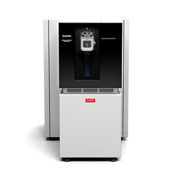 Orbitrap Astral™
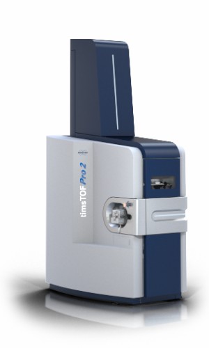 timsTOF Pro / HT
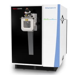 Orbitrap Exploris™ 480
Instrument Capability Overview
| Feature | Orbitrap Astral™ | timsTOF Pro / HT | Orbitrap Exploris™ 480 |
|---|---|---|---|
| Scan Speed | Up to 200 Hz | ~100 Hz (PASEF) | ~40 Hz |
| Ion Mobility Separation | No | Yes (TIMS) | No |
| Ideal Sample Input | Ultra-low input tissues | Low input / High complexity | Standard cell lines |
| Key Advantage | Maximum identification depth | Separation of isobaric peptides | Robust high-throughput |
Note: The integration of Trapped Ion Mobility Spectrometry (TIMS) in the timsTOF Pro allows for the separation of co-eluting, isobaric peptides based on their collisional cross-section, which is highly advantageous for deciphering dense immunopeptidomic samples.
Sample Requirements for Neoantigen Profiling
Proper sample preservation is absolutely vital for a successful discovery project. MHC-peptide complexes are highly susceptible to degradation by endogenous proteases if not handled correctly. Below are the recommended input amounts for optimal mass spectrometry depth.
| Sample Type | Recommended Minimum Amount | Preservation & Shipping | Notes |
|---|---|---|---|
| Fresh Frozen Tissue | 50 – 100 mg | Snap-frozen, Dry ice | Do not use fixatives; store at -80°C immediately upon resection. |
| Cultured Cell Lines | 1×10⁸ cells | Snap-frozen pellet, Dry ice | Wash thoroughly with cold PBS prior to freezing to remove serum proteins. |
| PBMCs | 5×10⁷ cells | Frozen in aliquots, Dry ice | Ensure high cell viability (>85%) prior to freezing. |
Please note: Formalin-Fixed Paraffin-Embedded (FFPE) tissues are generally incompatible with HLA ligandomics, as the crosslinking process irreversibly damages the native MHC-peptide complexes necessary for immunoaffinity purification.
Demo Results: High-Confidence Spectral Evidence
Our data packages provide highly transparent, publication-ready visualizations that confirm the mass spectrometry validity of the identified neoantigens. We do not just provide lists of sequences; we provide the spectral proof.
Peptide Length Distribution
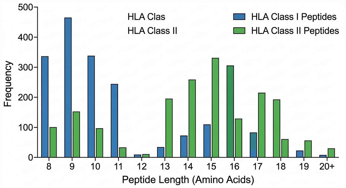
Data Interpretation: Ensures that the physical properties of the eluted molecules match the biological constraints of the HLA binding groove, confirming successful immunoprecipitation without proteolytic degradation.
HLA Binding Motif (Sequence Logo)
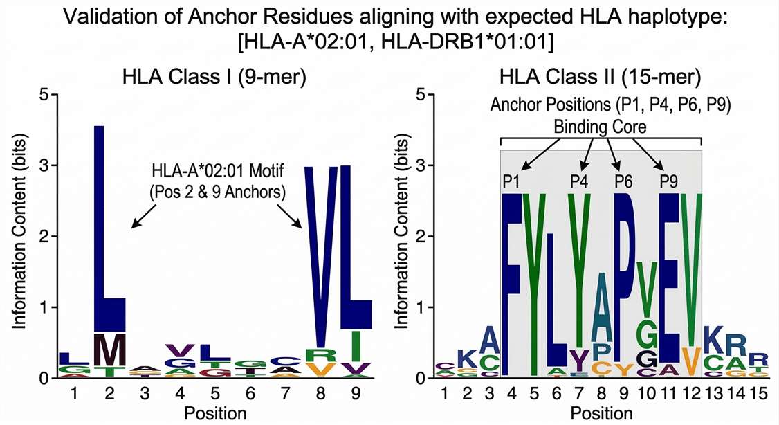
Data Interpretation: By analyzing the frequency of amino acids at specific positions (e.g., position 2 and position 9 for Class I), we confirm that the identified mutated peptides possess the correct biochemical anchors for the sample's known HLA alleles.
Annotated MS/MS Spectra
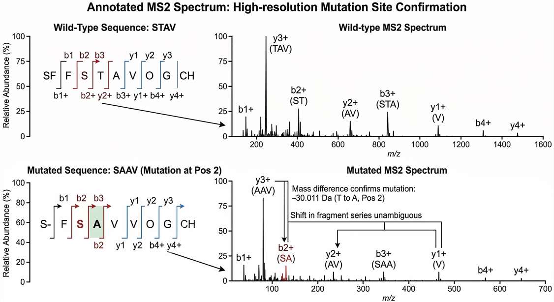
Data Interpretation: The ultimate proof of a mutation. The continuous series of b-ions and y-ions unambiguously pinpoints the mass shift caused by the somatic mutation, distinguishing it from the wild-type counterpart.
Prediction vs. MS Validation Overlap
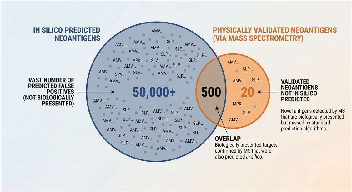
Data Interpretation: Clearly demonstrates the filtering power of mass spectrometry, highlighting the vast number of predicted false positives that were not biologically presented.
Strategic Selection: Choosing the Right Service Level
Depending on your current pipeline stage, we offer tailored mass spectrometry approaches. Once novel candidates are successfully identified and physically validated via mass spec, we strongly recommend evaluating their functional and structural engagement through our downstream TCR Binding Kinetics Analysis.
| Service Level | Objective | Methodology | Best Suited For |
|---|---|---|---|
| Discovery Profiling | Unbiased identification of mutated peptides from an unknown pool. | DDA/DIA LC-MS/MS searched against custom proteogenomic variant databases. | Early-stage research, identifying entirely novel targets from patient cohorts. |
| Targeted Validation | Confirming the physical presentation of AI-predicted candidates. | PRM / SureQuant™ targeted mass spectrometry utilizing heavy-isotope labeled internal standards. | Pre-clinical target validation, verifying specific epitopes with extreme sensitivity. |
Can you extract mutated HLA peptides from small research biopsies? +
Yes. Through highly optimized immunoaffinity purification protocols and the utilization of ultra-sensitive mass spectrometers like the Orbitrap Astral™, we can extract meaningful mass spectrometry data from input amounts as low as 50 mg of fresh frozen tissue. However, it is important to note that because mutant peptides often represent only a tiny fraction of the total cellular ligandome, providing higher input amounts (e.g., >100 mg) generally yields significantly deeper mutation coverage and higher confidence identifications.
How do you filter out wild-type peptides from the mass spec results? +
We utilize a rigorous proteogenomic approach. By building a custom FASTA database directly derived from your sample's Whole Exome Sequencing (WES) or RNA-seq data, we can strictly match the acquired MS/MS mass spectra against known somatic mutations present in that specific sample. Following the spectral matching process, our bioinformatics pipeline filters the results to remove any peptide sequences that share a 100% identity match with the wild-type human reference proteome, leaving only the mutant candidates.
Do I need to provide genomic data (WES/RNA-seq) for this service? +
For the true "discovery" of novel neoantigens via untargeted Data-Dependent Acquisition (DDA), providing sample-specific genomic data is highly recommended, as it is required to build the customized variant database. However, if your bioinformatics team has already established a predicted list of candidates, genomic data is not required. In that scenario, we can synthesize the predicted peptides as heavy-isotope internal standards and perform highly targeted PRM validation directly against your pre-defined list.
Case Study: Uncovering Intra-Patient HLA Peptide Diversity Using Tumor Organoids
Journal: Nature Communications
Published: Volume 11, Issue 1, 2020
Summary | Who needs this
This immunopeptidomics workflow is highly relevant for oncology research teams, immuno-oncology biotechs, and translational scientists developing multi-peptide cancer vaccines or TCR-T cell therapies. It is particularly valuable for researchers targeting low tumor-mutational burden (TMB) indications—such as microsatellite-stable (MSS) colorectal cancer—where identifying targetable, conserved tumor-specific antigens across heterogeneous tumor populations remains a major analytical bottleneck.
Methods
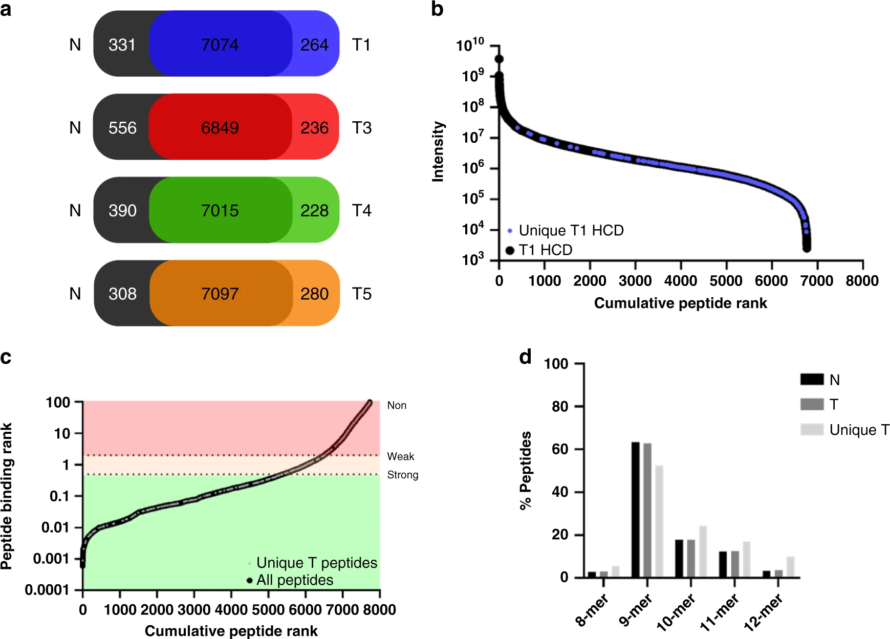
To investigate intra-patient clonal heterogeneity, researchers amplified single-cell patient material from an MSS colorectal cancer (CRC) patient into clonal tumor organoids. Normal tissue organoids from the same patient were cultured as a baseline control. The research team employed a coupled approach of organoid global proteomics and HLA class I peptide ligandomics.
Following lysis, HLA-A, -B, and -C complexes were isolated via immunoaffinity purification. The eluted naturally presented peptides were analyzed using high-resolution nanoLC-MS/MS on Orbitrap mass spectrometers (Q Exactive / Fusion Lumos). Raw mass spectrometry data underwent peptide identification and label-free quantification using MaxQuant against the human proteome database.
Results obtained
The deep immunopeptidomic profiling demonstrated that while single-cell derived organoids share core characteristics, there is profound intra-patient heterogeneity. The mass spectrometry analysis quantified a broad 15–25% inter-clone variability in HLA class I peptide presentation.
Crucially, the data provided direct physical evidence that tumor-specific ligands derived from highly conserved DNA damage control and tumor suppressor proteins were prominently and consistently presented by the tumor clones, which coincided with the silencing of their cytoprotective functions. These objective findings suggest that, rather than relying solely on highly variable mutational neoantigens, a multi-peptide vaccination strategy targeting universally presented tumor suppressor peptides may offer a robust alternative for invoking targeted anti-tumor responses in patients with low mutational burdens.
Reference
- Demmers, L. C., et al. "Uncovering Intra-Patient HLA Peptide Diversity Using Tumor Organoids." Nature Communications, Volume 11, Issue 1, 2020. https://doi.org/10.1038/s41467-020-19142-9