Why Conotoxin Characterization Is Critical for Drug Discovery
Cone snails (Conus genus) produce 50–200 distinct conopeptides per species — each stabilized by multiple disulfide bonds and extensively post-translationally modified. These conotoxins selectively target human ion channels, receptors, and transporters at nanomolar or picomolar potency. Ziconotide (Prialt, synthetic ω-MVIIA from Conus magus) is approved for severe chronic pain; multiple Nav1.7, Cav2.2, and nAChR-targeting candidates are in active development for pain, epilepsy, and cardiovascular disease.
Yet conotoxin structural characterization remains technically demanding. Each superfamily adopts a distinct cysteine framework — and the arrangement of disulfide bonds defines the three-dimensional fold and biological activity. Standard peptide sequencing workflows routinely fail to resolve disulfide connectivity or miss critical PTMs such as hydroxyproline, γ-carboxylation, C-terminal amidation, and bromination — leading to incorrect structural assignments that compromise downstream structure–activity relationship studies.
What We Offer — Conotoxin Structural Characterization Capabilities
Our conotoxin characterization service integrates multiple analytical modalities into a pipeline designed for the structural complexity of cone snail venom peptides. Every workflow is conotoxin-aware — informed by curated conotoxin databases and the recognized patterns of conopeptide signal sequences, cysteine frameworks, and post-translational modifications.
Detectable Conotoxin Families and Superfamily Classification
Conotoxin classification operates at three levels: gene superfamily (signal sequence), cysteine framework (number and arrangement of cysteine residues), and pharmacological family (molecular target). Our platform assigns characterized peptides to all three levels.
| Conotoxin Superfamily | Cysteine Framework | Typical Targets | Common PTMs |
|---|---|---|---|
| A (α, ρ, sConotoxin families) | Framework I (C–CC) — knotting motif | nAChR subtypes | Hydroxyproline, C-terminal amidation |
| I1, I2, I3 | Framework III (C–C–C–C) | nAChR, calcium channels | — |
| M (μ, m, ψ families) | Framework I (C–CC) | Nav channels | C-terminal amidation |
| O1, O2, O3 (δ, κ, γ families) | Framework III or VI/VII (knottin fold) | Nav, Kv, calcium channels | Hydroxyproline, γ-carboxylation |
| P (κM-RIIIK family) | Framework IV | Nav1.7, Kv channels | — |
| S (σ family) | Framework V | 5-HT receptors | — |
| T (τ, conomarphin families) | Framework V (4-Cys) | Neurotensin receptor, opioid receptors | Free C-terminal cysteine, amidation |
| Conomarphin | Non-cysteine (linear) | Mu opioid receptor | — |
| Framework VI/VII | Knottin fold (6–8 Cys) | Multiple | Bromination, sulfation |
- Key pharmaceutical targets: Nav1.7 and Nav1.8 (pain), N-type calcium channels Cav2.2 (chronic pain), α9α10 nAChR (neuropathic pain), NET and SERT transporters (antidepressants), NMDA receptors (analgesia).
- Custom superfamily panels for specific Conus species can be configured upon request.
Conotoxin Characterization Platform — Instrumentation and Analytical Methods
| Platform | Instrument | Primary Application | Key Capability |
|---|---|---|---|
| Primary screening | MALDI-TOF/TOF (AB Sciex 4800 or equivalent) | Rapid conotoxin screening; disulfide connectivity mapping | 8 keV CID produces clean b- and y-type ion series for de novo sequencing |
| Deep coverage | NanoLC-ESI-MS/MS (nano-flow LC + high-res ESI) | Complex conotoxin mixtures; PTM detection | Sub-nanomole sensitivity; eliminates isobaric interference in conotoxin-rich fractions |
| Disulfide mapping | Rapid partial reduction-alkylation (TCEP + IAA) | Native disulfide connectivity determination | Resolves ambiguous connectivities including non-canonical patterns (e.g., C1–C4/C2–C3) |
| Database search | ConoServer + SwissProt + open-search (custom pipeline) | Sequence ID; hypervariable loop resolution | >8,500 conopeptide sequences across 29 superfamilies (ConoServer, 2024) |
| Capability | Specification |
|---|---|
| Database | ConoServer (>8,500 sequences, 29 superfamilies) + SwissProt + open-search |
| Disulfide connectivity method | Rapid partial reduction-alkylation (TCEP pH 3, 2.5 min → IAA pH 8.0) + MS/MS |
| PTM panel breadth | 8 validated PTM types with site-localized MS/MS evidence; unmodified reference run in parallel |
| Minimum sample input | ≥10 µg purified conotoxin; ≥500 µg crude venom (≥100 µg for venom gland extract) |
| Reporting format | FASTA sequence file, disulfide connectivity diagram, PTM annotation, superfamily classification, raw mzML/RAW |
| Mass accuracy | <50 ppm (MALDI-TOF/TOF); <10 ppm (nanoLC-ESI-MS/MS) |
| Throughput | Sample receipt to preliminary report: 10–15 business days for standard projects |
Conotoxin Identification Workflow — From Venom to Confirmed Structure
Sample Receipt and Quality Control
Protein quantification and MALDI-TOF screening
Non-Reducing Enzymatic Digestion
Sequencing-grade protease, cysteine residues unmodified
LC-MS/MS Data Acquisition
MALDI-TOF/TOF and nanoLC-ESI-MS/MS dual-platform
Database Search and De Novo Sequencing
ConoServer + open-search + manual annotation
Disulfide Mapping, PTM Annotation and Reporting
Rapid partial reduction-alkylation + structured report
1
Sample Receipt and Quality Control
Venom or conotoxin sample is assessed for quantity, purity, and integrity upon arrival. Total protein concentration is measured; a preliminary MALDI-TOF screening determines the mass range and complexity of conopeptides present.
2
Non-Reducing Enzymatic Digestion
Sequencing-grade trypsin or chymotrypsin digests the sample under non-reducing conditions, with cysteine residues left unmodified. Parallel reducing digests (with DTT or TCEP) are prepared simultaneously for comparative analysis.
3
LC-MS/MS Data Acquisition
MALDI-TOF/TOF handles rapid screening and disulfide mapping; nanoLC-ESI-MS/MS delivers deep coverage of complex fractions. Data acquired in data-dependent acquisition (DDA) mode with dynamic exclusion to maximize peptide identification.
4
Database Search and De Novo Sequencing
Raw MS/MS data are searched against ConoServer conopeptide sequences, SwissProt entries, and species-specific transcriptomic data. Open-search algorithms identify peptides not in any database; these are then subjected to de novo sequencing.
5
Disulfide Connectivity Mapping, PTM Annotation, and Reporting
For each multi-cysteine conotoxin, the rapid partial reduction-alkylation protocol is applied to determine native disulfide pairings. PTM-modified peptides are identified from accurate mass shifts and verified by MS/MS fragmentation. All results are compiled into a structured report.
Sample Requirements and Sensitivity
| Sample Type | Minimum | Preferred | Required Purity | Storage | Notes |
|---|---|---|---|---|---|
| Crude venom (whole) | 500 µg total protein | 1–5 mg | — | −80°C, on dry ice | Lyophilized preferred; ship on dry ice |
| Venom gland extract | 100 µg total protein | 500 µg | — | −80°C | Homogenized in 8M urea or PBS buffer |
| Purified conotoxin | 10 µg | 50–100 µg | ≥85% (HPLC) | −20°C, dried | Provide known mass if available |
| Synthetic conotoxin analog | 5 µg | 20 µg | ≥90% (HPLC) | Room temperature | Specify sequence if available |
| Conopeptide mixture | 20 µg total | 100 µg | — | −80°C | Desalt if salt content is elevated |
- For non-model Conus species, submit at least 500 µg of crude venom or 100 µg of venom gland extract to ensure sufficient coverage of low-abundance novel conotoxins.
- Contact our team for species-specific recommendations before sample submission.
Representative Results — Conotoxin Structural Elucidation
The following representative results illustrate the analytical output for a typical novel T1-conotoxin characterization project — from de novo sequencing through disulfide connectivity resolution.
De Novo Sequence Coverage from MALDI-TOF/TOF MS/MS
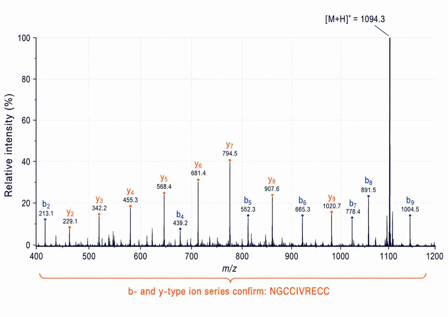
Annotated MALDI-TOF/TOF MS/MS spectrum (8 keV CID) of a 10-residue T1-conotoxin from Conus bandanus. Continuous b- and y-type ion series (b₂ through b₉; y₂ through y₉) confirm the complete sequence NGCCIVRECC. Mass accuracy: <50 ppm with external calibration.
Disulfide Connectivity Mapping by Rapid Partial Reduction-Alkylation
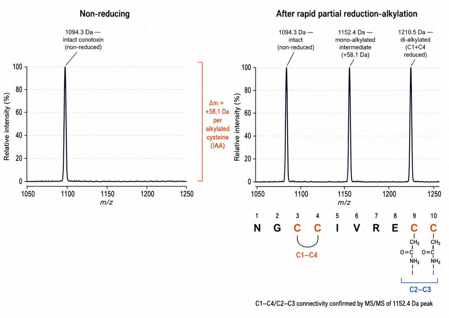
Non-reducing (top) and reducing (bottom) MALDI-TOF MS comparison following rapid partial reduction-alkylation. Three diagnostic peaks: intact (1,094.3 Da), mono-alkylated intermediate (+58.1 Da), and di-alkylated product (+116.2 Da). MS/MS of the mono-alkylated intermediate identifies C2–C3 as the reduced pair, confirming C1–C4/C2–C3 connectivity.
PTM Detection — Hydroxyproline and C-Terminal Amidation
Extracted ion chromatogram (XIC) from nanoLC-ESI-MS/MS comparing modified and unmodified fractions. Unmodified: [M+H]⁺ m/z 1,078.3. Hydroxyproline-modified: m/z 1,110.3 (+32.0 Da). y₁ ion shift of 17.027 Da confirms C-terminal amidation.
Conotoxin Superfamily Classification Output
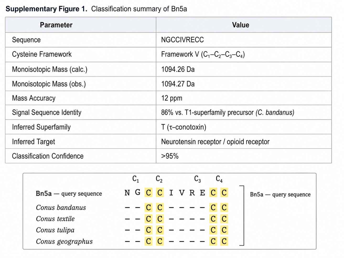
Classification for a novel conotoxin from Conus striolatus: Framework V (C₁–C₂–C₃–C₄), 86% signal sequence identity to T1-superfamily precursor, pharmacological inference consistent with neurotensin receptor or opioid receptor targeting. Mass accuracy: 12 ppm. Classification confidence: >95%.
Conotoxin Drug Discovery and Marine Toxin Research Applications
- Non-opioid pain therapeutics — Conotoxin-derived candidates targeting Nav1.7, N-type calcium channels (Cav2.2), and nAChRs offer alternatives to opioid analgesics for severe chronic pain
- Neurological and psychiatric disorders — Conotoxin scaffolds targeting nAChR subtypes (α9α10), NMDA receptors, and neurotransmitter transporters (NET, SERT) are in development for epilepsy, neuropathic pain, and depression
- Cardiovascular drug discovery — κ-conotoxin targeting Kv channels and other conotoxin families with cardiovascular targets
- Marine natural product research — Venom peptidomics and structural elucidation of novel conotoxins from unexplored Conus species
- Venom peptide lead optimization — Structural characterization of disulfide connectivity and PTM state as the foundation for SAR studies and analog design
- Academic venom biology — De novo conotoxin sequencing and superfamily classification from venom gland extracts or partially characterized venom samples
Deliverables
- Conotoxin sequence file — annotated FASTA format, including post-translational modification positions
- Disulfide connectivity report — diagram showing native Cys–Cys pairings for each multi-cysteine conotoxin, with MS evidence summary
- PTM annotation report — list of all detected modifications with site localization confidence scores
- Superfamily classification summary — assignment of each conotoxin to its gene superfamily, cysteine framework, and inferred pharmacological family
- Raw mass spectrometry data — mzML or RAW format files for all LC-MS/MS runs, suitable for deposit as supplementary data or re-analysis
- Structural confidence score — per-conotoxin rating (high / medium / low confidence) based on sequence coverage, mass accuracy, and PTM localization
Why Choose Our Conotoxin Characterization Platform
*All services are for research use only (RUO). Not for diagnostic or clinical use.*
How much venom do you need for conotoxin characterization? +
Minimum requirements depend on sample type. For crude venom, we recommend at least 500 µg of total protein. For a venom gland extract, 100 µg total protein is sufficient. If you have already purified a specific conotoxin, as little as 10 µg is adequate for complete structural characterization including disulfide mapping and PTM analysis. Contact our team for species-specific guidance before submission, as venom complexity varies significantly between Conus species.
Can you characterize conotoxins from non-model Conus species? +
Yes. Our workflow is specifically designed for species whose venom has not been previously characterized. We combine de novo sequencing — which does not rely on prior sequence knowledge — with conotoxin-aware open database searching. Hypervariable loop regions are handled through manual annotation by scientists with conotoxin-specific expertise, rather than by fully automated pipelines that discard sequences not present in existing databases.
How do you determine disulfide bond connectivity in conotoxins? +
We use a rapid partial reduction-alkylation strategy. The conotoxin is briefly treated with TCEP under mildly acidic conditions (pH 3, 2.5 minutes) to partially reduce the disulfide bonds, then immediately alkylated with iodoacetamide at pH 8.0. The mass difference between non-reduced, mono-alkylated, and di-alkylated peptides is measured by MS/MS, and the positions of the alkylated residues are identified from b- and y-ion mass shifts. This resolves ambiguous connectivities that cannot be determined from a simple non-reducing versus reducing mass comparison alone.
What post-translational modifications can you detect? +
Our validated PTM detection panel for conotoxins includes: hydroxyproline (+15.995 Da), γ-carboxylation of glutamate to Gla (+43.990 Da per residue), C-terminal amidation (17.027 Da mass difference from free acid), tryptophan bromination (+77.911 Da for dibromo-Trp), tyrosine sulfation (+79.956 Da), phosphorylation (+79.966 Da), N-terminal pyroglutamation (+17.027 Da), and methionine oxidation (+15.995 Da). For each PTM detected, we provide site-localized MS/MS evidence. Unmodified reference peptides are run in parallel with each batch to eliminate false-positive assignments.
Is the data suitable for peer-reviewed publication? +
Yes. We provide raw mass spectrometry data files (mzML or RAW format) for all LC-MS/MS runs, annotated spectra with sequence assignments and PTM evidence, disulfide connectivity diagrams, and superfamily classification reports. These deliverables have supported publications in peer-reviewed journals including those focused on venom biology, marine natural products, and drug discovery.
How do I submit a venom or conotoxin sample? +
Contact us via the inquiry form or by email to initiate a project consultation. Our team will provide a sample submission kit that includes shipping instructions, sample documentation forms, and chain-of-custody requirements for international shipments. All venom samples should be shipped on dry ice or liquid nitrogen dry shippers. We will confirm sample receipt, assess initial quality, and begin the characterization workflow within five business days of receipt.
Case Study — Novel T1-Conotoxin from Conus bandanus with Non-Canonical Disulfide Connectivity
Journal: Journal of Venomous Animals and Toxins Including Tropical Diseases
Authors: Nguyen Bao, Jean-Pière Lecaer, Ngo Dang Nghia, Phan Thi Khanh Vinh
Published: 2020
Why This Case Matters for Drug Discovery
T1-conotoxins are a pharmacologically significant subfamily — typically 9–13 residues with a four-cysteine framework (C–C–C–C), targeting neurotensin receptors, opioid receptors, and sodium channels. They are prime scaffolds for non-opioid pain therapeutics and neurological disease programs. But a critical challenge in conotoxin-based drug discovery is this: the primary amino acid sequence does not uniquely determine the biological activity. The disulfide connectivity — which cysteine pairs form bonds — defines the three-dimensional fold and pharmacophore orientation. Getting it wrong has downstream consequences for every SAR study and analog design decision.
This case illustrates what happens when a conotoxin defies the canonical pattern, and how experimental disulfide mapping — rather than inference from sequence alone — changes the picture entirely.
The Challenge
Standard conotoxin characterization pipelines face two systematic failure modes when encountering novel T1-conotoxins:
- Sequence misassignment in hypervariable loop regions. Generic peptide ID algorithms discard sequences that don't match existing database entries. In novel T1-conotoxins, the 4-residue hypervariable loop (positions 4–7 in a 10-residue peptide) is precisely the region that determines target selectivity — and precisely the region most likely to be misread or dropped.
- Incorrect disulfide connectivity inference. Most automated pipelines assume the canonical C1–C3/C2–C4 pattern for four-cysteine conotoxins, because that is overwhelmingly the most common. But conotoxin superfamilies can harbor non-canonical connectivities that change the fold entirely. No automated disulfide predictor can reliably detect this.
In this study, previous characterization attempts on related species had either failed to resolve disulfide connectivity, or had assumed the canonical pattern — leading to structural models that were subsequently revised.
Analytical Approach
Venom ducts from Conus bandanus (a molluscivorous species found in Vietnamese waters) were dissected and extracted with 0.1% TFA. The extract was fractionated by RP-HPLC on a C18 column. MALDI-TOF/TOF MS (AB Sciex 4800) was used to screen fractions for conotoxin candidates based on mass range. Fractions containing candidate peptides were selected for MS/MS at 8 keV CID. Edman degradation was run in parallel to confirm the N-terminal sequence independently.
For disulfide connectivity, the rapid partial reduction-alkylation method was applied: TCEP treatment at pH 3.0 for 2.5 minutes (partial reduction), followed immediately by IAA alkylation at pH 8.0. This generates a diagnostic mixture of non-reduced, mono-alkylated, and di-alkylated species. MS/MS of each species identifies which cysteine residues carry the alkyl tag — and therefore which were reduced — revealing the native connectivity.
Key Findings
A novel T1-conotoxin, designated Bn5a, was isolated:
| Property | Value |
|---|---|
| Sequence | NGCCIVRECC (10 amino acids) |
| Monoisotopic mass | 1,094.26 Da |
| Cysteine framework | Framework V: C₁–C₂–x(4)–C₃–C₄ |
| Disulfide connectivity | C1–C4 / C2–C3 (non-canonical for T1-superfamily) |
| C-terminal status | Free cysteine — not amidated |
| Signal sequence identity | 87% match to T1-superfamily precursor (ConoServer) |
| Inferred pharmacological target | Norepinephrine transporter (NET) — based on homology to chi-MrIA |
The most critical finding: despite clearly belonging to the T1-superfamily (87% signal sequence identity), Bn5a exhibits C1–C4/C2–C3 connectivity — the same pattern seen in T2-conotoxins and the U-superfamily toxin MrIA. This is structurally distinct from the canonical T1 pattern (C1–C3/C2–C4). Edman degradation confirmed all 10 residues including isoleucine at position 5, which had been misassigned as leucine in homologs from other species. The C-terminal residue was confirmed as a free cysteine rather than an amide — a finding with implications for synthetic analog design, since amidated and non-amidated forms can differ in protease stability and receptor affinity.
What This Means for Your Conotoxin Drug Discovery Program
- Sequence alone is not enough. The same 10-residue sequence with a different disulfide connectivity folds into a completely different 3D structure. Any SAR study based solely on sequence — without experimental connectivity confirmation — risks drawing wrong conclusions.
- Non-canonical connectivities are not rare exceptions. Within the T1-superfamily alone, multiple non-canonical patterns have been documented across species. When characterizing a novel conotoxin from a non-model Conus species, assuming the canonical pattern is a material risk for drug discovery programs.
- The C-terminal status matters for synthesis. Free C-terminal cysteine versus amidation changes the synthetic route and affects downstream activity. Bn5a's free C-terminus was only confirmed by MS/MS of the unmodified peptide — it would have been missed by any workflow that only analyzed the amidated form.
- Signal sequence identity does not guarantee canonical connectivity. Bn5a's 87% signal sequence match to T1-superfamily precursors did not predict its non-canonical connectivity. Only the experimental rapid partial reduction-alkylation protocol revealed it.
Conclusion
Accurate determination of disulfide connectivity is not an optional add-on in conotoxin-based drug discovery — it is a prerequisite. The C1–C4/C2–C3 connectivity in Bn5a would have been misassigned by any automated pipeline relying on the canonical T1-conotoxin pattern, and any downstream analog design or SAR study built on that incorrect structure would have been compromised from the start. This case underscores why experimental disulfide mapping, combined with expert manual annotation, is essential before proceeding to any conotoxin lead optimization program.
Read the full paper (DOI: 10.1590/1678-9199-JVATITD-2019-0095)